
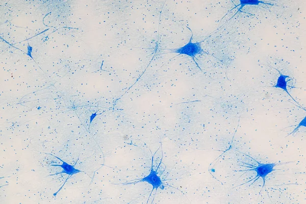
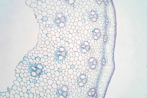
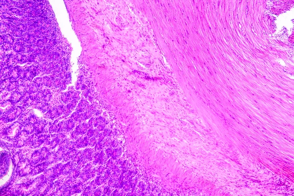
Stock image Histological

The Study Plant Tissue Of Under The Microscope For Classroom Education.
Image, 17.11MB, 5760 × 3840 jpg
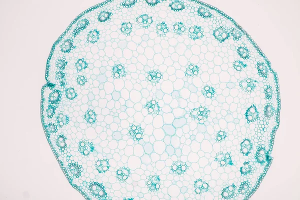
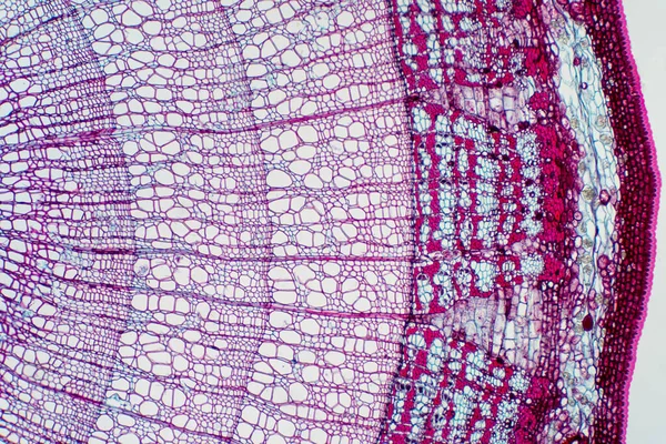
The Study Plant Tissue Of Under The Microscope For Classroom Education.
Image, 20.8MB, 6720 × 4480 jpg
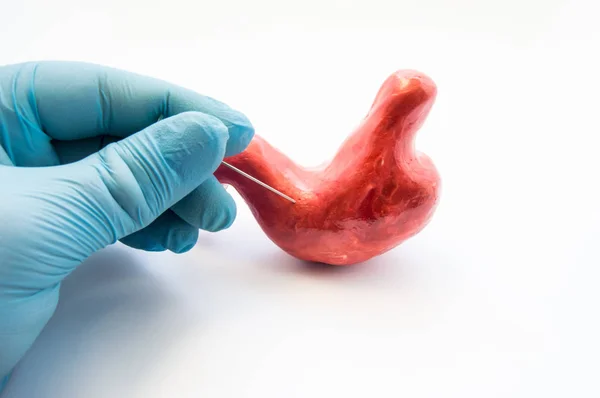
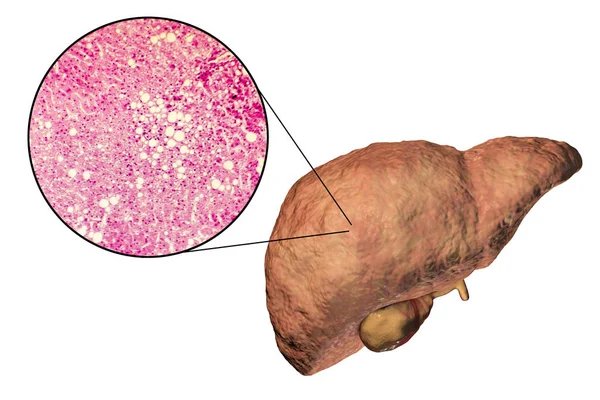
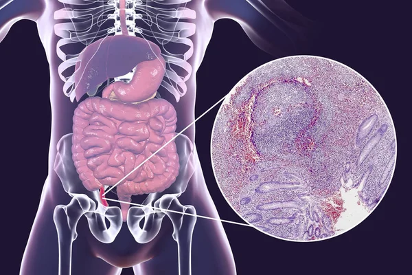
Concept Of Stomach Puncture Or Gastrointestinal Perforation. Hand Of Surgeon Pierces Wall Of Model Of Human Stomach For Therapeutic Purposes Or For Biopsy Tissue Analysis By Histology Or Cytology
Image, 5.08MB, 6016 × 4000 jpg

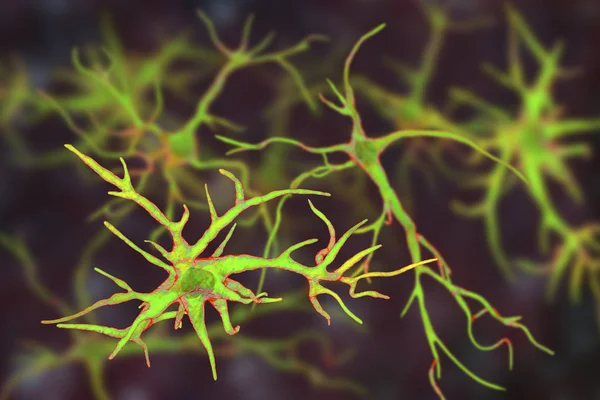
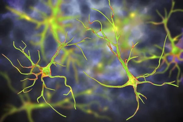
Spinal Cord, Cross-section. 3D Illustration Which Shows The White And The Grey Matter With Dorsal And Ventral Horns
Image, 3.85MB, 4500 × 3000 jpg


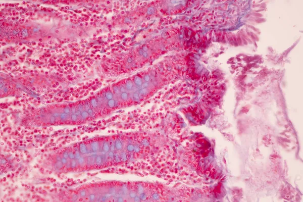
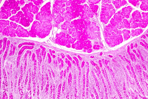
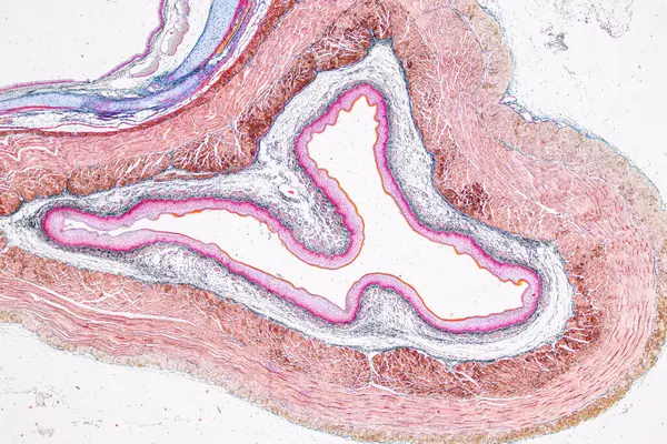
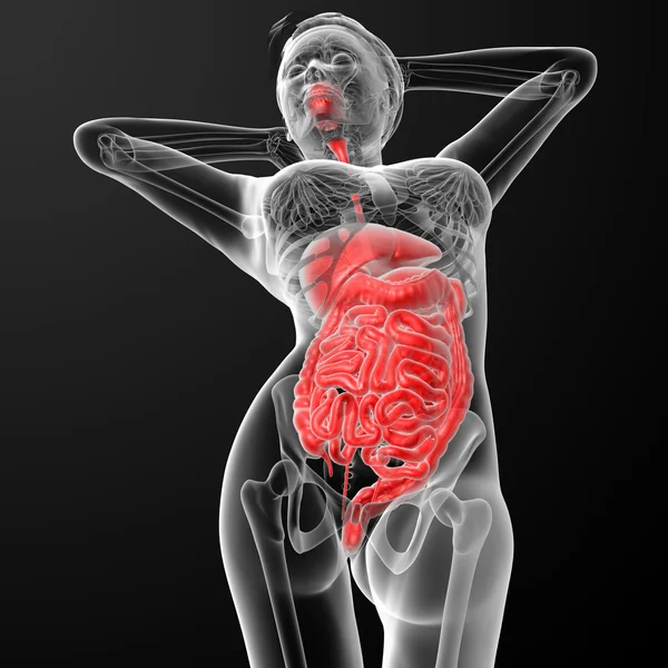
Tissue Of Small Intestine (Duodenum), Large Intestine Human And Stomach Human Under The Microscope In Lab.
Image, 17.29MB, 8192 × 5461 jpg


Echinococcal Cyst Under The Microscope (400x). Echinococcus Granulosus. Dog Tapeworm Parasite.
Image, 6.99MB, 2592 × 1944 jpg

Mammalogist Doctor Examines A Woman Breasts And Lymph Nodes During Appointment. Skillful Oncologist Puncture Of Mammary Glands Of Young Patient Under Review Ultrasound For Diagnosis Of Breast Cancer.
Image, 2.11MB, 3030 × 2020 jpg
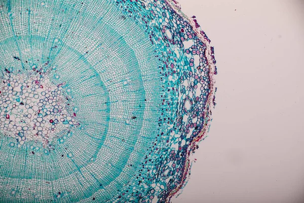
Cross Sections Of Plant Stem Under Microscope View For Education Plant Physiology.
Image, 13.32MB, 6000 × 4000 jpg
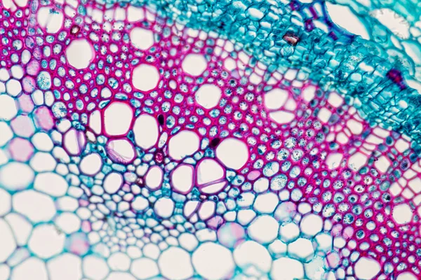
Cross Section - Xylem Is A Type Of Tissue In Vascular Plants That Transports Water And Some Nutrients. Scientific Research. Plant Tissue Structure.
Image, 17.52MB, 5845 × 3897 jpg

Light Microscopic Of Human Ovary Showing Primary And Secondary Follicles. Human Physiology Education.
Image, 26.67MB, 6000 × 4000 jpg
Tissue Of Small Intestine (Duodenum) And Vermiform Appendix Human Under The Microscope In Lab.
Image, 22.69MB, 6000 × 4000 jpg

Tissue Of Small Intestine (Duodenum) And Vermiform Appendix Human Under The Microscope In Lab.
Image, 20.31MB, 6000 × 4000 jpg

Osteon, Highlighting Osteocytes, Haversian Canal, And The Lacunocanalicular Network In Bone Tissue Structure Diagram Hand Drawn Schematic Raster Illustration. Medical Science Educational Illustration
Image, 5.56MB, 6000 × 6000 jpg

SMA Disease, Doctor Holding Wooden Blocks With Word SMA Genetic. Spinal Muscular Atrophy. Health Concept
Image, 13.03MB, 6720 × 4480 jpg

Pathology And Histology Tissue Of Mouse, Rabbit, Cat And Cow Under Microscope.
Image, 32.75MB, 6000 × 4000 jpg

Pathology And Histology Tissue Of Mouse, Rabbit, Cat And Cow Under Microscope.
Image, 6.85MB, 2667 × 4000 jpg
Pathology And Histology Tissue Of Mouse, Rabbit, Cat And Cow Under Microscope.
Image, 18.37MB, 6000 × 4000 jpg

Pathology And Histology Tissue Of Mouse, Rabbit, Cat And Cow Under Microscope.
Image, 8.03MB, 6000 × 3245 jpg

Slices Of The Tumor Under Glass. Histological Examination Of Tumor Cells For The Presence Of Cancer
Image, 0.79MB, 1650 × 2475 jpg
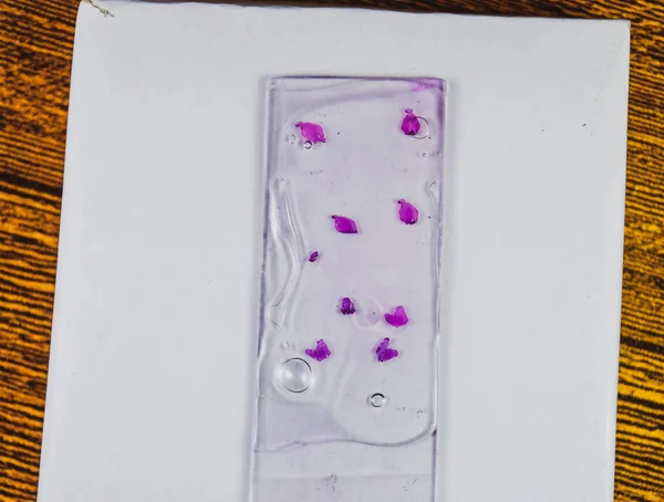
Slices Of The Tumor Under Glass. Histological Examination Of Tumor Cells For The Presence Of Cancer
Image, 1.41MB, 2475 × 1873 jpg

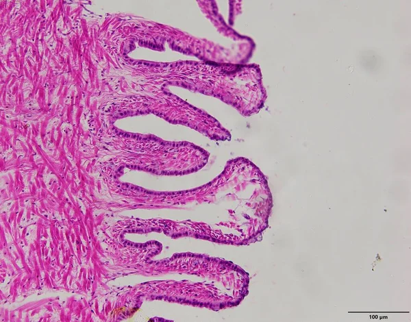
Characteristics Of Columnar Epithellum Cell (Cell Structure) Of Human Under Microscope View For Education In Laboratory.
Image, 16.38MB, 6720 × 4480 jpg

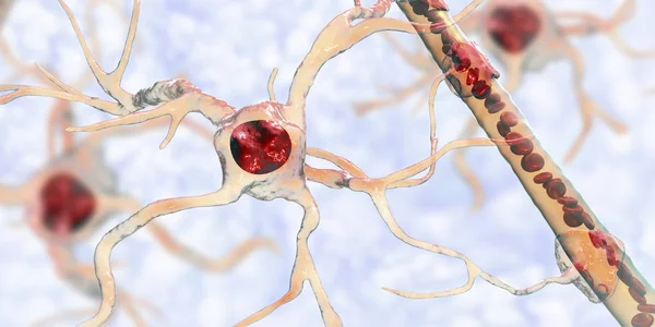
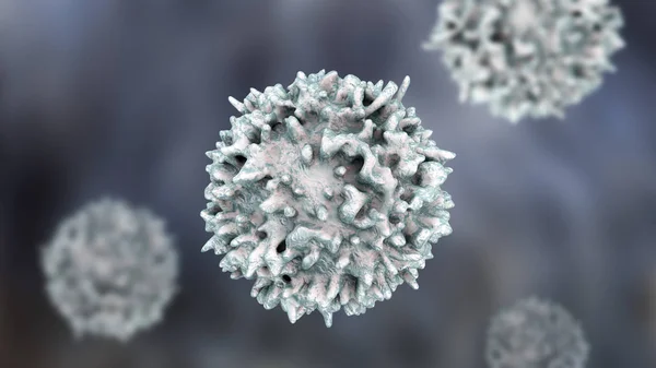
Bone Marrow, Stem Cells Of Human Bone Marrow Stem Cells. These Cells Are Known As Multipotential Stem Cells Because They Form The Precursors To Every Type Of Blood Cell. During Blood Cell Development The Multipotential Stem Cell Develops By A Process
Image, 0.75MB, 4000 × 3000 jpg
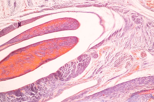
Education Anatomy And Histological Sample Of Human Under The Microscope.
Image, 21.93MB, 6720 × 4480 jpg
The Study Plant Tissue Of Under The Microscope For Classroom Education.
Image, 15.03MB, 6000 × 4000 jpg
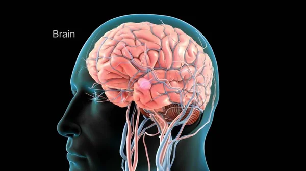
Digital Medical Illustration: Human Brain With Blood Vessels. Anatomically Correct, Isolated On Black. 3D Rendering
Image, 0.72MB, 3840 × 2160 jpg
Anatomy And Histological Bone, Elastic Cartilage Human And Joint Of Human Foetus Under The Microscope For Education.
Image, 8.38MB, 4254 × 2836 jpg
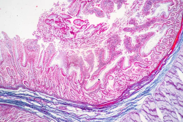
Tissue Of Small Intestine (Duodenum) And Vermiform Appendix Human Under The Microscope In Lab.
Image, 21.54MB, 6000 × 4000 jpg
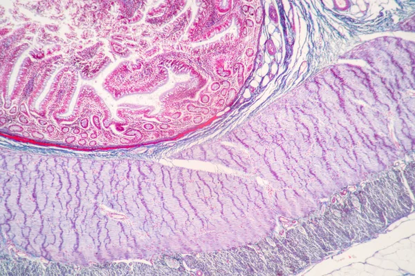
Tissue Of Small Intestine (Duodenum) And Vermiform Appendix Human Under The Microscope In Lab.
Image, 18.99MB, 6000 × 4000 jpg
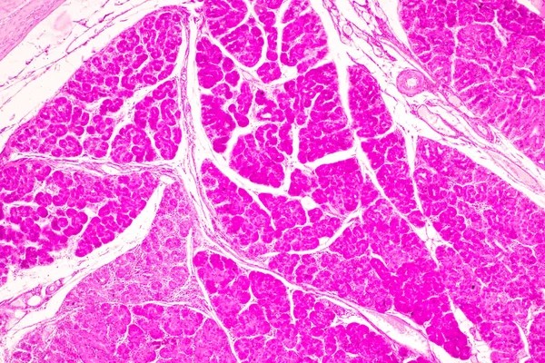
Backgrounds Of Characteristics Tissue Of Stomach Human, Small Intestine Human, Pancreas Human And Large Intestine Human Under The Microscope In Lab.
Image, 20.16MB, 6720 × 4480 jpg
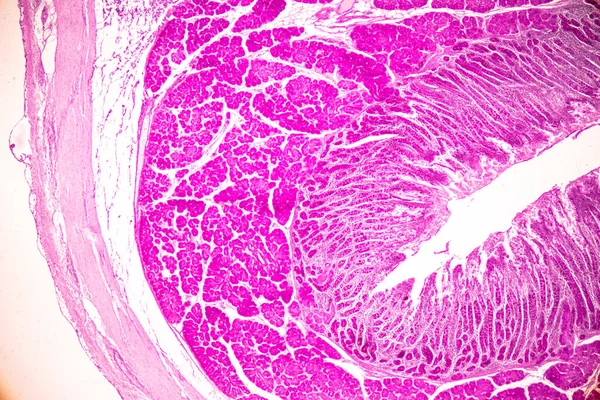
Backgrounds Of Characteristics Tissue Of Stomach Human, Small Intestine Human, Pancreas Human And Large Intestine Human Under The Microscope In Lab.
Image, 19.31MB, 6720 × 4480 jpg
Laboratory Interior Out Of Focus, Template For A Poster, Webpage Or Leaflet
Image, 2.28MB, 4146 × 2774 jpg

Cross Section Through Cells Of A Stem From A Maize Plant Under The Microscope
Image, 1.45MB, 4500 × 3000 jpg
Page 1 >> Next