
Stock image Immunohistochemistry
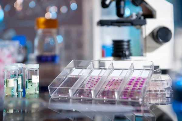
Blurred Background For Histopathology-related Presentation. Stained Histological Tissue Samples On Glass, Fixed Tissue, Automatic Pipette And Other Work-related Tools. Toned Image, Text Space.
Image, 2.1MB, 4550 × 3034 jpg
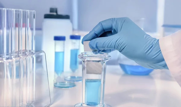
Scientific Or Medical Background. Cancer Diagnosis: Histopathology, Cytology And Analysis Of Tumour Markers. Histochemical Staining Of Tissue Samples. Gloved Hands Hold A Slide Glass With Tissue Samples For Histological Staining.
Image, 3.61MB, 4603 × 2737 jpg
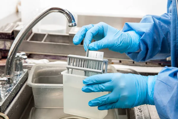
Scientist Preparing Slides With Paraffin Embedded Tissue Samples For Immunohistochemistry Assay In The Laboratory.
Image, 7.39MB, 5616 × 3744 jpg
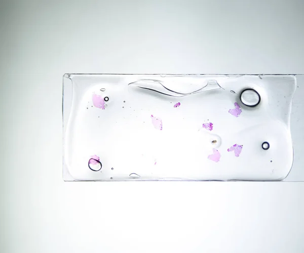
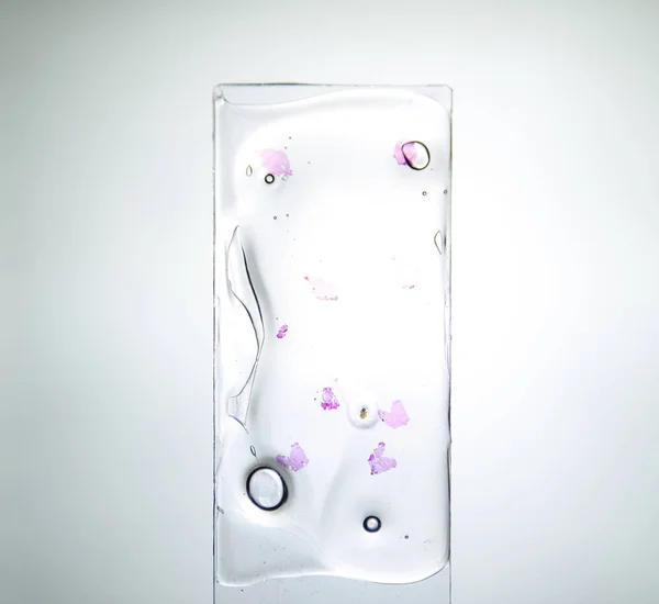
Slices Of The Tumor Under Glass. Histological Examination Of Tumor Cells For The Presence Of Cancer
Image, 1.3MB, 2800 × 2340 jpg
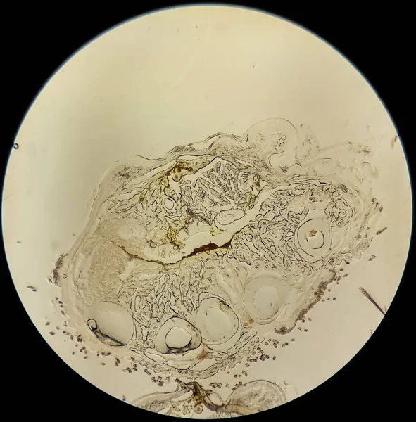
Histological Cross-section For Immunohistochemistry Of The Cellular Tissue Of The Leg Of A Mouse Seen Under An Optical Microscope In The Form Of Microphotography For Scientific Studies And Preclinical Experimentation With Animals
Image, 2.81MB, 2706 × 2744 jpg
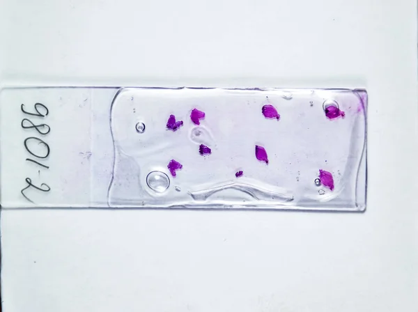
Slices Of The Tumor Under Glass. Histological Examination Of Tumor Cells For The Presence Of Cancer
Image, 3.54MB, 4000 × 2989 jpg
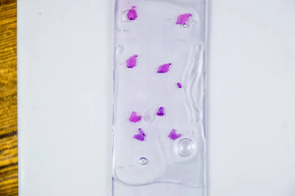
Slices Of The Tumor Under Glass. Histological Examination Of Tumor Cells For The Presence Of Cancer
Image, 2.7MB, 4000 × 2667 jpg

Slices Of The Tumor Under Glass. Histological Examination Of Tumor Cells For The Presence Of Cancer
Image, 2.65MB, 4000 × 3484 jpg
Slices Of The Tumor Under Glass. Histological Examination Of Tumor Cells For The Presence Of Cancer
Image, 2.63MB, 4000 × 3666 jpg
Slices Of The Tumor Under Glass. Histological Examination Of Tumor Cells For The Presence Of Cancer
Image, 2.73MB, 4000 × 2667 jpg

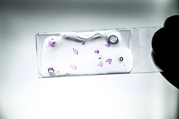
Slices Of The Tumor Under Glass. Histological Examination Of Tumor Cells For The Presence Of Cancer. Samples Of Tumor Cells Under The Sleek Against The Background Of The Lamp.
Image, 0MB, 4000 × 2989 jpg
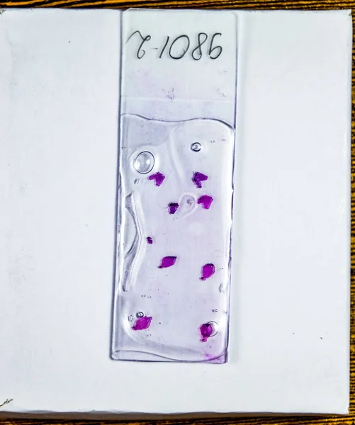
Slices Of The Tumor Under Glass. Histological Examination Of Tumor Cells For The Presence Of Cancer
Image, 2.4MB, 2340 × 2800 jpg

Paraffin Blocks With Cancer Samples, Samples For Histological Examination Of The Tumor. Pieces Of A Tumor For Deep Research Of Cancer Cells.
Image, 2.47MB, 3000 × 2925 jpg

Brain Mapping As Neuroscience Spatial Representations Tiny Person Concept
Vector, 0.49MB, 6000 × 3140 eps

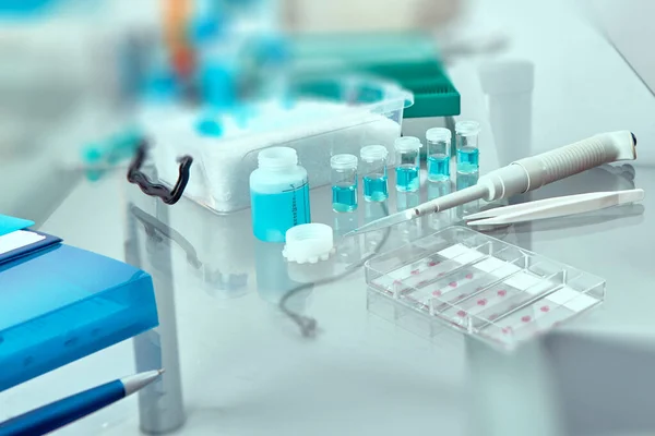
Cancer Diagnosis: Histopathology, Cytology And Analysis Of Tumour Markers. Histochemical Staining Of Tissue Samples. Automatic Pipette, Dye And Slide Glasses In A Coplin Jar.
Image, 3.78MB, 4256 × 2841 jpg
Page 1 >> Next