
Stock image Immunological Response

Coronavirus Under The Microscope Attacked By Microorganisms Evoking An Immunological Response. Vaccine Against The Covid-19 Coronavirus Virus
Image, 9.6MB, 3840 × 3904 jpg
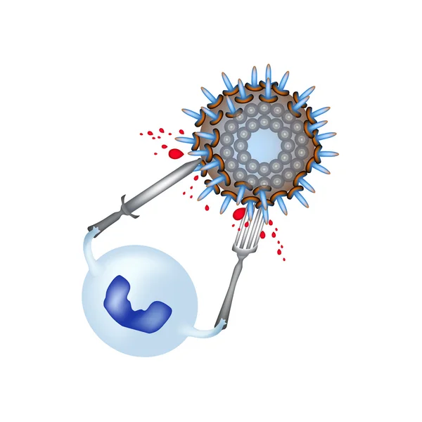
Phagocytosis. Leukocyte Absorbs The Virus. Infographics. Leukocyte Eats Bacteria. Vector Illustrations In Cartoon Style On Isolated Background
Vector, 6.91MB, 5000 × 5000 eps


Phagocytosis. Leukocyte Absorbs The Virus. Infographics. Leukocyte Eats Bacteria. Vector Illustrations In Cartoon Style On Isolated Background
Vector, 6.91MB, 5000 × 5000 eps
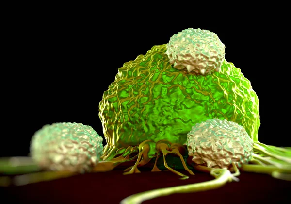
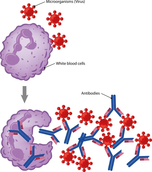
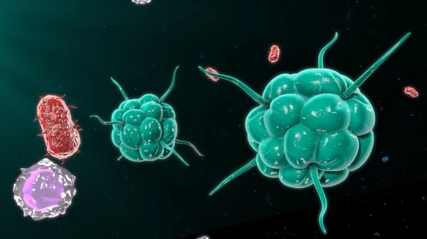

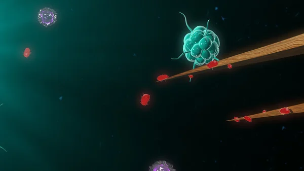

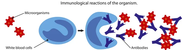


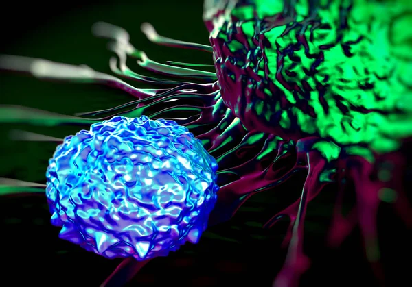

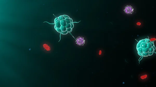

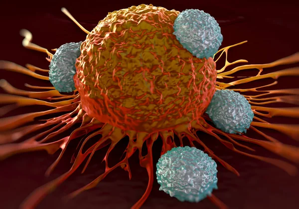
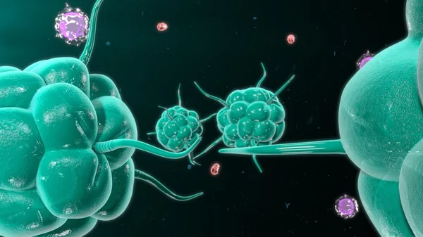
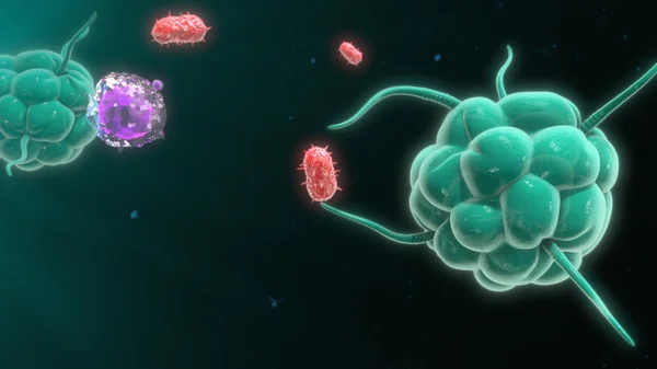


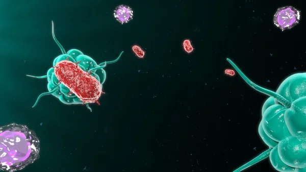
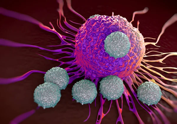
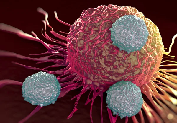


Illustration Showing A Type Of White Blood Cell Known As A Macrophage (purple) Releasing Tattoo Ink (black) Which It Had Engulfed. During Tattooing, Ink Is Injected Into The Second Skin Layer, Known As The Dermis, And Triggers An Immune Response.
Image, 5.64MB, 7200 × 4050 jpg

Illustration Of Natural Killer T (NKT) Cells (grey) Attacking Cancer Cells (orange). NKT Cells Are Lymphocytes (white Blood Cells) That Act As Part Of The Body's Innate (non-specific) Immune Response.
Image, 10.63MB, 5340 × 3354 jpg

Illustration Showing The Immune Response To Tattoo Ink (black). During Tattooing, Ink Is Injected Into The Second Skin Layer, Known As The Dermis, And Triggers An Immune Response.
Image, 5.37MB, 7200 × 4050 jpg


Illustration Showing The Immune Response To Tattoo Ink (black). During Tattooing, Ink Is Injected Into The Second Skin Layer, Known As The Dermis, And Triggers An Immune Response. Certain Types Of White Blood Cell Such As Dendritic Cells (blue) And T
Image, 8.16MB, 7200 × 4050 jpg

Illustration Showing Bacteria (green) Entering The Body Through A Breach In The Skin's Outer Layer (epidermis, Pink) Caused By A Needle (top Centre) Wound. Dendritic Cells (blue), A Type Of White Blood Cell, Are Responding To The Infection.
Image, 7.19MB, 7200 × 4050 jpg


Illustration Showing The Immune Response To Tattoo Ink (black). During Tattooing, Ink Is Injected Into The Second Skin Layer, Known As The Dermis, And Triggers An Immune Response.
Image, 5.49MB, 7200 × 4050 jpg

Illustration Of Macrophages (purple) Approaching Bacteria (green). Macrophages Are White Blood Cells Which Mainly Function To Engulf And Destroy Pathogens, Dead Cells, Cancerous Cells, Cellular Debris And Other Substances.
Image, 7.54MB, 7200 × 4050 jpg


Illustration Showing A Type Of White Blood Cell Known As A Macrophage (purple) Containing Tattoo Ink (black). During Tattooing, Ink Is Injected Into The Second Skin Layer, Known As The Dermis, And Triggers An Immune Response.
Image, 6.29MB, 7200 × 4050 jpg


Illustration Of Three Types Of White Blood Cell Beginning An Immune Response: A Macrophage (purple), Dendritic Cell (blue) And T Helper Cells (red).
Image, 5.87MB, 7200 × 4050 jpg

Illustration Of Three Types Of White Blood Cell Responding To Infection By Bacteria (green): Macrophages (purple), Dendritic Cells (blue) And T Helper Cells (red).
Image, 7.18MB, 7200 × 4050 jpg

Illustration Showing The Immune Response To Tattoo Ink (black). During Tattooing, Ink Is Injected Into The Second Skin Layer, Known As The Dermis, And Triggers An Immune Response.
Image, 6.41MB, 7200 × 4050 jpg

Lab Technician Or Medical Scientist Wearing Protective Suit,face Maks And Goggles, Holding COVID-19 Patient Blood Sample Container,serological Testing Procedure,detection Of Antibodies And Immunity
Image, 8.48MB, 3744 × 5616 jpg


Illustration Showing Enzymes (small Pink Spheres) And Tattoo Ink (black) Within A Vacuole (fluid-filled Sac) Inside A Type Of White Blood Cell Known As A Macrophage (purple).
Image, 6.43MB, 7200 × 4050 jpg

Page 1 >> Next