
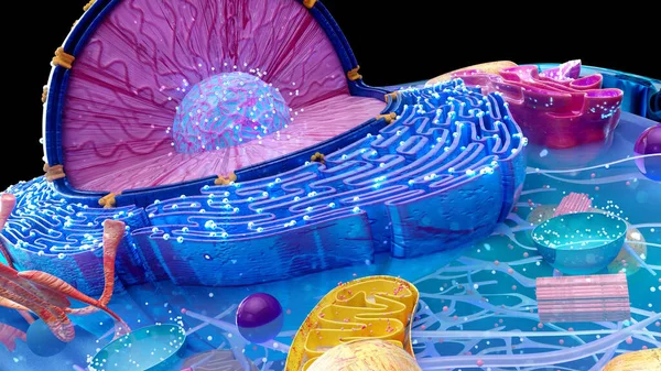
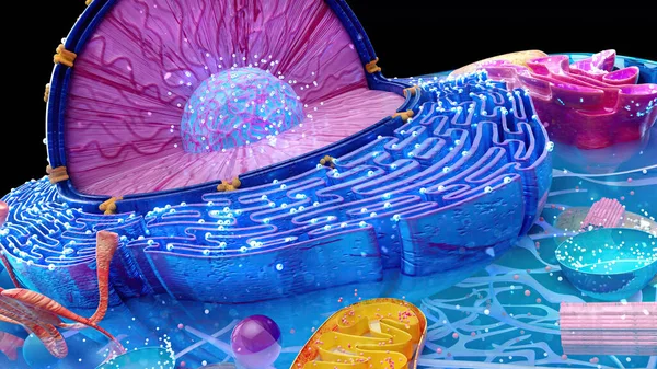
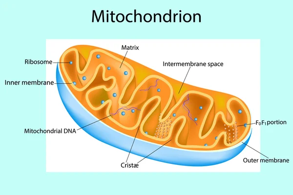
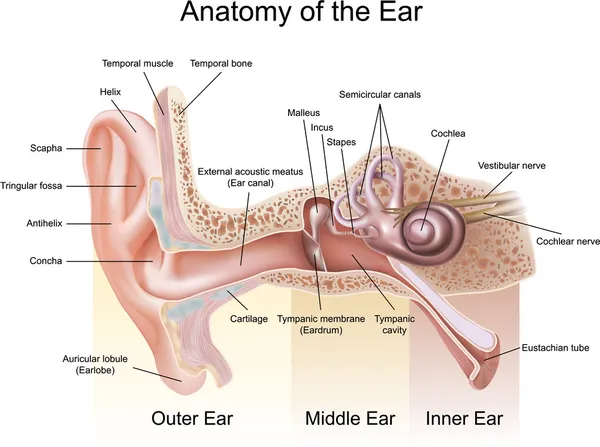
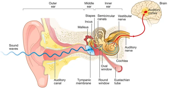
Stock image Inner Membrane
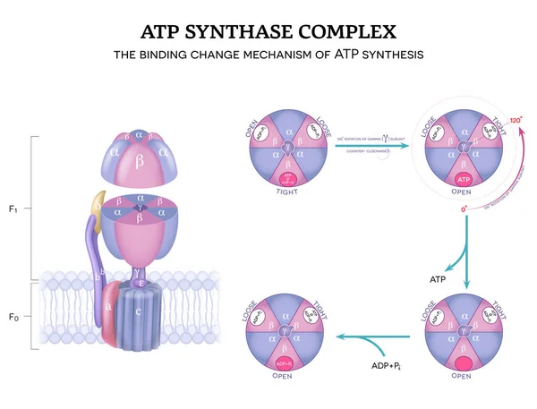
ATP Synthase Complex Structure And Mechanism Of ATP Synthase. The Binding Change Mechanism. 120-degree Rotation Of Gamma Subunit Counter-clockwise.
Vector, 9.35MB, 5512 × 4202 eps
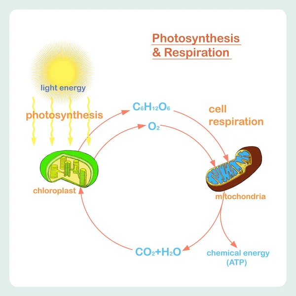
Scheme Of Photosynthesis And Respiration, Hand Drawn Biology Stock Vector Illustration
Vector, 4.41MB, 5000 × 5000 eps
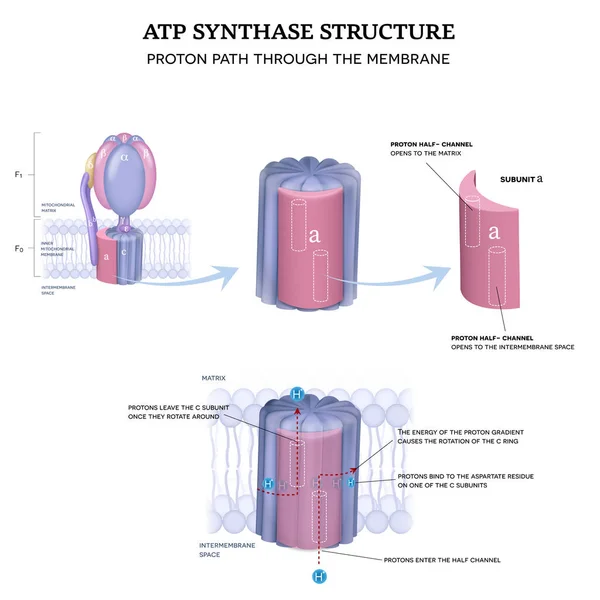
ATP Synthase Structure. Proton Path Through The Inner Mitochondrial Membrane.
Vector, 12.62MB, 5000 × 5000 eps


Mechanism Of ATP Synthase. The Binding Change Mechanism For ATP Synthesis. 120-degree Rotation Of Gamma () Subunit Counter-clockwise.
Vector, 5.41MB, 7000 × 2840 eps


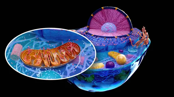

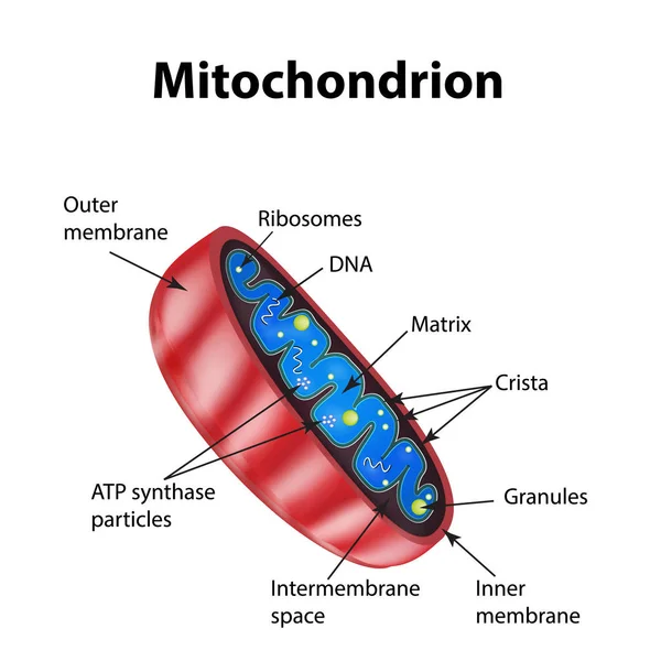
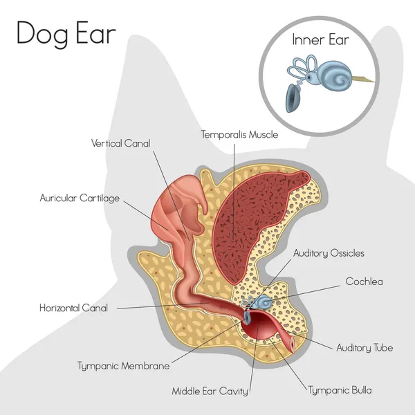
Mitochondrion. Components Of A Typical Mitochondrion. Structure. Interactive Diagram
Vector, 3.76MB, 5001 × 3334 eps

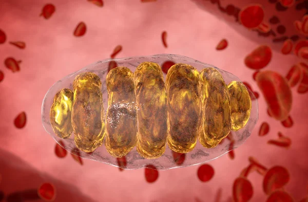
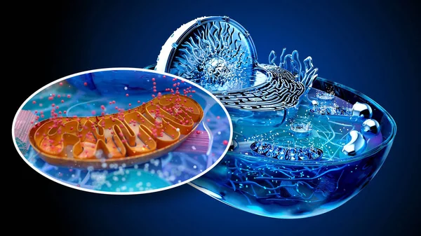
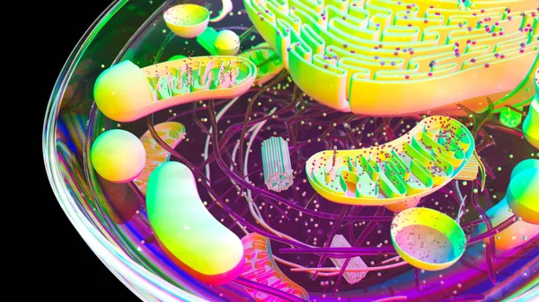
Mitochondrion, Mitochondrial. Medical Concept . Inside Human Organism
Image, 8.04MB, 6000 × 3937 jpg
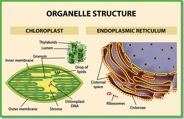
Cell Organelles Structure. Chloroplast And Endoplasmic Reticulum - Vector Illustration.
Vector, 4.37MB, 6000 × 3872 eps
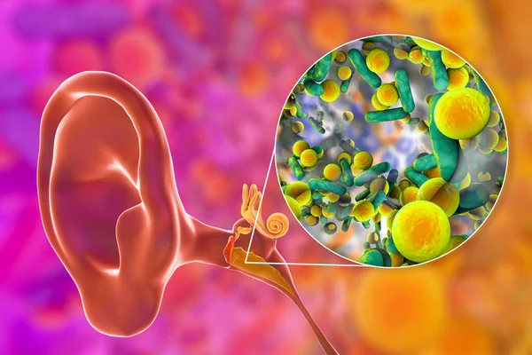
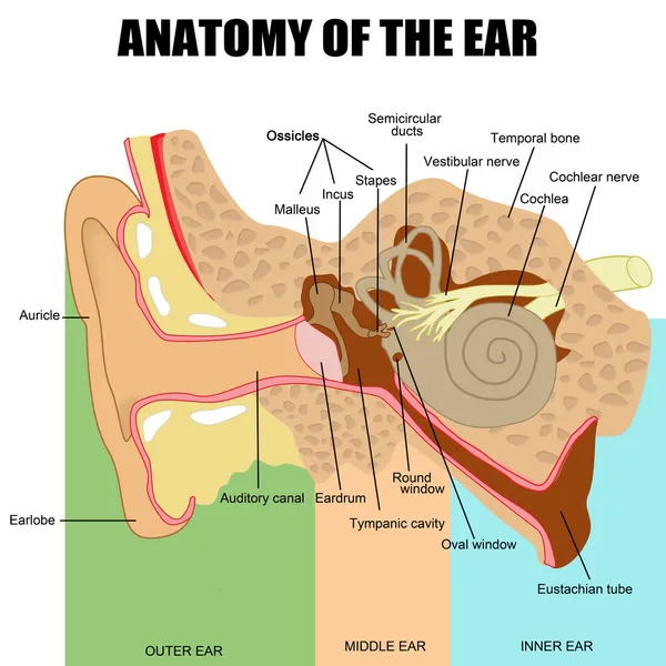
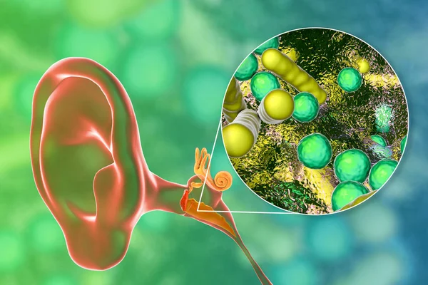
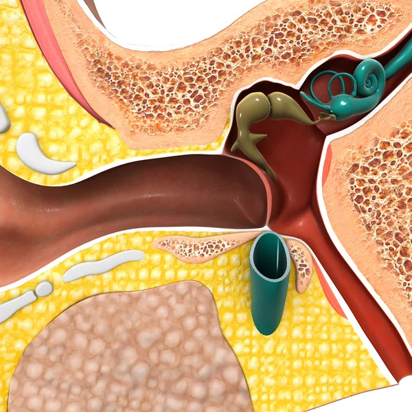
Otitis Media, An Inflammatory Disease Of The Middle Ear, And Close-up View Of Bacteria, The Causative Agent Of Otitis, 3D Illustration
Image, 10.65MB, 7500 × 5000 jpg

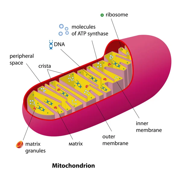
Mitochondria Structure. Mitochondrial Function. Vector Illustration On Isolated Background
Vector, 7.6MB, 5000 × 5000 eps
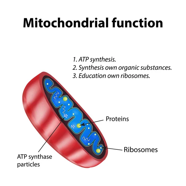
Mitochondria Structure. Mitochondrial Function. Vector Illustration On Isolated Background
Vector, 7.6MB, 5000 × 5000 eps

Anatomy Of A Birds Egg. Labeled Egg Structure Chart With Names Of The Components. Isolated Vector Illustration On White Background.
Vector, 3.58MB, 8115 × 6000 eps
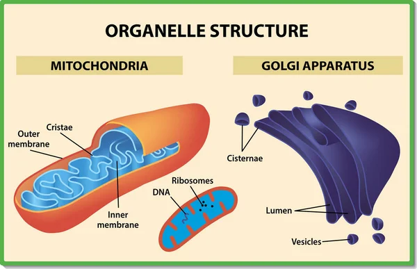
Cell Organelles Structure. Mitochondria And Golgi Apparatus - Vector Illustration.
Vector, 3.05MB, 6000 × 3872 eps

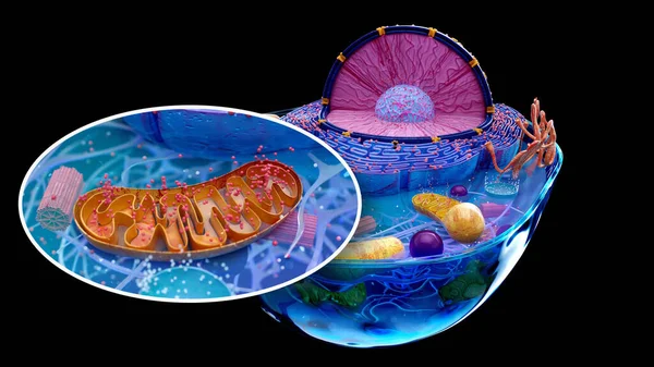
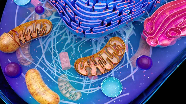
Mitochondrion, Mitochondrial. Medical Concept . Inside Human Organism
Image, 12.92MB, 6000 × 3937 jpg
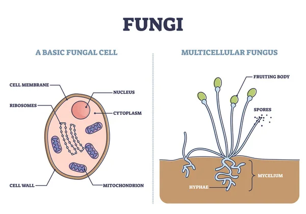
Fungi As Basic Fungal Cell And Multicellular Fungus Structure Outline Diagram
Vector, 5.81MB, 5000 × 3500 eps
Otitis Media, An Inflammatory Disease Of The Middle Ear, And Close-up View Of Bacteria, The Causative Agent Of Otitis, 3D Illustration
Image, 14.82MB, 7500 × 5000 jpg


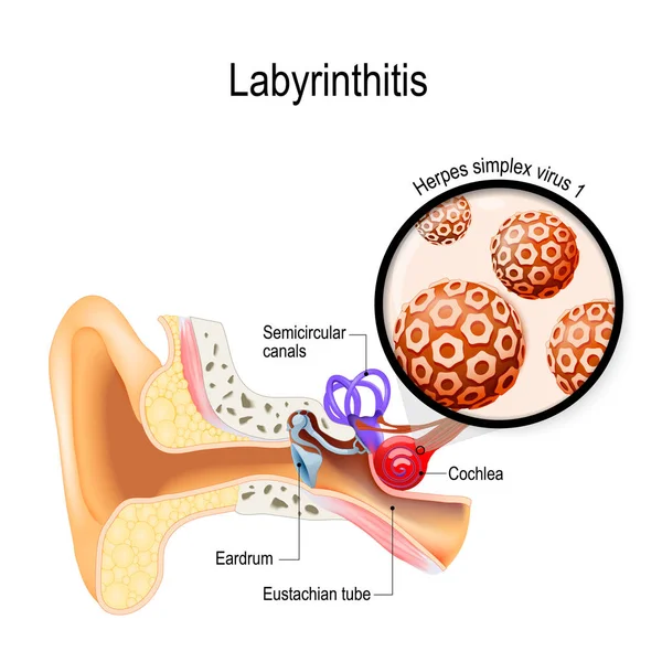
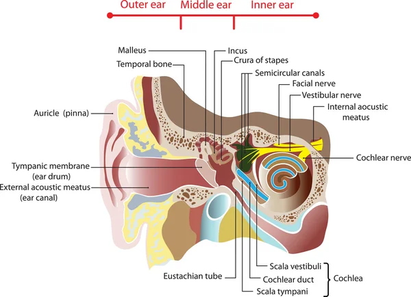
Labyrinthitis. Vestibular Neuritis. Inflammation Of The Inner Ear And Virus That Caused This Disease. Herpes Simplex Virus. Human Anatomy. Vector Illustration For Medical Use
Vector, 10.75MB, 4951 × 4951 eps

Labyrinthitis. Vestibular Neuritis. Inflammation Of The Inner Ear And Virus That Caused This Disease. Herpes Simplex Virus. Human Anatomy. Vector Illustration For Medical Use
Vector, 10.74MB, 4951 × 4951 eps
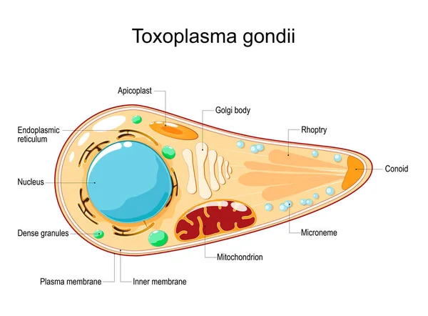
Toxoplasma Gondii. Cell Structure And Anatomy. Vector Illustration. Flat Style. Toxoplasma Is An Obligate Intracellular, Parasitic Protozoan That Causes The Disease Toxoplasmosis
Vector, 0.91MB, 5000 × 3685 eps

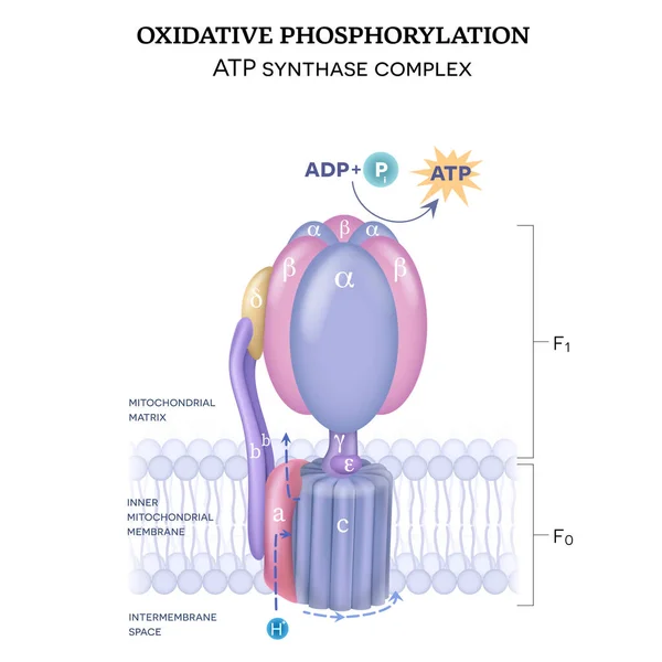
The ATP Synthase Structure (complex V) Consists Of Two Components F0 And F1. The Formation Of ATP Using Adenosine Diphosphate (ADP) And Inorganic Phosphate (Pi)
Vector, 11.37MB, 5000 × 5000 eps

Egg Anatomy Cross Section. Structure Of A Birds Egg, Labeled Chart With Names Of The Components. Isolated Vector Diagram Illustration On Black Background.
Vector, 3.8MB, 9000 × 5527 eps

Otitis Media Caused By Bacteria Haemophilus Influenzae, Inflammatory Disease Of The Middle Ear, 3D Illustration
Image, 7.26MB, 7138 × 4759 jpg
Page 1 >> Next