
Stock image Intracellular
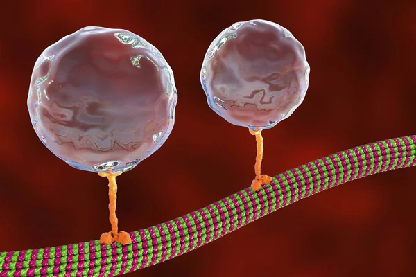
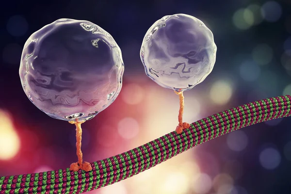
Intracellular Transport, Kinesin Proteins Transport Molecules Moving Across Microtubules
Image, 1.92MB, 4500 × 3000 jpg

Intracellular Transport, Kinesin Proteins Transport Molecules Moving Across Microtubules
Image, 4.16MB, 4500 × 4500 jpg
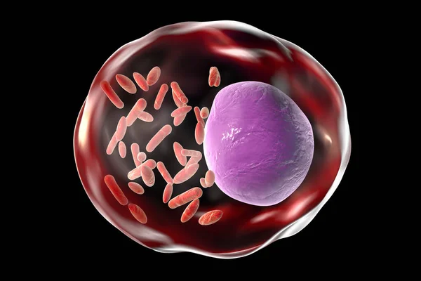
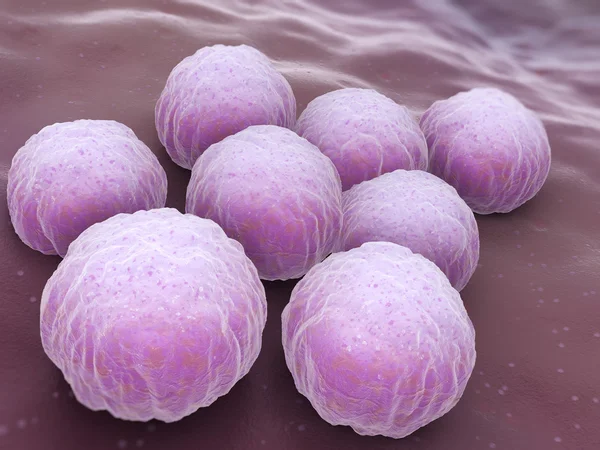
Chlamydophila Psittaci, Intracellular Bacteria That Cause Psittacosis
Image, 1.33MB, 4500 × 3000 jpg
Intracellular Transport, Kinesin Motor Proteins Transport Molecules Moving Across Microtubules
Image, 1.99MB, 4500 × 3000 jpg
Bacteria Rickettsia Inside Human Cell, 3D Illustration. Gram-negative Bacteria Which Cause Epidemic Typhus, Murine Typhus Other Rickettsioses And Are Transmitted By Arthropods
Image, 1.75MB, 6000 × 4000 jpg
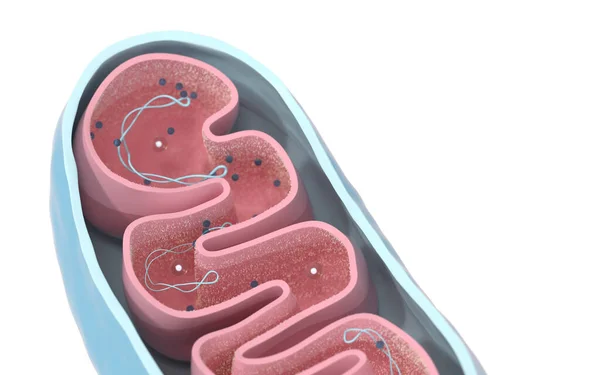
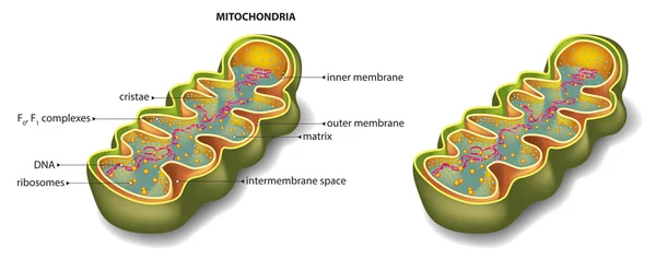
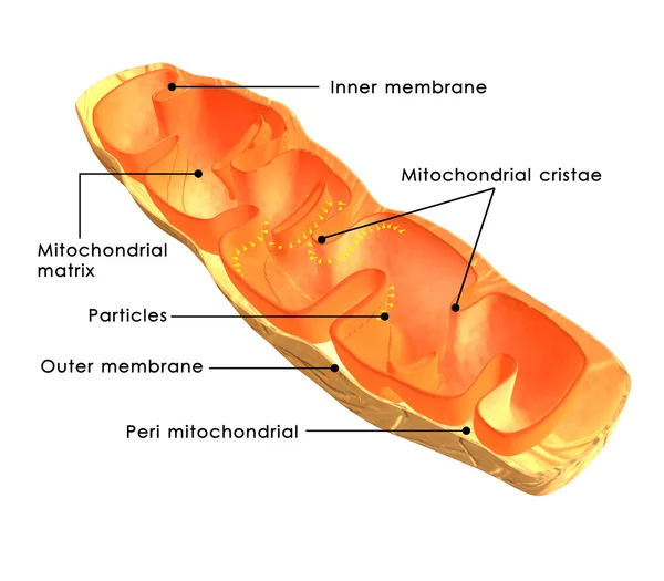
Cross-section View Of Mitochondria. Medical Info Graphics On White Background, 3d Rendering. Computer Digital Drawing.
Image, 10.47MB, 8000 × 5000 jpg
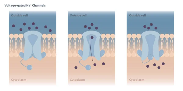
Neuronal Charged Membranes. Voltage-gated Ion Channels Are Closed At The Resting Potential And Open In Response To Changes In Membrane Voltage.
Vector, 7.67MB, 8334 × 4167 eps
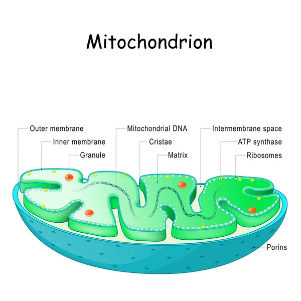
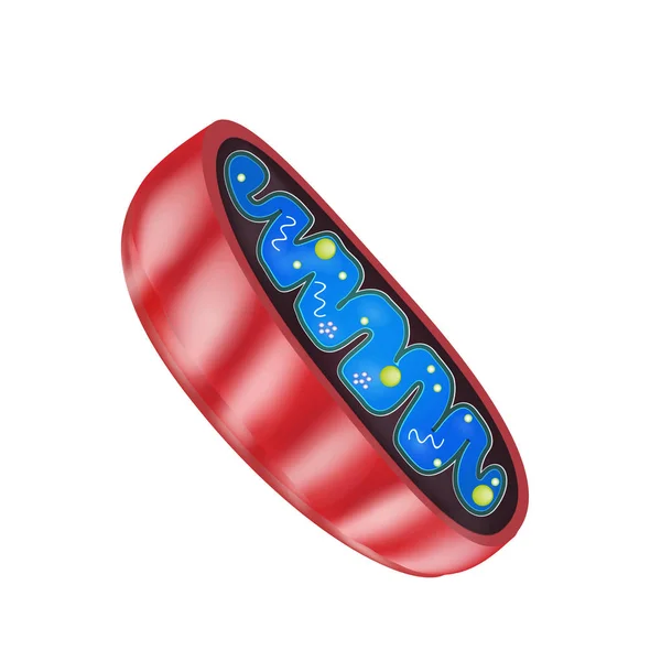
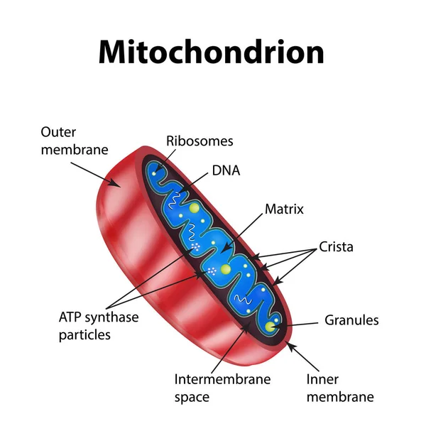
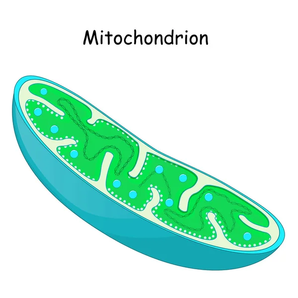
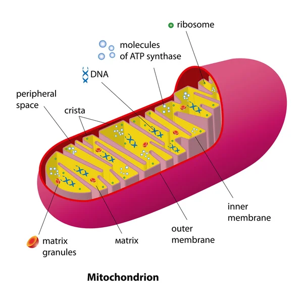
Mitochondrion Anatomy. Structure, Components And Organelles. Cross-section Of Mitochondria. Vector Illustration
Vector, 5.1MB, 4444 × 4444 eps

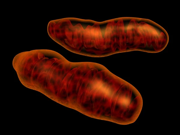
Mitochondria, Cellular Organelles, Produce Energy, Cell Energy And Cellular Respiration, DNA , 3D Rendering
Image, 1.39MB, 2688 × 2016 jpg
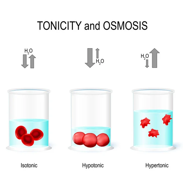
Isotonic, Hypotonic And Hypertonic Solutions Effects On Animal Cells. Tonicity And Osmosis. This Diagram Shows The Effects Of Hypertonic, Hypotonic And Istonic Solutions To Red Blood Cells. Vector Illustration For Biological, Medical, Science Use
Vector, 5.63MB, 4685 × 4685 eps

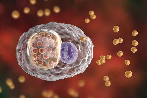
Toxoplasma Gondii Parasites In Blood, The Causative Agent Of Toxoplasmosis Disease, 3D Illustration
Image, 1.63MB, 4500 × 3000 jpg

3D Image Of Cyclic Guanosine Monophosphate Skeletal Formula - Molecular Chemical Structure Of Second Messenger CGMP Isolated On White Background
Image, 3.96MB, 8514 × 4080 jpg

Lymphogranuloma Venereum. An Infectious Disease Caused By Chlamydial Infection. During The Disease, The Lymph Nodes Are Affected, Purulent And Inflammatory Processes Occur.
Vector, 0.41MB, 5810 × 2684 eps

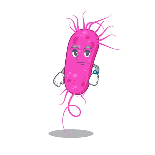
A Sweet Rickettsia Cartoon Character Style With A Heart. Vector Illustration
Vector, 5.53MB, 5000 × 5000 eps
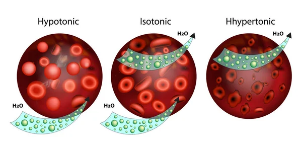
Effect Of Different Solutions On Blood Cells.The Effect Of Osmosis On Cells. Hypotonic, Isotonic, And Hypertonic Solution. Tonicity
Vector, 4.44MB, 6840 × 3420 eps

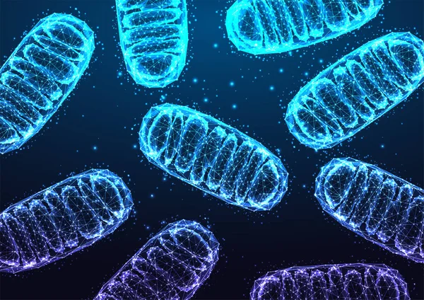
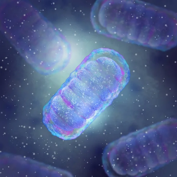
Mitochondria Under Microscope On Dark Blue Backgound In Futuristic Glowing Low Polygonal Style. Medical Research Concept, Science, Biology Research Banner. Modern Abstract Design Vector Illustration
Vector, 10.07MB, 5893 × 4168 eps


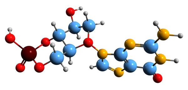
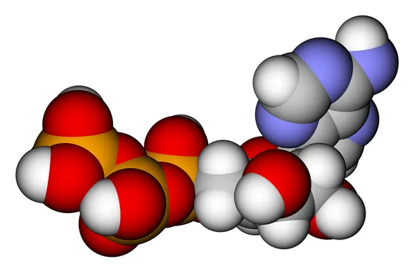
Cyclic Adenosine Monophosphate (cAMP) Molecule, It Is A Derivative Of Adenosine Triphosphate (ATP) And Used For Intracellular Signal Transduction . Structural Chemical Formula And Molecule Model. Vector Illustration
Vector, 0.47MB, 5000 × 3409 eps

Chlamydophila Psittaci, Intracellular Bacteria That Cause Psittacosis
Image, 1.34MB, 4500 × 3000 jpg

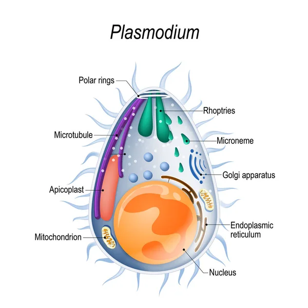
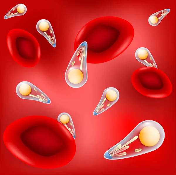
Plasmodium Is The Malaria Parasite, Is A Large Genus Of Parasitic Protozoa. Infection With These Protozoans Is Known As Malaria, A Deadly Disease. Diagram Of Plasmodium Merozoites Structure. Vector Illustration For Medical, Educational And Science
Vector, 8.41MB, 4649 × 4649 eps
Medical Background, Mitochondrion Is A Two-membrane Spherical Or Ellipsoid Organelle, Supplies Energy To The Cell, Electrical Potential Generation, 3d Rendering
Image, 3.01MB, 3300 × 3300 jpg
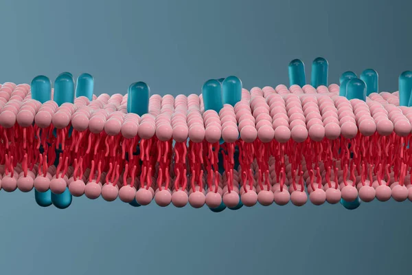
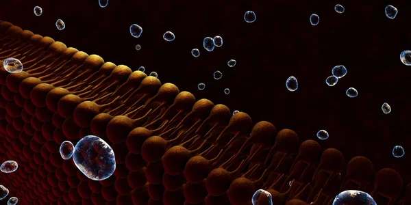
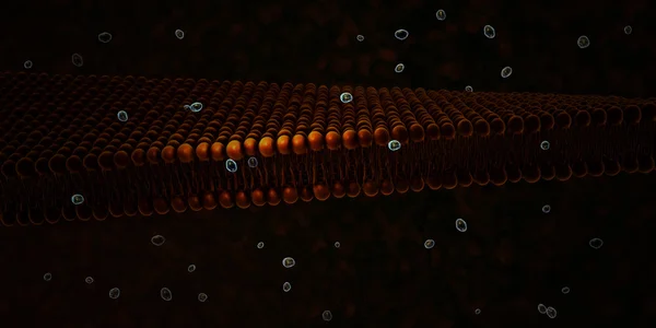
Cell Membrane And Biology, Biological Concept, 3d Rendering. Computer Digital Drawing.
Image, 8.25MB, 6000 × 4000 jpg

CAMP Cyclic Adenosine MonoPhosphate - Second Messenger Important In Many Biological Processes, Acronym Text On Blackboard
Image, 11.5MB, 5760 × 3840 jpg


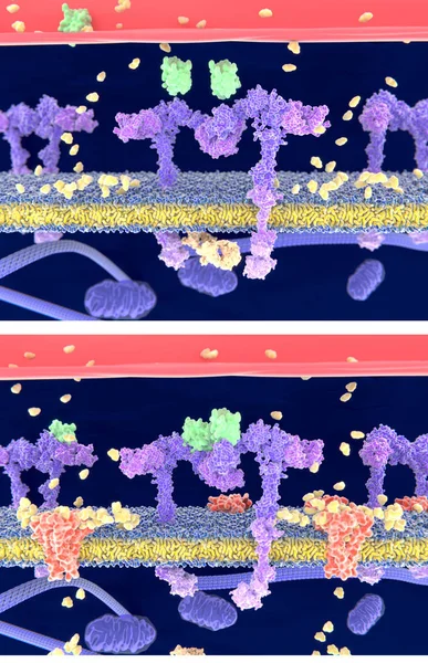
Insulin (green) Binding To The Insulin Receptor (violet) Activates The Transport Of Glucose (yellow) Into The Cell (depicted In 2 Phases) - Illustration
Image, 4.57MB, 4000 × 6200 jpg

Intracellular Transport, Kinesin Proteins Transport Molecules Moving Across Microtubules
Image, 1.92MB, 4500 × 3000 jpg
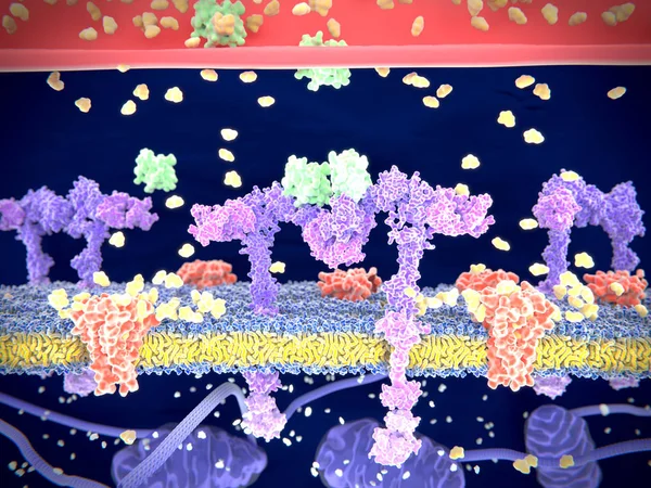
Insulin (green) Binding To The Insulin Receptor (violet) Activates The Transport Of Glucose (yellow) Into The Cell. Illustration
Image, 6.21MB, 8000 × 6000 jpg
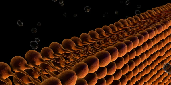
Cell Membrane And Biology, Biological Concept, 3d Rendering. Computer Digital Drawing.
Image, 8.61MB, 6000 × 4000 jpg
Page 1 >> Next