
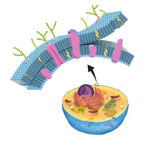
Stock image Intracellular Transport
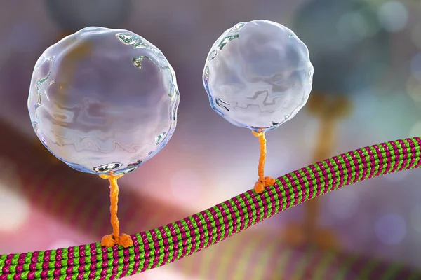
Intracellular Transport, Kinesin Proteins Transport Molecules Moving Across Microtubules
Image, 1.92MB, 4500 × 3000 jpg

Intracellular Transport, Kinesin Proteins Transport Molecules Moving Across Microtubules
Image, 4.16MB, 4500 × 4500 jpg

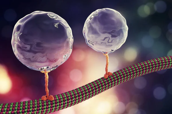
Intracellular Transport, Kinesin Proteins Transport Molecules Moving Across Microtubules
Image, 4.28MB, 4500 × 4500 jpg
Intracellular Transport, Kinesin Proteins Transport Molecules Moving Across Microtubules
Image, 2.61MB, 4500 × 3000 jpg
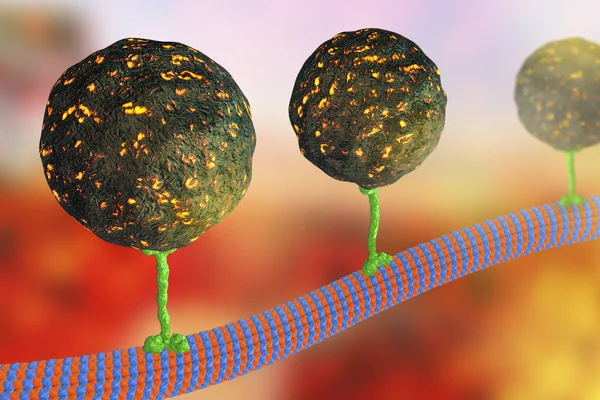
Intracellular Transport, Kinesin Motor Proteins Transport Molecules Moving Across Microtubules
Image, 1.99MB, 4500 × 3000 jpg
Intracellular Transport, Kinesin Motor Proteins Transport Molecules Moving Across Microtubules
Image, 2.08MB, 4500 × 3000 jpg

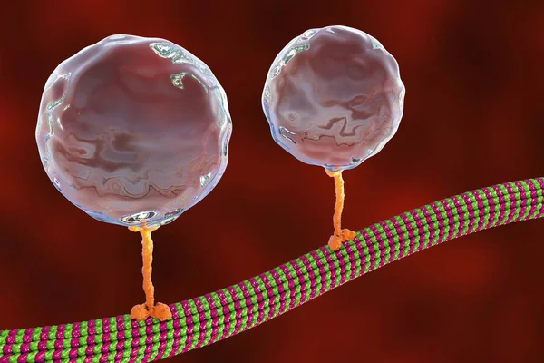
Intracellular Transport, Kinesin Proteins Transport Molecules Moving Across Microtubules
Image, 1.92MB, 4500 × 3000 jpg
The Cell Membrane, Also Called The Plasma Membrane, Is Found In All Cells And Separates The Interior Of The Cell From The Outside Environment
Image, 12.29MB, 8268 × 8268 jpg
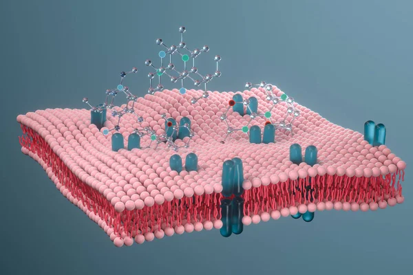
Cell Membrane And Biology, Biological Concept, 3d Rendering. Computer Digital Drawing.
Image, 8.59MB, 6000 × 4000 jpg
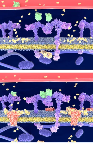
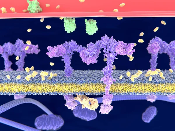
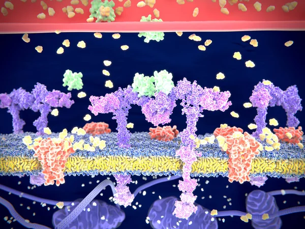
Insulin (green) Binding To The Insulin Receptor (violet) Activates The Transport Of Glucose (yellow) Into The Cell (depicted In 2 Phases) - Illustration
Image, 4.57MB, 4000 × 6200 jpg
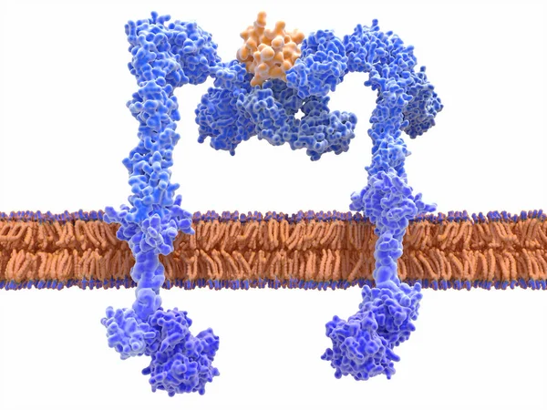
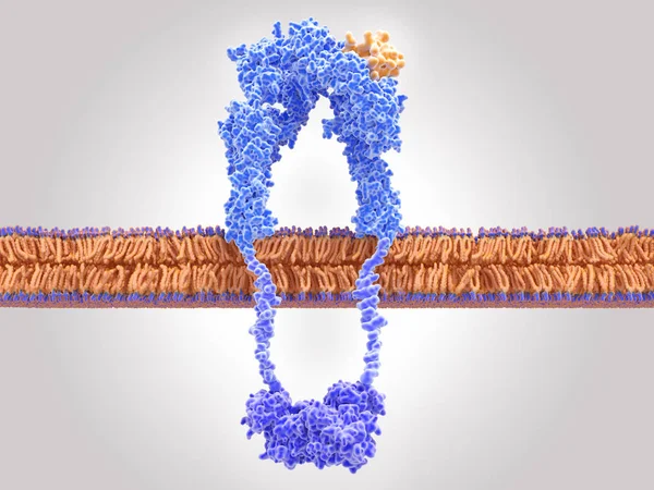
The Insulin Receptor (blue) Is A Transmembrane Protein, That Is Activated By Insulin (orange). Insulin Binding Induces Structural Changes Within The Receptor That Finally Leads To The Activation Of The Glucose Transporter Protein.
Image, 12.2MB, 8000 × 6000 jpg
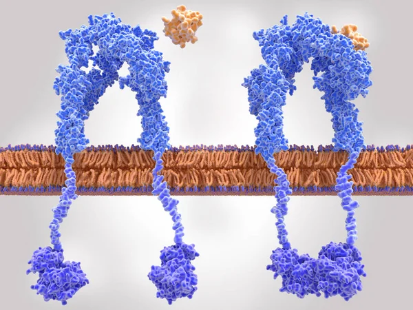
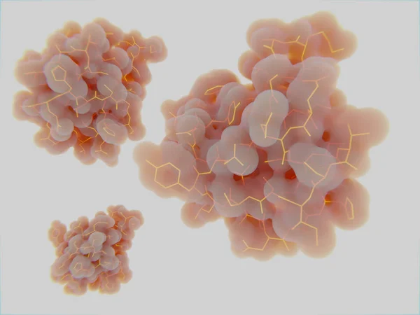
Insulin Receptor Inactivated (left) And Activated (right) After Insulin Binding
Image, 9MB, 8000 × 6000 jpg

Cell Membrane And Biology, Biological Concept, 3d Rendering. Computer Digital Drawing.
Image, 8.17MB, 6000 × 4000 jpg
Insulin (green) Binding To The Insulin Receptor (violet) Activates The Transport Of Glucose (yellow) Into The Cell. Illustration
Image, 3.71MB, 8000 × 6000 jpg

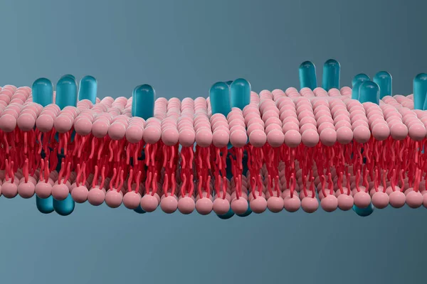
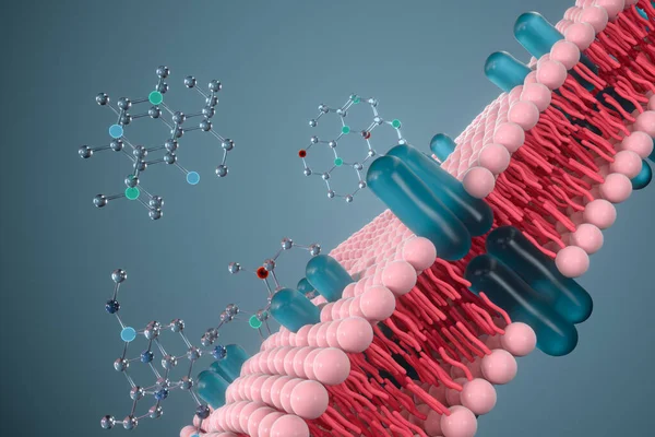
Cell Membrane And Biology, Biological Concept, 3d Rendering. Computer Digital Drawing.
Image, 8.51MB, 6000 × 4000 jpg
Cell Membrane And Biology, Biological Concept, 3d Rendering. Computer Digital Drawing.
Image, 8.25MB, 6000 × 4000 jpg

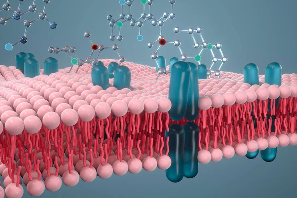
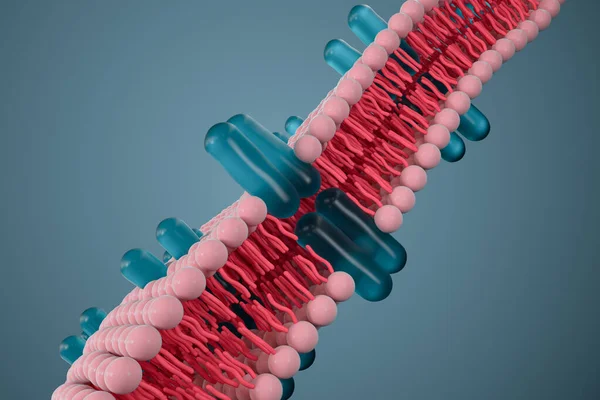
Cell Membrane And Biology, Biological Concept, 3d Rendering. Computer Digital Drawing.
Image, 8.48MB, 6000 × 4000 jpg
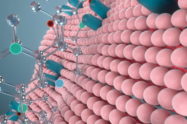
Cell Membrane And Biology, Biological Concept, 3d Rendering. Computer Digital Drawing.
Image, 9.32MB, 6000 × 4000 jpg


Insulin (green) Binding To The Insulin Receptor (violet) Activates The Transport Of Glucose (yellow) Into The Cell (phase 1). Illustration
Image, 3.96MB, 8000 × 6000 jpg

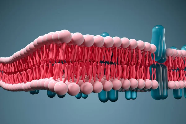
Cell Membrane And Biology, Biological Concept, 3d Rendering. Computer Digital Drawing.
Image, 8.42MB, 6000 × 4000 jpg
Insulin (green) Binding To The Insulin Receptor (violet) Activates The Transport Of Glucose (yellow) Into The Cell. Illustration
Image, 6.21MB, 8000 × 6000 jpg

Cell Membrane And Biology, Biological Concept, 3d Rendering. Computer Digital Drawing.
Image, 8.08MB, 6000 × 4000 jpg
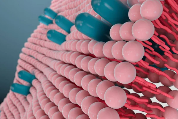
Cell Membrane And Biology, Biological Concept, 3d Rendering. Computer Digital Drawing.
Image, 8.67MB, 6000 × 4000 jpg
Cell Membrane And Biology, Biological Concept, 3d Rendering. Computer Digital Drawing.
Image, 8.83MB, 6000 × 4000 jpg
Cell Membrane And Biology, Biological Concept, 3d Rendering. Computer Digital Drawing.
Image, 8.61MB, 6000 × 4000 jpg

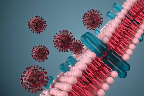
Cell Membrane And Coronavirus, Medical Concept, 3d Rendering. Computer Digital Drawing.
Image, 10.38MB, 6000 × 4000 jpg
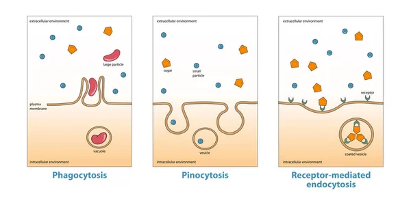
Variations Of Endocytosis: Phagocytosis, Pinocytosis, Receptor-mediated Endocytosis. Various Types Of Endocytosis, Uptake Of Matter Through Plasma Membrane Invagination And Vacuole, Vesicle Formation
Vector, 5.9MB, 12500 × 6245 eps

Cell Membrane And Coronavirus, Medical Concept, 3d Rendering. Computer Digital Drawing.
Image, 10.24MB, 6000 × 4000 jpg
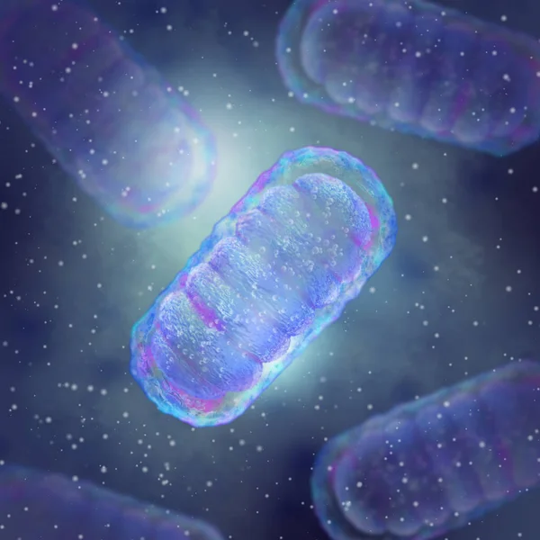
Medical Background, Mitochondrion Is A Two-membrane Spherical Or Ellipsoid Organelle, Supplies Energy To The Cell, Electrical Potential Generation, 3d Rendering
Image, 3.01MB, 3300 × 3300 jpg
Page 1 >> Next