
Stock image Joint Degeneration page 2


Knee Joint Damage With Osteoarthritis, Flat Vector Illustration Isolated.
Vector, 0.48MB, 6340 × 5000 eps
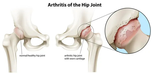
Knee Osteoarthritis And Normal Joint Detailed Anatomy. Osteoarthritis. Arthritis Or Pain Within A Joint. Degenerative Joint Disease. Cartilage Becomes Worn. Vector Flat Design
Vector, 1.6MB, 6000 × 4000 eps

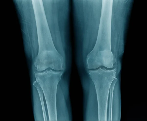
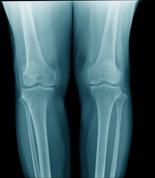
High Quality X-ray Knee Joint Of Old Man, OA Knee Of Old Man On Black Background In Blue Tone
Image, 1.35MB, 2448 × 1682 jpg


Vector Banner With Medical Anatomy With Knee Osteoarthritis And Normal Joint
Vector, 0.77MB, 7000 × 3874 eps
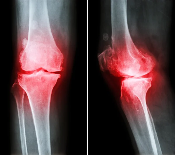
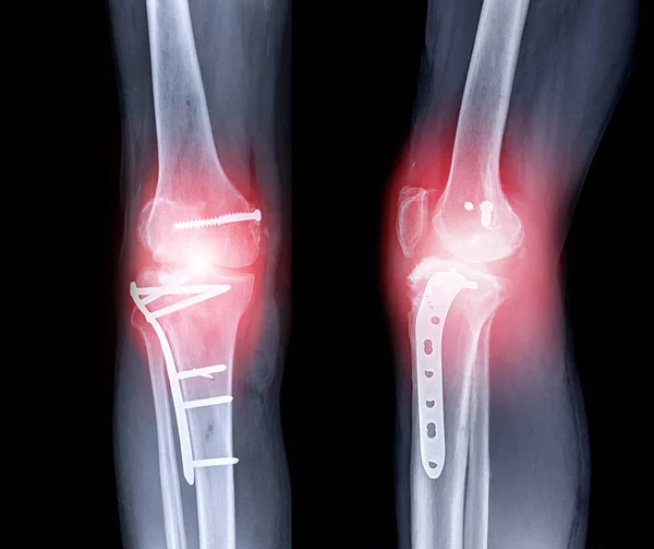
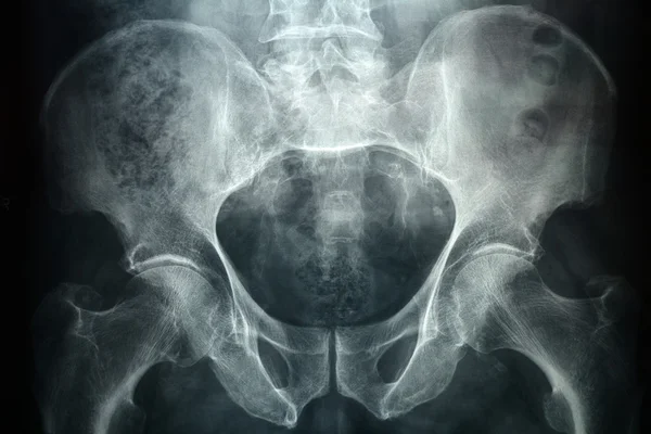
Osteoarthritis Knee . Film X-ray Knee ( Anterior - Posterior And Lateral View ) Show Narrow Joint Space , Osteophyte ( Spur ) , Subcondral Sclerosis Due To Degenerative Change
Image, 4.98MB, 3692 × 3263 jpg


High Quality X-ray Knee Joint Of Old Man, OA Knee Of Old Man On Black Background In Blue Tone
Image, 1.64MB, 2428 × 1994 jpg

Healthy Joint And Damaged With Osteoarthritis, Flat Vector Illustration.
Vector, 0.78MB, 6000 × 4921 eps
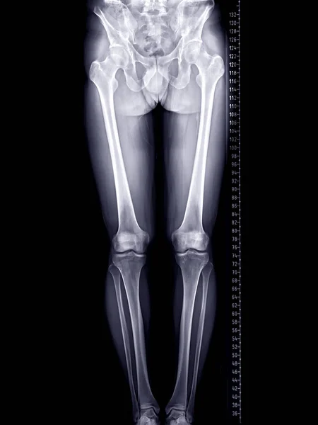
Scanogram Of Lower Limb Or X-ray Image Of Total Lower Extremity With Scale.
Image, 3.15MB, 3280 × 4375 jpg
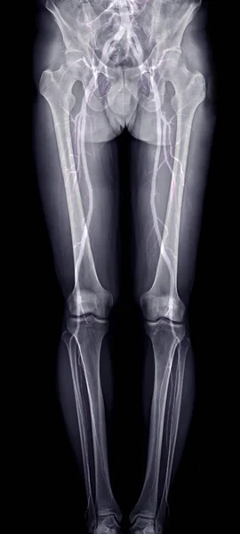
Scanogram Of Lower Limb Or X-ray Image With Merge CTA Femoral Run Off Showing Bone And Vessel Of Lower Limb.
Image, 2.19MB, 1936 × 4303 jpg

High Quality X-ray Knee Joint Of Old Man, OA Knee Of Old Man On Black Background In Blue Tone
Image, 1.41MB, 1996 × 2290 jpg
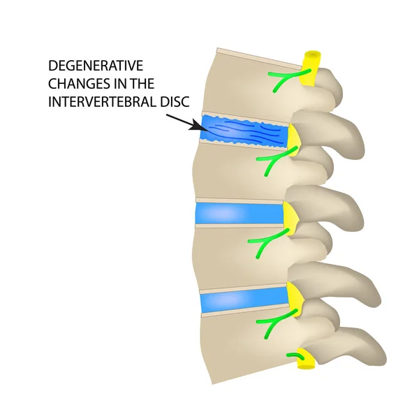
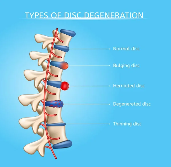
Degenerative Changes In The Intervertebral Disc. Vector Illustration On Isolated Background
Vector, 0.99MB, 5000 × 5000 eps
X-ray Of Dog Lateral View Closed Up In Thorax Standard And Chest With Red Highlight In Neck Bone To Back Bone Pain Areas Or Spinal Disease In Dog- Veterinary Medicine- Veterinary Anatomy Concept
Image, 6.74MB, 5500 × 5500 jpg

Hand Of Asian Old Elderly Touch Lower Back Muscles,senior Patient With Walker Stick,suffering From Back Pain,backache From Spinal Joint Problems,myositis,painful Of Lumbago,health Care,medical Concept
Image, 10.95MB, 6309 × 4206 jpg

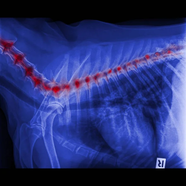
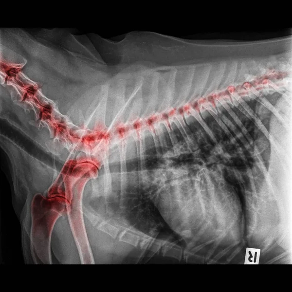
X-ray Of Dog Lateral View Closed Up Thorax And Chest Red Highlight Foreleg Bone In Shoulder Joint And Neck Bone To Back Bone- Degenerative Joint Disease In Dog- Veterinary Medicine- Veterinary Anatomy
Image, 6.73MB, 5500 × 5500 jpg
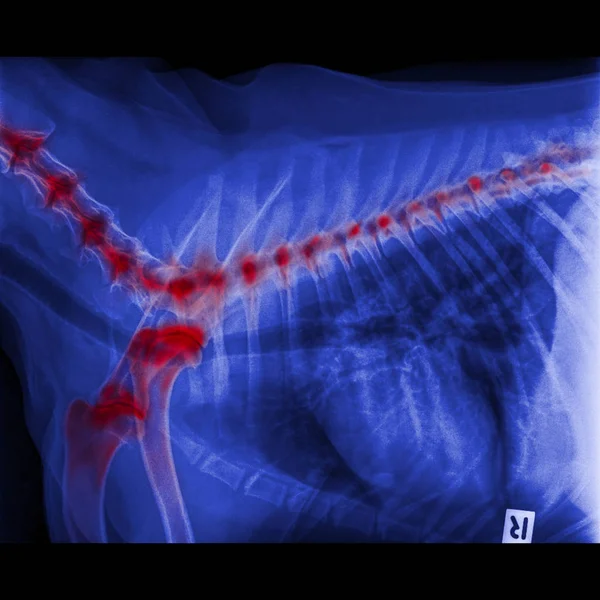
X-ray Of Dog Lateral View Closed Up Thorax And Chest Red Highlight Foreleg Bone In Shoulder Joint And Neck Bone To Back Bone- Degenerative Joint Disease In Dog- Veterinary Medicine- Veterinary Anatomy
Image, 5.39MB, 5500 × 5500 jpg

High Quality X-ray Knee Joint Of Old Man, OA Knee Of Old Man On Black Background In Blue Tone
Image, 1.75MB, 1996 × 2428 jpg

X-ray Image Of Spine Show Degeneration Of Lumbar Spine With Red High Light, Spur Or Calcification At Body Of Lumbar Spine
Image, 1.81MB, 1996 × 2428 jpg
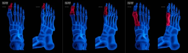
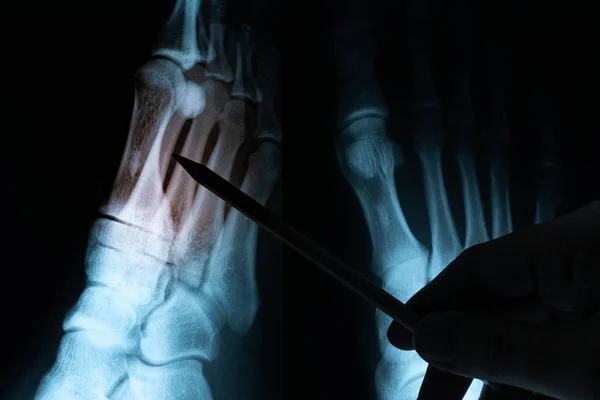
X-ray Blue Film Collection Of Big Toe Foot Bone With Red Highlights On Different Pain And Joint Area-top And Side View-Healthcare-Human Anatomy And Medical Concept-Isolated On Black Background.
Image, 17.43MB, 16051 × 4578 jpg
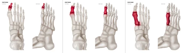
X-ray Collection Of Big Toe Foot Bone With Red Highlights On Different Pain And Joint Area-top And Side View-Healthcare-Human Anatomy And Medical Concept-Isolated On White Background.
Image, 16.69MB, 16051 × 4578 jpg

Medical Illustration Of The Femoroacetabular Impingement. Different Of The Cam Impingement And Pincer Impingement. Healthy Hip
Vector, 17.99MB, 6960 × 3600 eps
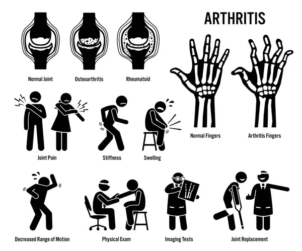
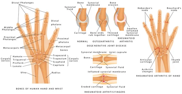
Arthritis, Joint Pain, And Joint Disease Icons. Pictograms Depict Arthritis Signs, Symptoms, Diagnosis, And Treatment. Icons Include Bones For Osteoarthritis And Rheumatoid Arthritis.
Vector, 3.2MB, 7200 × 6000 eps

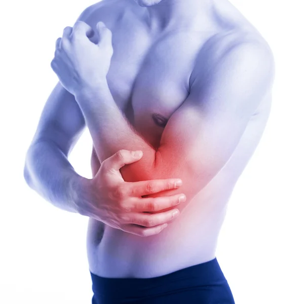
Asian Senior Woman With Knee Joint Pain,female Patient Having Aching Patella,arthritis,osteoarthritis Of The Knee,elderly People Touch On The Knee With Hand,pain In The Kneecap,physical Injury Concept
Image, 12.02MB, 6469 × 4313 jpg
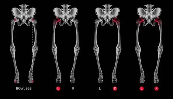
Varus Alignment Of Leg Or Bowlegs Bone X-ray Film Collection With Red Highlights On Hip Arthritis And Hip Joint Area-Healthcare-Human Anatomy And Medical Concept-Isolated On Black Background.
Image, 5.33MB, 10000 × 5721 jpg
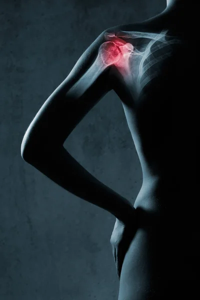
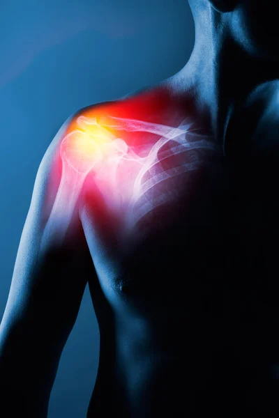
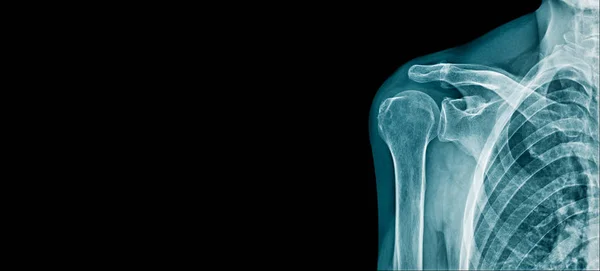
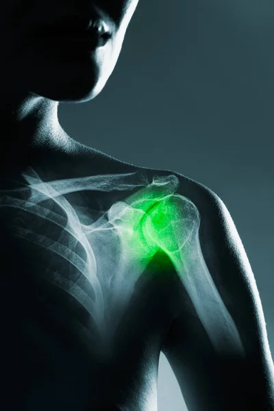
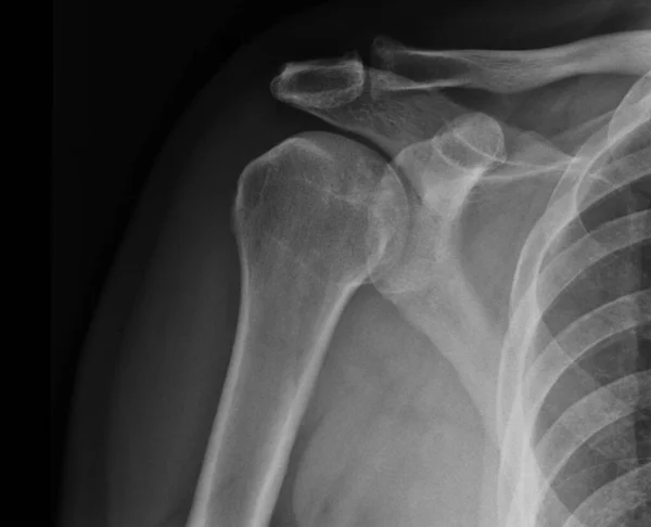
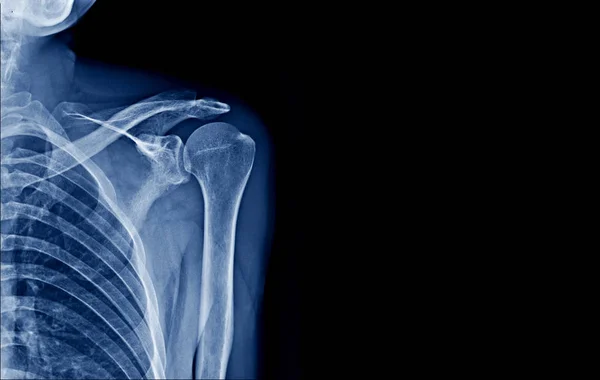
Human Shoulder Joint In X-ray, On Gray Background. The Hand Is Highlighted By Red Colour.
Image, 6.81MB, 3744 × 5616 jpg

Legs And Angles Of The Knees, Different Types Of Leg Shapes. Front View Girl, Body Anatomy. Normal Varus And Valgus
Vector, 0.8MB, 4689 × 3640 eps
Previous << Page 2 >> Next