
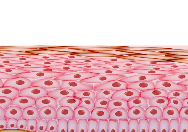
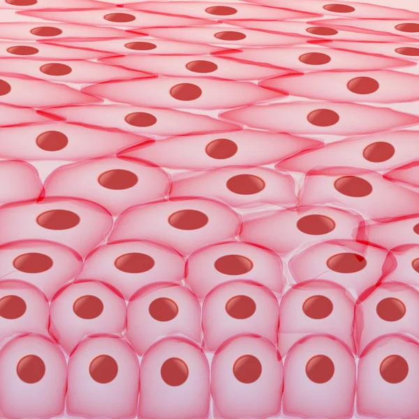
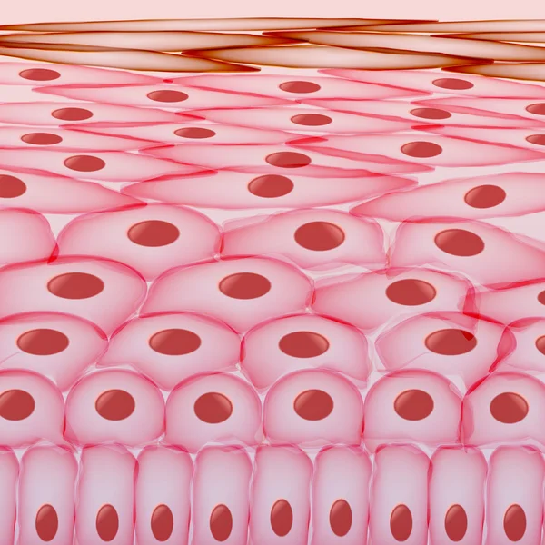
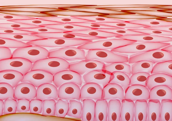
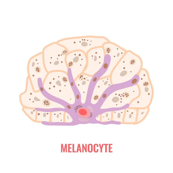
Stock image Keratinocyte
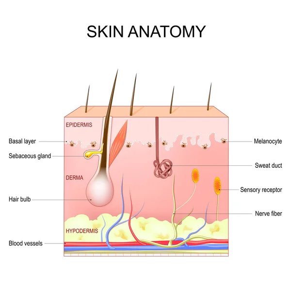
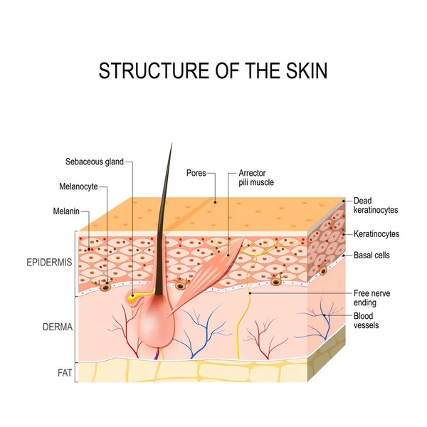
Skin Anatomy. Structure And Layers Of Skin: Epidermis, Dermis, Hypodermis, Melanocytes And Basal Layer. Cross Section Of The Human Skin With Sebaceous Gland, Sweat Duct, Sensory Receptor, And Hair Bulb. Vector Illustration. Poster For Medical And Ed
Vector, 14.88MB, 4444 × 4444 eps
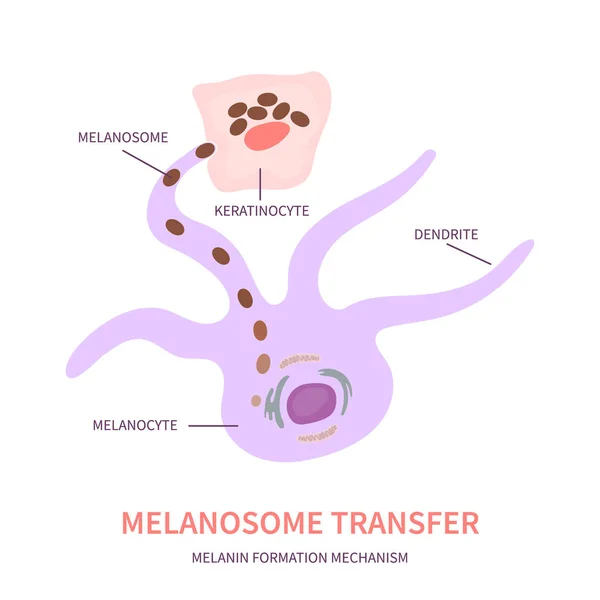
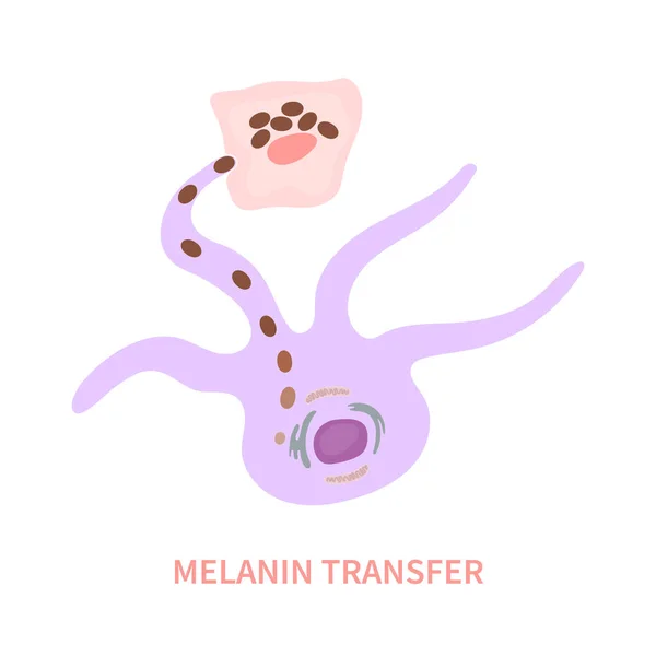
Melanosome Transfer To Keratinocytes Scheme. Melanocyte Cell Biology And Skin Pigmentation Diagram. Melanin Pigment Production And Distribution Process. Vector Illustration.
Vector, 0.32MB, 4500 × 4500 eps
Melanin Color Palette Scheme From Light To Dark Brown. Skin Tanning Process Diagram. Skin Complexion Diversity. Fitzpatrick Skin Type Classification Scale. Beauty Concept Design. Vector Illustration
Vector, 0.25MB, 4500 × 4500 eps
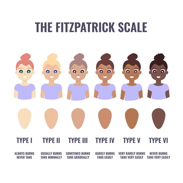
Fitzpatrick Skin Type Classification Scale Shown In Women. Human Skin Tone Pigmentation Diversity Infographics. Six Phototypes From Fair To Dark Complexion Variations. Vector Cartoon Illustration.
Vector, 0.47MB, 4500 × 4500 eps
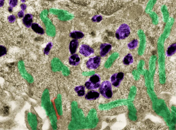
Structure Of Intermediate Filaments Of Keratin Protein. It Is One Of A Family Of Fibrous Structural Proteins
Image, 5.64MB, 6722 × 2957 jpg

Stem Cell Application. Undifferentiated Or Partially Differentiated Cells. Using Stem Cells To Treat Disease. Vector Illustration
Vector, 9.5MB, 4444 × 4443 eps
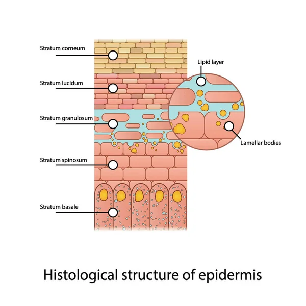
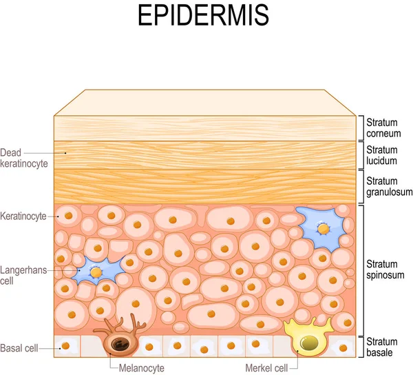
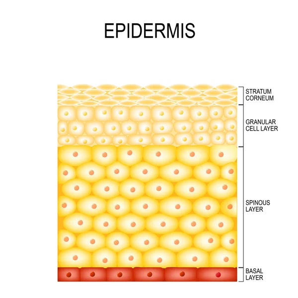
Histological Structure Of Epidermis - Skin Layers Shcematic Vector Illustration Showing Stratum Basale, Spinosum, Granulosum, Lucidum And Corneum And Lamellar Bodies
Vector, 7.07MB, 3090 × 3090 eps

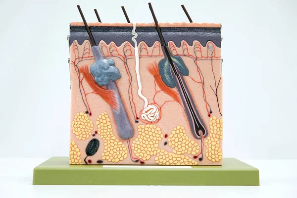
Doctor Dermatologist Or Trichologist Holding And Showing Human Skin Cross-section Plastic Model.Detailed Skin Structure With Hair In Clinic For Education.Diagnosis Of Hair Diseases, Hair Loss Problem
Image, 4.31MB, 4000 × 2667 jpg
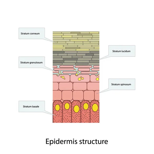
Histological Structure Of Epidermis - Skin Layers Shcematic Vector Illustration Showing Stratum Basale, Spinosum, Granulosum, Lucidum And Corneum
Vector, 5.89MB, 3090 × 3090 eps

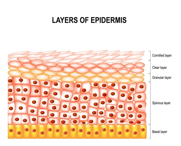
Epidermis Anatomy. Layers And Cell Structure Of The Human Skin. Close-up Of Epidermis. Vector Illustration
Vector, 2.34MB, 4444 × 4444 eps

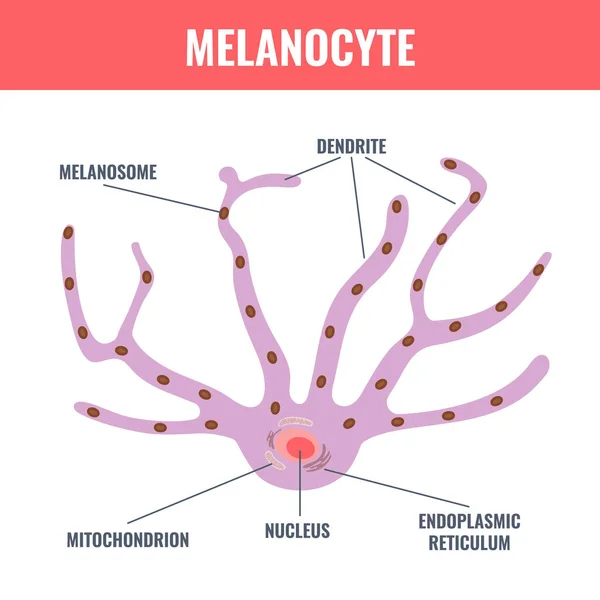
Melanocyte Cell Biology And Skin Tone Pigmentation Diagram. Melanin Pigment Production And Distribution Process. Melanosome Transfer To Keratinocytes Scheme. Vector Illustration.
Vector, 0.3MB, 4500 × 4500 eps

Melanin Color Palette Scheme. Eumelanin And Pheomelanin Pigment Grades Of Skin, Hair And Eyes. Skin Complexion Diversity. Fitzpatrick Skin Type Classification Scale. Vector Illustration
Vector, 0.27MB, 4500 × 4500 eps

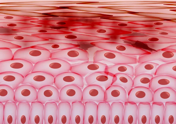
Cross Section Human Skin Tissue Under Microscope View For Physiology Education.
Image, 17.18MB, 6000 × 4000 jpg
Melanosome Cells And Skin Pigmentation Mechanism. Melanin Pigment Content And Distribution Diagram In Different Ethnic Skin Types. Vector Illustration.
Vector, 0.39MB, 4500 × 4500 eps

Melanin Color Palette Scheme From Light To Dark Brown. Skin Tanning Process Diagram. Skin Complexion Diversity. Fitzpatrick Skin Type Classification Scale. Beauty Concept Design. Vector Illustration
Vector, 0.3MB, 4500 × 4500 eps
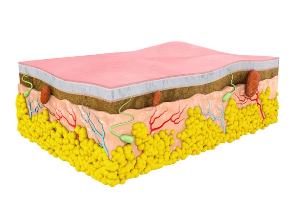
Vector Illustration With Structure Of Dermis For Medical And Educational Pictures Isolated
Vector, 4.14MB, 6250 × 4321 eps
Melanocyte Cell Biology And Skin Tone Pigmentation Diagram. Melanin Pigment Production And Distribution Process. Melanosome Transfer To Keratinocytes Scheme. Vector Illustration.
Vector, 0.47MB, 4500 × 4500 eps
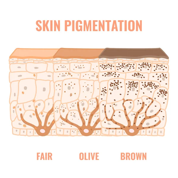
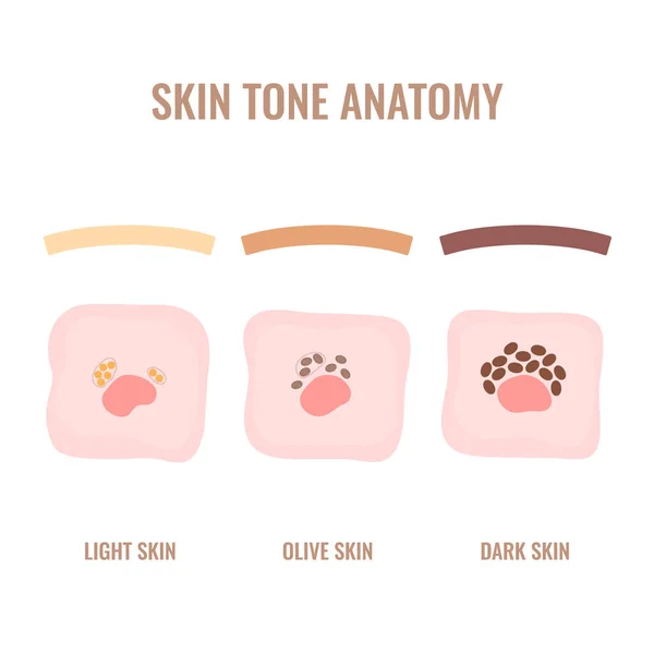
Melanin Content And Distribution In Different Skin Tone Phototypes. Pigmentation Mechanism In Dark, Olive And Light Skin. Epidermis Cross-section Infographic Medical Diagram. Vector Illustration.
Vector, 1.59MB, 4500 × 4500 eps
Epidermis Structure. Skin Anatomy. Cell, And Layers Of A Human Skin. Cross Section Of The Epidermis. Skin Care. Vector Illustration.
Vector, 2.11MB, 4444 × 4444 eps

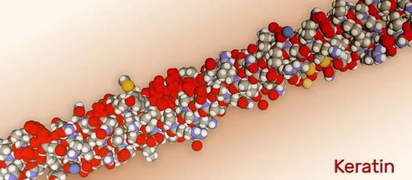
Keratin Intermediate Filament, Chemical Structure. 3D Illustration. Keratin Is One Of The Main Components Of Human Skin, Hair And Nails.
Image, 2.04MB, 8000 × 1838 jpg
Melanin Content And Distribution In Dark Skin Phototype. Pigmentation Mechanism Infographic Diagram. Epidermis Cross-section In Closeup. Vector Illustration.
Vector, 0.8MB, 4500 × 4500 eps

Comparison Of Melanosomes Distribution In Dark And Light Skin. Pigmentation Mechanism In Different Phototypes. Close Up Of Epidermis Cross-section. Infographic Medical Diagram. Vector Illustration.
Vector, 5.74MB, 4500 × 4500 eps

Histological Structure Of Epidermis - Skin Layers Shcematic Vector Illustration Showing Stratum Basale, Spinosum, Granulosum, Lucidum And Corneum
Vector, 7.1MB, 3090 × 3090 eps

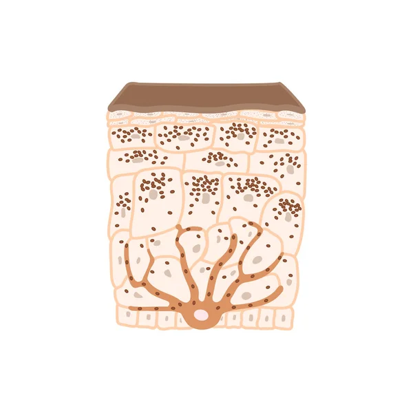
Cross-section Of The Epidermis Of A Dark Phototype Skin With Many Grains Of Melanin.
Image, 2.2MB, 3844 × 3020 jpg
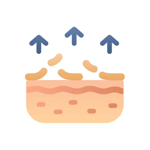
Dead Skin Protection Dermatology Epidermis Remoce Care Single Isolated Icon With Smooth Style Vector Illustration
Vector, 0.23MB, 5340 × 5340 eps
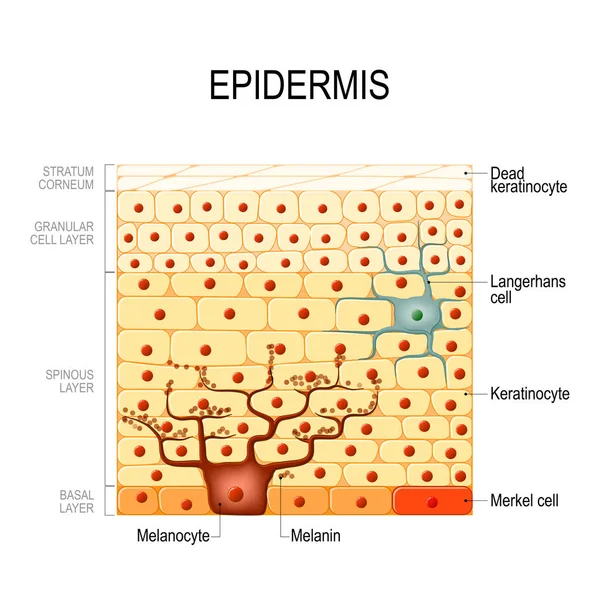
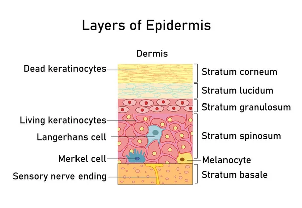
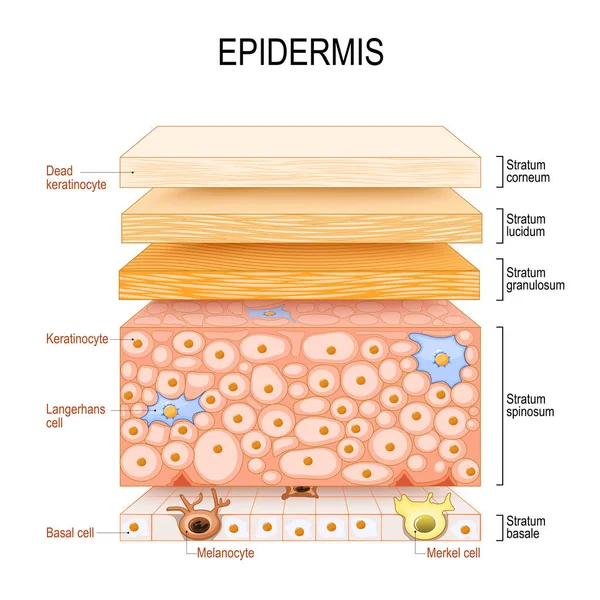
Layers Of Epidermis. Epithelial Cells: Keratinocytes, Melanocyte, Langerhans, Merkel And Basal Cells. Poster For Medical And Educational Use. Structure Of The Humans Skin. Vector Illustration
Vector, 1.67MB, 8717 × 7916 eps
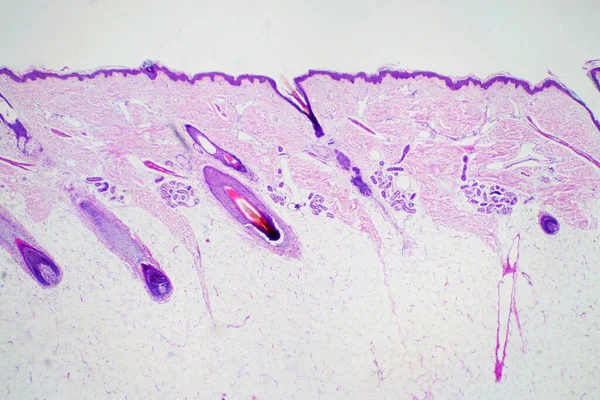
Cross Section Human Skin Head Under Microscope View For Education Histology. Histological For Human Physiology.
Image, 11.8MB, 6000 × 4000 jpg
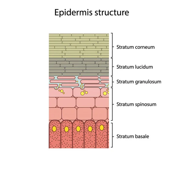
Histological Structure Of Epidermis - Skin Layers Shcematic Vector Illustration Showing Stratum Basale, Spinosum, Granulosum, Lucidum And Corneum
Vector, 5.89MB, 3090 × 3090 eps

Melanosome Transfer To Keratinocytes Scheme. Melanocyte Cell Biology And Skin Pigmentation Diagram. Melanin Pigment Production And Distribution Process. Vector Illustration.
Vector, 0.28MB, 4500 × 4500 eps
Melanocyte Cell Biology And Skin Tone Pigmentation Diagram. Melanin Pigment Production And Distribution Process. Melanosome Transfer To Keratinocytes Scheme. Vector Illustration.
Vector, 0.38MB, 4500 × 4500 eps
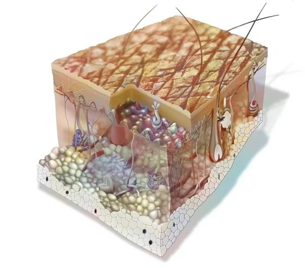
Skin Section Showing All The Structures: The Layers Of The Epidermis, The Structure Of The Dermis. .
Image, 1.78MB, 3937 × 3475 jpg
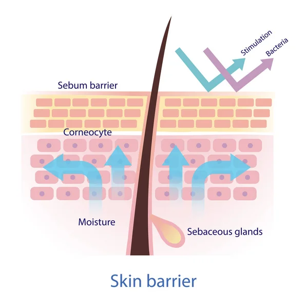
Healthy Protective Skin Barrier Vector On White Background. The Mechanical Protection Skin Barrier To Water Loss. The Sebaceous Glands Produce Sebum. The Sebum Protect Skin From Stimulation And Bacteria. Skin Care And Beauty Concept Illustration.
Vector, 5.82MB, 4583 × 4583 eps

Melanocyte Cell Biology And Skin Tone Pigmentation Diagram. Melanin Pigment Production. Eumelanin And Pheomelanin Distribution Process. Melanosome Transfer To Keratinocytes Scheme. Vector Illustration
Vector, 0.34MB, 4500 × 4500 eps
Page 1 >> Next