
Stock image Keratoplasty
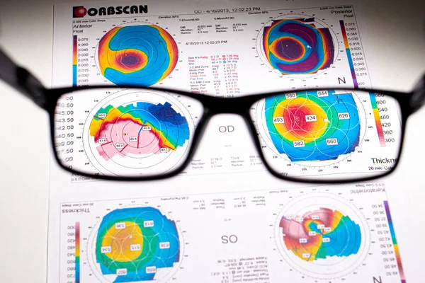
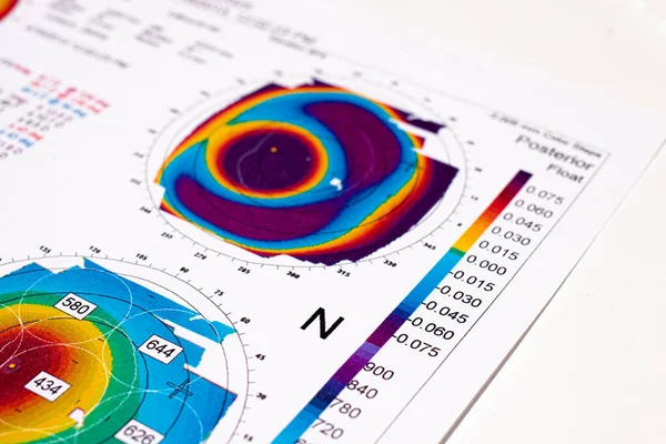
25.07.2020, Zarorizhzhya. Topography Of The Cornea Of The Eye, Layout. Keratoconus And Glasses. Keratotopography - Ophthalmological Examination.
Image, 8.75MB, 4720 × 3147 jpg

25.07.2020, Zarorizhzhya. Topography Of The Cornea Of The Eye, Layout. Keratoconus And Glasses. Keratotopography - Ophthalmological Examination.
Image, 10.41MB, 6000 × 4000 jpg
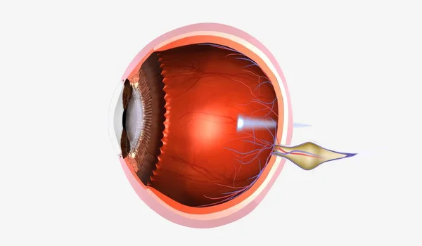
Presbyopia Is A Degenerative Eye Condition Characterized By The Gradual Inability To See Objects Up Close. 3D Rendering
Image, 5.08MB, 7191 × 4200 jpg


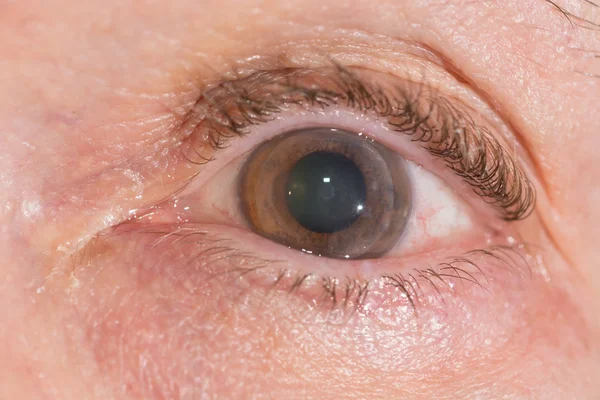
Thinning Corneal Dystrophy, Eye Keratotopography. A Rare Eye Disease.
Image, 13.04MB, 5939 × 3959 jpg

25.07.2020, Zarorizhzhya. Topography Of The Cornea Of The Eye, Layout. Keratoconus Of The Right Eye Is 3rd Degree And The Left Eye Is 2 Degrees.
Image, 13.44MB, 6000 × 4000 jpg
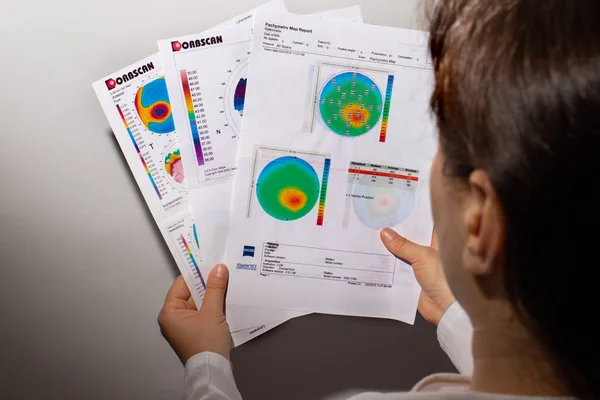
07.25.2020, Zarorizhzhya, Ukraine. Doctor Ophthalmologist Holds A Corneal Topography Examination For Keratoconus.
Image, 11.83MB, 6000 × 4000 jpg
25.07.2020, Zarorizhzhya. Topography Of The Cornea Of The Eye, Layout. Keratoconus Of The Right Eye Is 3rd Degree And The Left Eye Is 2 Degrees.
Image, 13.72MB, 6000 × 4000 jpg

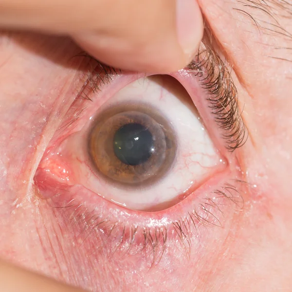
07.25.2020, Zarorizhzhya, Ukraine. A Patient With Keratoconus Holds An Ophthalmologic Examination Report. Keratometry, Corneal Dystrophy, Visual Impairment. A Rare Eye Disease.
Image, 11.49MB, 6000 × 4000 jpg
Page 1 >> Next