
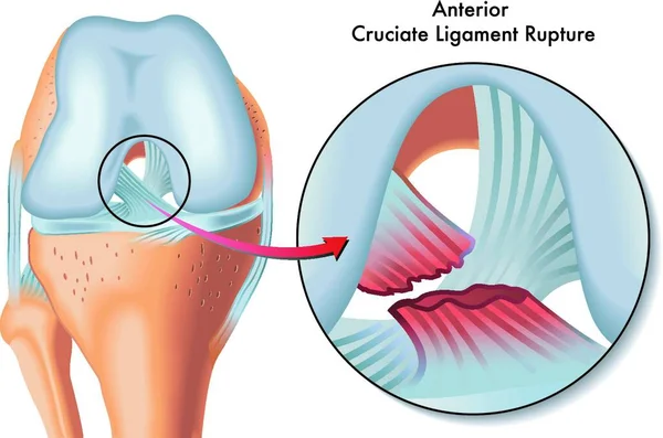
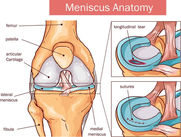
Stock image Ligament Tear
Man Suffering From Pain In Leg Injury After Sport Exercise Running Jogging And Workout Outdoor
Image, 7.46MB, 5500 × 3432 jpg

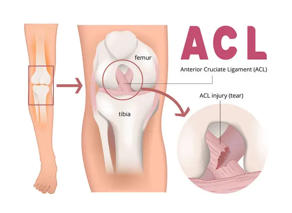
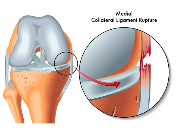
ACL Injury Or Trauma As Tear Or Sprain Of Anterior Cruciate Outline Concept
Vector, 5.83MB, 4200 × 3818 eps
Plantar Fasciitis, Plantar Fasciosis. Inflammatory And Degenerative Changes In Plantar Fascia. Pain During Walking. Vector 3D Rendering Of The Human Foot In Flexed Position
Vector, 18.4MB, 2000 × 2000 eps


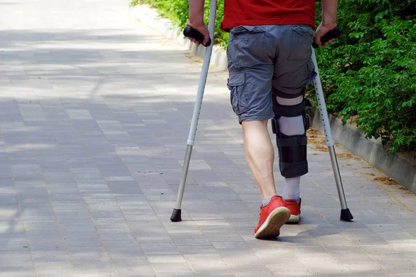
Violation Of The Musculoskeletal System. Sports Injury. Fixing Arch Support On The Leg. The Man Has Crutches In His Hands
Image, 10.3MB, 4500 × 3000 jpg


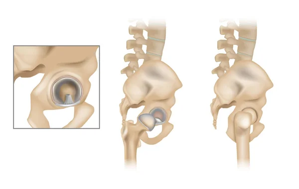
Anatomy Of The Hip And Acetabular Labrum. Ligamentum Teres And Articular Cartilage. Lateral View With Femur Removed Of Right Hip.
Vector, 25.08MB, 6000 × 3800 eps
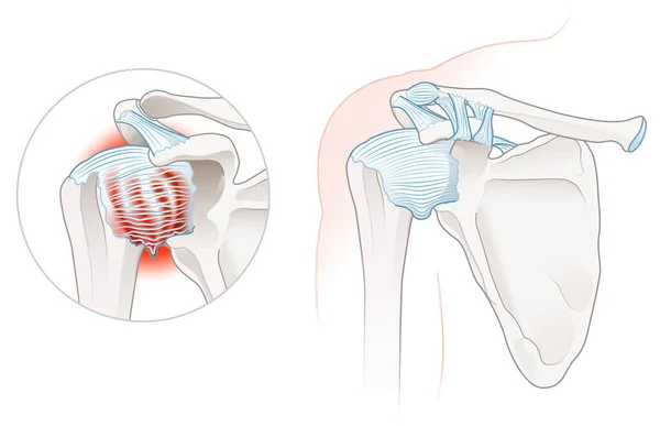
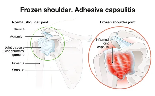
Illustration Showing Frozen Shoulder Adhesive Capsulitis Illustration. Labeled
Image, 2.51MB, 4988 × 3219 jpg

Anatomy Of The Hip And Acetabular Labrum. Ligamentum Teres And Articular Cartilage. Lateral View With Femur Removed Of Right Hip.
Vector, 21.36MB, 6000 × 3800 eps


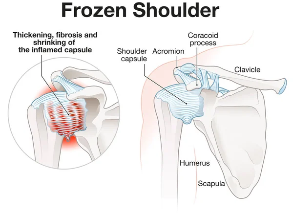
Illustration Showing Frozen Shoulder Adhesive Capsulitis Illustration. Labeled
Image, 3.01MB, 5000 × 3689 jpg
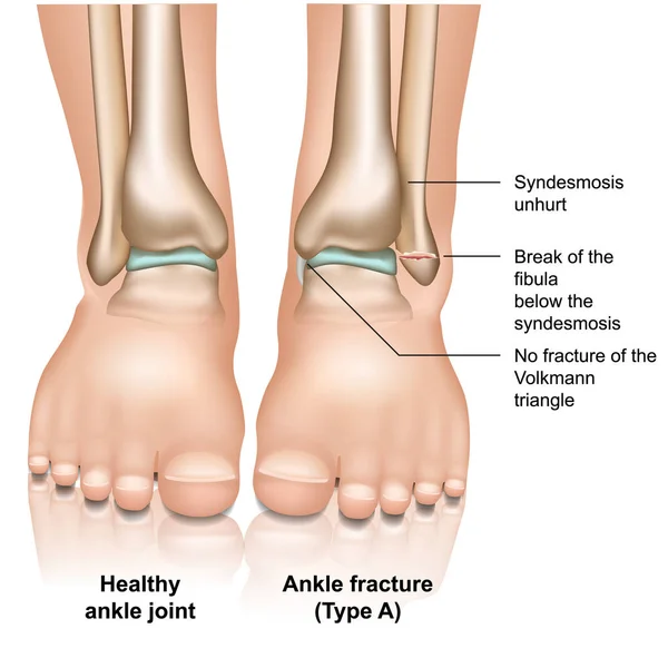
Ankle Joint Fracture Type A Medical Vector Illustration On White Background
Vector, 11.29MB, 5000 × 5000 eps
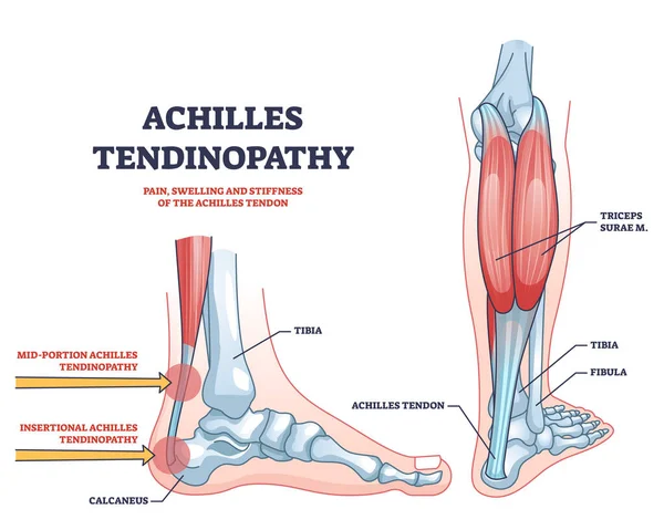
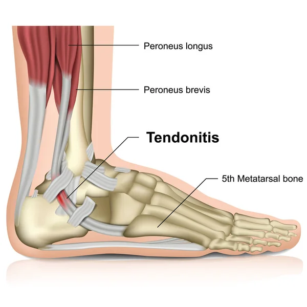
Achilles Tendinopathy As Injury To Tendon In Heel Outline Diagram. Labeled Educational Scheme With Anatomical Leg And Foot Skeleton And Muscles Vector Illustration.Trauma And Band Of Tissue Problem.
Vector, 9.54MB, 4750 × 3800 eps
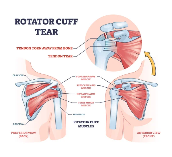
Rotator Cuff Tear As Shoulder Muscle Trauma Or Arm Injury Outline Diagram. Labeled Educational Upper Body Anatomy With Medical Tendon Torn Away From Bone Vector Illustration. Painful Joint Condition.
Vector, 16.95MB, 4200 × 3850 eps

Cuboid Syndrome As Orthopedic Trauma After Foot Torn Injury Outline Diagram. Labeled Educational Scheme With Feet Bone Dislocation And Free Movement Vector Illustration. Cuboid Subluxation Problem.
Vector, 6.4MB, 5000 × 3500 eps

Achilles Tendon Rupture As Painful Injury And Leg Trauma Outline Diagram. Labeled Educational Anatomical Scheme With Orthopedic Problem Explanation Vector Illustration. Medical Body Muscle Condition.
Vector, 10.37MB, 4706 × 4000 eps
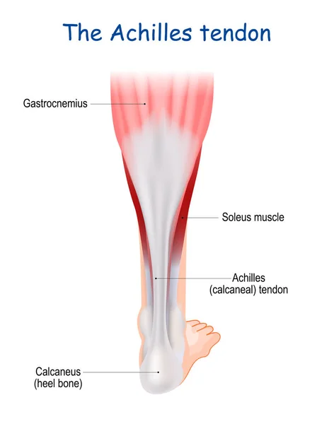
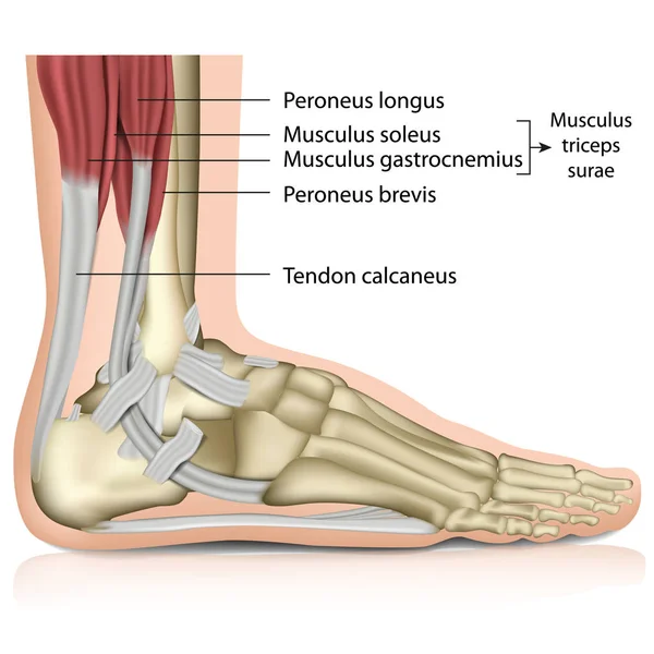
The Achilles Tendon Serves To Attach The Plantaris, Gastrocnemius (calf) And Soleus Muscles To The Calcaneus (heel) Bone. Heel Cord Or Calcaneal Tendon. Human Leg. Medical Infographic
Vector, 2.92MB, 3936 × 5000 eps
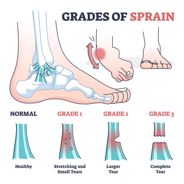
Grades Of Sprain As Ankle Or Foot Medical Injury Levels Outline Diagram
Vector, 6.03MB, 4000 × 4000 eps
Page 1 >> Next