photo free
Home Images Vectors Videos

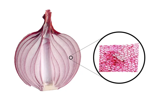
Stock image Light Micrograph

stock image Cuboidal epithelium of a mouse under the microscope.

Cuboidal Epithelium Of A Mouse Under The Microscope.

Image, 1.23MB, 4500 × 3000 jpg
stock image Tilia stem micrograph

Tilia Stem Micrograph

Image, 1.98MB, 2350 × 1763 jpg
stock image Tilia stem micrograph

Tilia Stem Micrograph

Image, 1.96MB, 2350 × 1763 jpg
stock image Tilia stem micrograph

Tilia Stem Micrograph

Image, 2.02MB, 2350 × 1763 jpg
stock image Tilia stem micrograph

Tilia Stem Micrograph

Image, 2.11MB, 2350 × 1763 jpg
stock image Cucurbita stem micrograph

Cucurbita Stem Micrograph

Image, 10.06MB, 6823 × 6325 jpg
stock image Tilia stem micrograph

Tilia Stem Micrograph

Image, 2.02MB, 2350 × 1763 jpg
stock image Tilia stem micrograph

Tilia Stem Micrograph

Image, 2.08MB, 2350 × 1763 jpg
stock image Tilia stem micrograph

Tilia Stem Micrograph

Image, 2.09MB, 2350 × 1763 jpg
stock image Tilia stem micrograph

Tilia Stem Micrograph

Image, 5.09MB, 3500 × 3500 jpg
stock image Tilia stem micrograph

Tilia Stem Micrograph

Image, 4.59MB, 3310 × 3310 jpg
stock image Cotton stem micrograph

Cotton Stem Micrograph

Image, 2.79MB, 3000 × 3000 jpg
stock image Light photomicrograph of an Onion epidermus cells seen through a microscope

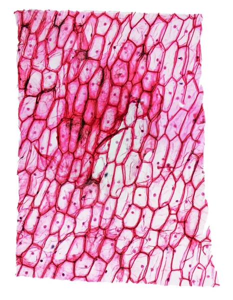
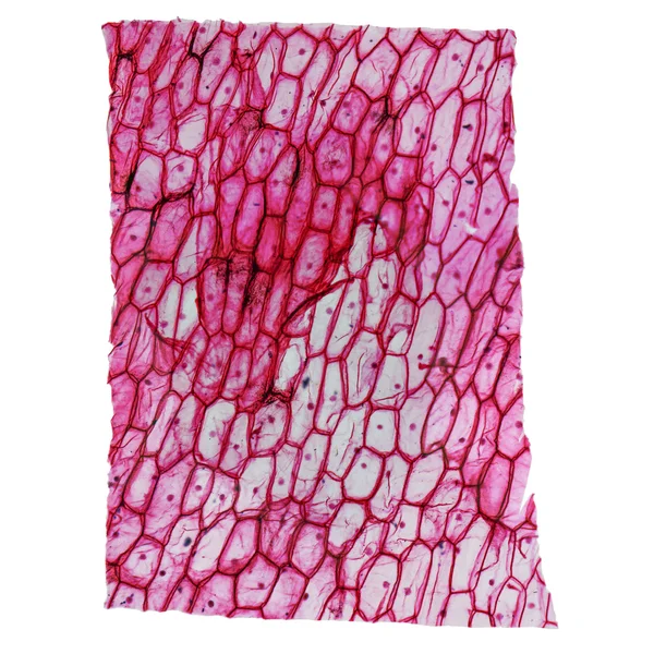
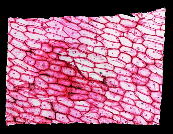
Light Photomicrograph Of An Onion Epidermus Cells Seen Through A Microscope

Image, 1.06MB, 2900 × 2172 jpg
stock image Cerebellum cross section tissue under the light microscope for pathology education. Haematoxylin and eosin staining technique for human tissue.

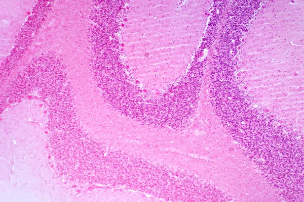
Cerebellum Cross Section Tissue Under The Light Microscope For Pathology Education. Haematoxylin And Eosin Staining Technique For Human Tissue.

Image, 22.42MB, 8192 × 5464 jpg
stock image Heliansthus stem micrograph

Heliansthus Stem Micrograph

Image, 4.09MB, 3000 × 3000 jpg
stock image Reticulosarcoma. Light micrograph from a lymph node in a case of reticulosarcoma, a malignant lymphoma type. The normal structure of the lymph node is totally occupied by sarcoma malignant cells.

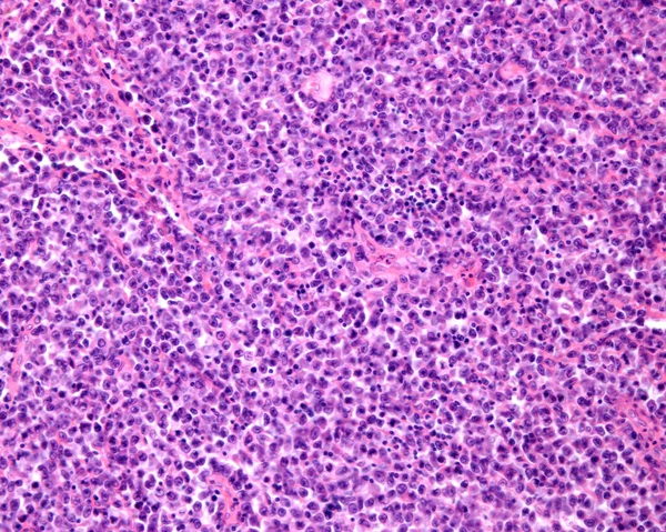
Reticulosarcoma. Light Micrograph From A Lymph Node In A Case Of Reticulosarcoma, A Malignant Lymphoma Type. The Normal Structure Of The Lymph Node Is Totally Occupied By Sarcoma Malignant Cells.

Image, 12.07MB, 3840 × 3072 jpg
stock image Tilia stem micrograph

Tilia Stem Micrograph

Image, 14.18MB, 5000 × 5000 jpg
stock image Onion epidermus micrograph

Onion Epidermus Micrograph

Image, 2.55MB, 3111 × 4000 jpg
stock image Endemic goiter, light micrograph, abnormal enlargement of the thyroid gland due to dietary iodine deficiency. Photomicrograph shows follicles of varying size, abundant colloid, lymphocytic infiltrate

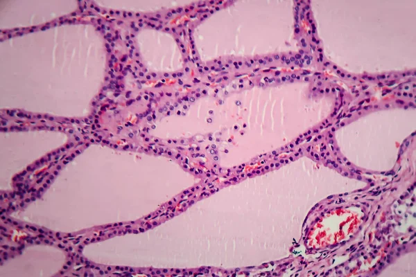
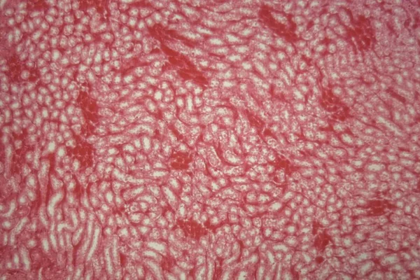
Endemic Goiter, Light Micrograph, Abnormal Enlargement Of The Thyroid Gland Due To Dietary Iodine Deficiency. Photomicrograph Shows Follicles Of Varying Size, Abundant Colloid, Lymphocytic Infiltrate

Image, 6.63MB, 4197 × 2798 jpg
stock image Onion epidermus micrograph

Onion Epidermus Micrograph

Image, 5.65MB, 5000 × 5000 jpg
stock image Onion epidermus micrograph

Onion Epidermus Micrograph

Image, 1.51MB, 3000 × 2333 jpg
stock image Corn root tip micrograph

Corn Root Tip Micrograph

Image, 4.74MB, 8500 × 4000 jpg
stock image Onion epidermus micrograph

Onion Epidermus Micrograph

Image, 1.68MB, 3000 × 3000 jpg
stock image Cells mitosis micrograph

Cells Mitosis Micrograph

Image, 1.82MB, 5500 × 3000 jpg
stock image Cells mitosis micrograph

Cells Mitosis Micrograph

Image, 0.56MB, 2000 × 2750 jpg
stock image Freshwater Closterium algae (unicellular charophyte green algae) - optical microscope x200 magnification

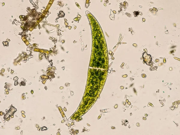
Freshwater Closterium Algae (unicellular Charophyte Green Algae) - Optical Microscope X200 Magnification

Image, 3.66MB, 2400 × 1799 jpg
stock image Black and white Onion epidermus micrograph

Black And White Onion Epidermus Micrograph

Image, 1.38MB, 2800 × 2178 jpg
stock image Black and white Tilia stem micrograph

Black And White Tilia Stem Micrograph

Image, 2.76MB, 2469 × 2469 jpg
stock image Onion epidermus micrograph

Onion Epidermus Micrograph

Image, 1.8MB, 3600 × 2400 jpg
stock image Microscope perfect detail

Microscope Perfect Detail

Image, 13.45MB, 3533 × 5300 jpg
stock image Modern microscope in laboratory

Modern Microscope In Laboratory

Image, 3.71MB, 2962 × 3663 jpg
stock image Beautiful microscope in hospital or laboratory interior

Beautiful Microscope In Hospital Or Laboratory Interior

Image, 5.44MB, 2333 × 3500 jpg
stock image A 3D scientific illustration showcasing a human body with transparent skin, revealing a tumor in his thyroid gland, along with a micrograph image of papillary thyroid carcinoma.

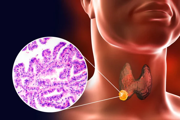
A 3D Scientific Illustration Showcasing A Human Body With Transparent Skin, Revealing A Tumor In His Thyroid Gland, Along With A Micrograph Image Of Papillary Thyroid Carcinoma.

Image, 7.63MB, 6750 × 4500 jpg
stock image An X-ray image of a bone, revealing intricate internal structures, representing medical precision and the human body's complexity

An X-ray Image Of A Bone, Revealing Intricate Internal Structures, Representing Medical Precision And The Human Body's Complexity

Image, 6.7MB, 5824 × 3264 jpg
stock image 3D illustration and light micrograph depicting a man with lungs affected by silicosis, revealing dark silicotic nodules, emphasizing respiratory health issues due to silica exposure.

3D Illustration And Light Micrograph Depicting A Man With Lungs Affected By Silicosis, Revealing Dark Silicotic Nodules, Emphasizing Respiratory Health Issues Due To Silica Exposure.

Image, 14.42MB, 6000 × 4000 jpg
stock image Tilia stem, cross section, 8X light micrograph. Stem of basswood, also known as linden, under the light microscope. Hematoxylin-eosin stained for better visualization. Isolated, over white, photo.

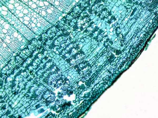
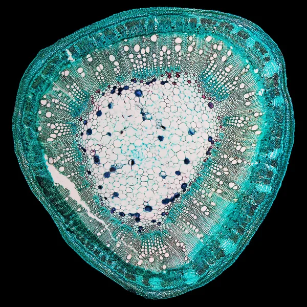
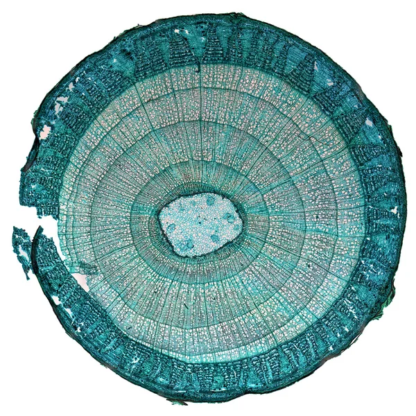
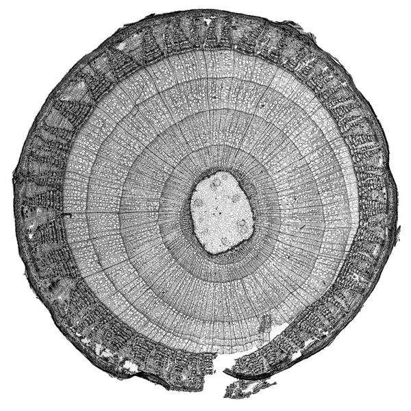
Tilia Stem, Cross Section, 8X Light Micrograph. Stem Of Basswood, Also Known As Linden, Under The Light Microscope. Hematoxylin-eosin Stained For Better Visualization. Isolated, Over White, Photo.

Image, 13.33MB, 5288 × 5288 jpg
stock image 3D illustration and light micrograph depicting a man with lungs affected by silicosis, revealing dark silicotic nodules, emphasizing respiratory health issues due to silica exposure.

3D Illustration And Light Micrograph Depicting A Man With Lungs Affected By Silicosis, Revealing Dark Silicotic Nodules, Emphasizing Respiratory Health Issues Due To Silica Exposure.

Image, 14.93MB, 6000 × 4000 jpg
stock image Close-up x-ray of a spine in vibrant blue and pink hues, capturing the delicate vertebrae.

Close-up X-ray Of A Spine In Vibrant Blue And Pink Hues, Capturing The Delicate Vertebrae.

Image, 10.26MB, 5824 × 3264 jpg
stock image Retina layers, light micrograph. From top to bottom: pigment epithelium, rods and cones, outer nuclear, outer plexiform, inner nuclear, inner plexiform, ganglion cell, and nerve fibre layers. A dilated artery and a vein appear in the ganglion cell la

Retina Layers, Light Micrograph. From Top To Bottom: Pigment Epithelium, Rods And Cones, Outer Nuclear, Outer Plexiform, Inner Nuclear, Inner Plexiform, Ganglion Cell, And Nerve Fibre Layers. A Dilated Artery And A Vein Appear In The Ganglion Cell La

Image, 9.2MB, 3840 × 3072 jpg
stock image 3D illustration and light micrograph depicting lungs affected by silicosis, revealing dark silicotic nodules, emphasizing respiratory health issues due to silica exposure.

3D Illustration And Light Micrograph Depicting Lungs Affected By Silicosis, Revealing Dark Silicotic Nodules, Emphasizing Respiratory Health Issues Due To Silica Exposure.

Image, 7.9MB, 7111 × 4000 jpg
stock image Alpine Squill (Scilla bifolia) in early spring forest

Alpine Squill (Scilla Bifolia) In Early Spring Forest

Image, 19.47MB, 5984 × 3707 jpg
stock image Willow stem, cross section, under the light microscope. Stem of Salix, also known as sallows and osiers, 8X light micrograph. Hematoxylin-eosin stained for better visualization. Isolated, over white.

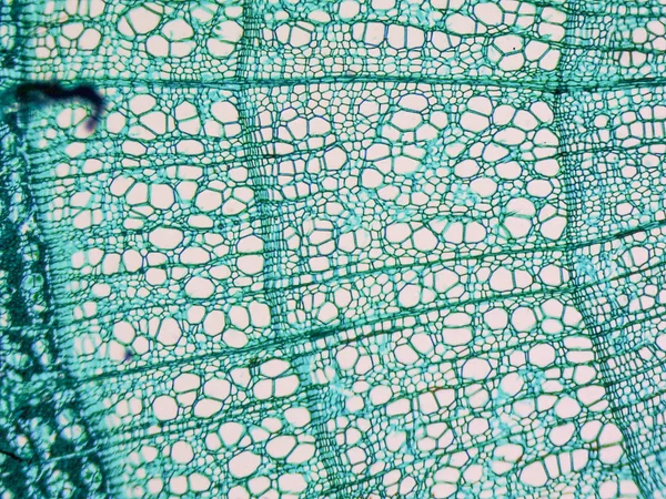
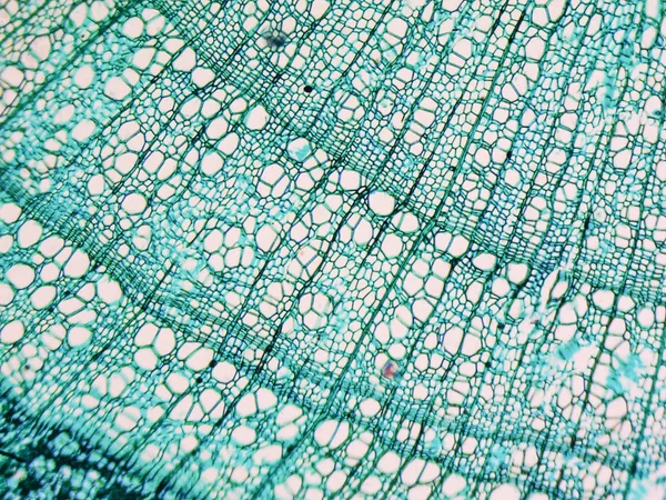
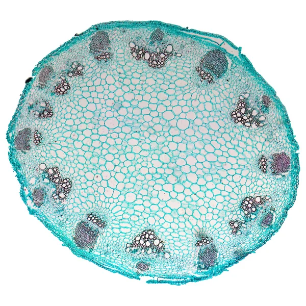
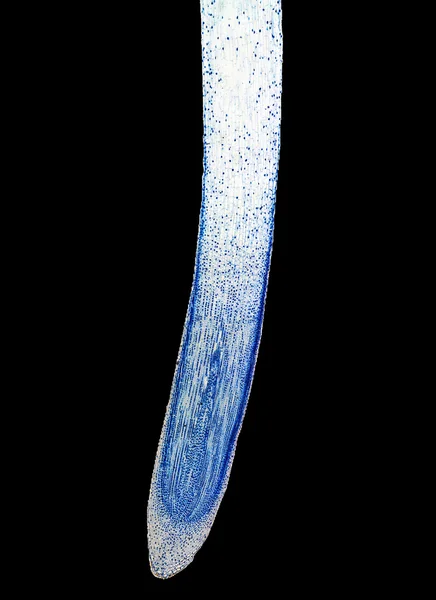
Willow Stem, Cross Section, Under The Light Microscope. Stem Of Salix, Also Known As Sallows And Osiers, 8X Light Micrograph. Hematoxylin-eosin Stained For Better Visualization. Isolated, Over White.

Image, 8.49MB, 3791 × 3791 jpg
stock image 3d illustration - Closterium Desmid

3d Illustration - Closterium Desmid

Image, 2.82MB, 5000 × 5000 jpg
stock image Mushroom gills, section, 8X light micrograph. Transverse section of a fungus cap with gills, stained for better visualization, under a light microscope. Three combined photos.

Mushroom Gills, Section, 8X Light Micrograph. Transverse Section Of A Fungus Cap With Gills, Stained For Better Visualization, Under A Light Microscope. Three Combined Photos.

Image, 11.07MB, 7848 × 5232 jpg
stock image 3d illustration - Freshwater Alga

3d Illustration - Freshwater Alga

Image, 0.38MB, 4000 × 3000 jpg
stock image Light micrograph revealing liver tissue with fatty infiltration, indicative of hepatic steatosis or fatty liver disease.

Light Micrograph Revealing Liver Tissue With Fatty Infiltration, Indicative Of Hepatic Steatosis Or Fatty Liver Disease.

Image, 29.69MB, 7178 × 4786 jpg
stock image Spirogyra, whole mount, under a light microscope. 20X light micrographs, 2 combined photos of stained water silk, also known as blanket weed and mermaids tresses, a filamentous charophyte green algae.

Spirogyra, Whole Mount, Under A Light Microscope. 20X Light Micrographs, 2 Combined Photos Of Stained Water Silk, Also Known As Blanket Weed And Mermaids Tresses, A Filamentous Charophyte Green Algae.

Image, 5.71MB, 9000 × 4521 jpg
stock image Clostridium Botulinum Bacteria closeup

Clostridium Botulinum Bacteria Closeup

Image, 1.64MB, 4000 × 2750 jpg
stock image 3d illustration - Amoeba proteus

3d Illustration - Amoeba Proteus

Image, 0.2MB, 4000 × 2750 jpg
stock image 3d illustration - Pronuclear Egg Cells

3d Illustration - Pronuclear Egg Cells

Image, 0.97MB, 5000 × 3500 jpg
Page 1 >> Next