photo free
Home Images Vectors Videos

Stock image Light Microscope

stock image optical microscope on white tablet in front of tiled wall

Optical Microscope On White Tablet In Front Of Tiled Wall

Image, 9.34MB, 4827 × 7233 jpg
stock image Laboratory Microscope background

Laboratory Microscope Background

Image, 1.76MB, 6271 × 4180 jpg
stock image modern optical microscope on white tablet in front of tiled wall

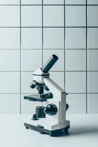
Modern Optical Microscope On White Tablet In Front Of Tiled Wall

Image, 9.21MB, 4912 × 7360 jpg
FREE IMAGE
stock image Close-up shot of Scientific Microscope in a Bright Modern Labora

Close-up Shot Of Scientific Microscope In A Bright Modern Labora

Image, 2.12MB, 5760 × 3240 jpg
stock image blood sample and optical microscope on white tablet in front of tiled wall

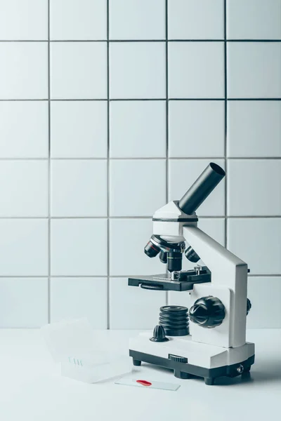
Blood Sample And Optical Microscope On White Tablet In Front Of Tiled Wall

Image, 7.93MB, 4718 × 7070 jpg
FREE IMAGE
stock image Microscope on a light background. Black and white microscope. Search for cancer cells. in the medical department of medicine. Salvation concept.

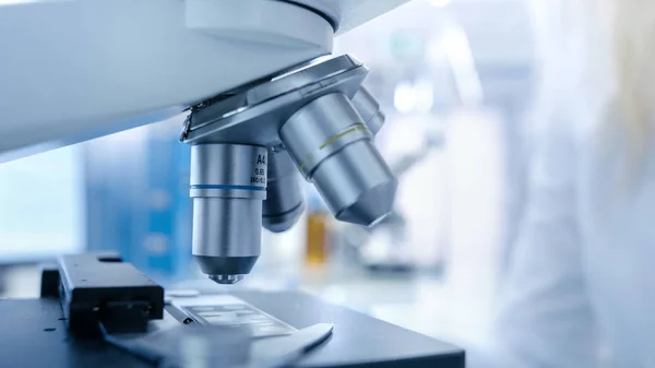
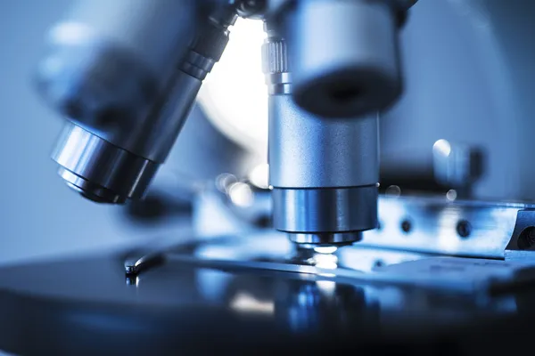
Microscope On A Light Background. Black And White Microscope. Search For Cancer Cells. In The Medical Department Of Medicine. Salvation Concept.

Image, 9.25MB, 6708 × 4472 jpg
stock image young biologist in medical mask and latex gloves working with microscope in laboratory

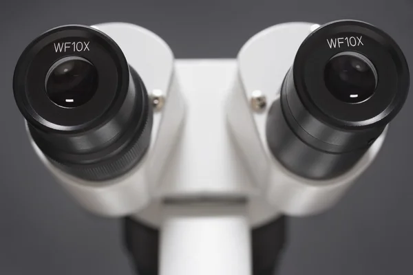
Young Biologist In Medical Mask And Latex Gloves Working With Microscope In Laboratory

Image, 12.27MB, 7360 × 4912 jpg
stock image Polarizing Light Microscope

Polarizing Light Microscope

Image, 2.13MB, 4493 × 3694 jpg
stock image cropped shot of doctor putting blood sample into microscope for examination

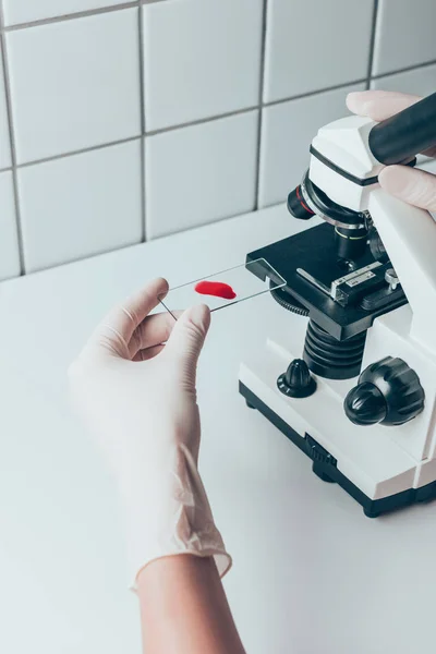
Cropped Shot Of Doctor Putting Blood Sample Into Microscope For Examination

Image, 10.43MB, 4912 × 7360 jpg
stock image Microscope Lens On Slide

Microscope Lens On Slide

Image, 16.77MB, 7365 × 4915 jpg
stock image Microscope and computer

Microscope And Computer

Image, 9.23MB, 7261 × 4846 jpg
stock image Scientific background with closeup on light microscope and labor

Scientific Background With Closeup On Light Microscope And Labor

Image, 2.58MB, 4470 × 2957 jpg
stock vector Colored background with different accessories

Colored Background With Different Accessories

Vector, 0.65MB, 5000 × 3000 eps
stock image female doctor with cap and face mask using light microscope observing virus on computer

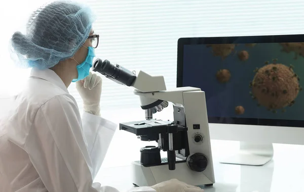
Female Doctor With Cap And Face Mask Using Light Microscope Observing Virus On Computer

Image, 12.96MB, 6587 × 4160 jpg
stock image pensive and sexy nurse in glasses sitting near women in white coats in laboratory

Pensive And Sexy Nurse In Glasses Sitting Near Women In White Coats In Laboratory

Image, 12.3MB, 4912 × 7360 jpg
stock vector Microscope isometric illustration with light microscope parts infographic elements isolated on white background.

Microscope Isometric Illustration With Light Microscope Parts Infographic Elements Isolated On White Background.

Vector, 6.2MB, 4168 × 4167 eps
stock image selective focus of sexy nurse reading science magazine while sitting near women and biting pencil

Selective Focus Of Sexy Nurse Reading Science Magazine While Sitting Near Women And Biting Pencil

Image, 12.33MB, 7360 × 4912 jpg
stock vector Pack Of Medical Equipment line Icons

Pack Of Medical Equipment Line Icons

Vector, 5.83MB, 8000 × 8000 eps
stock image cropped view of smiling african american nurse holding teddy bear in light clinic with microscope

Cropped View Of Smiling African American Nurse Holding Teddy Bear In Light Clinic With Microscope

Image, 14.09MB, 7360 × 4912 jpg
FREE IMAGE
stock image Heart muscle, light micrograph

Heart Muscle, Light Micrograph

Image, 4.46MB, 1918 × 4384 jpg
stock vector A conceptual education and e learning icons are here for your ease. The use of this set is not restricted to online education only, this is a perfect fit for any education related field. Enjoy!

A Conceptual Education And E Learning Icons Are Here For Your Ease. The Use Of This Set Is Not Restricted To Online Education Only, This Is A Perfect Fit For Any Education Related Field. Enjoy!

Vector, 5.9MB, 8000 × 8000 eps
stock image Adipose tissue

Adipose Tissue

Image, 10.4MB, 3840 × 3072 jpg
stock image Histopathology of prostate gland hyperplasia

Histopathology Of Prostate Gland Hyperplasia

Image, 11.54MB, 4928 × 2772 jpg
stock image Adipose tissue

Adipose Tissue

Image, 10.61MB, 3840 × 3072 jpg
stock image Heart muscle, light micrograph

Heart Muscle, Light Micrograph

Image, 4.33MB, 4121 × 2747 jpg
stock image Photomicrograph of prostate hyperplasia

Photomicrograph Of Prostate Hyperplasia

Image, 5.11MB, 3876 × 2584 jpg
stock image Onion epidermis under light microscope. Purple colored, large epidermal cells of an onion, Allium cepa, in a single layer. Each cell with wall, membrane, cytoplasm, nucleus and large vacuole.

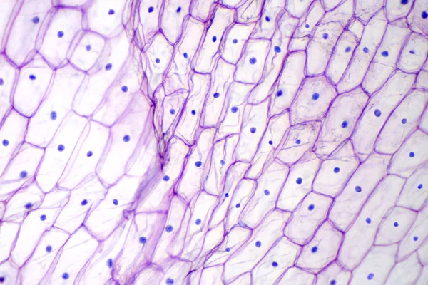
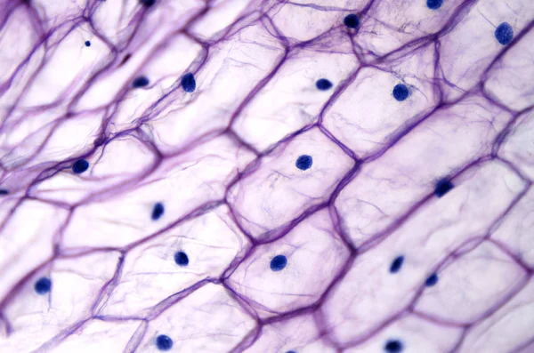
Onion Epidermis Under Light Microscope. Purple Colored, Large Epidermal Cells Of An Onion, Allium Cepa, In A Single Layer. Each Cell With Wall, Membrane, Cytoplasm, Nucleus And Large Vacuole.

Image, 3.62MB, 3786 × 2524 jpg
stock image Histopathology of prostate gland hyperplasia

Histopathology Of Prostate Gland Hyperplasia

Image, 13.07MB, 4928 × 2772 jpg
stock image Histopathology of prostate gland hyperplasia

Histopathology Of Prostate Gland Hyperplasia

Image, 11.13MB, 4928 × 2772 jpg
stock image Pine mature wood cross section. Light microscope slide with microsection of an evergreen conifer in the genus Pinus. Plant anatomy. Biology. Photo.

Pine Mature Wood Cross Section. Light Microscope Slide With Microsection Of An Evergreen Conifer In The Genus Pinus. Plant Anatomy. Biology. Photo.

Image, 16.74MB, 4928 × 3264 jpg
stock vector The working cycle of doctors, nurses, laboratory staff who work as a team, starting from the doctor, examining the patients, getting in the car and checking in the laboratory.vector illustration.

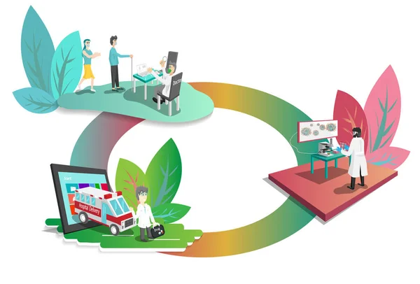
The Working Cycle Of Doctors, Nurses, Laboratory Staff Who Work As A Team, Starting From The Doctor, Examining The Patients, Getting In The Car And Checking In The Laboratory.vector Illustration.

Vector, 14.46MB, 9390 × 6354 eps
stock image Heart muscle, light micrograph

Heart Muscle, Light Micrograph

Image, 4.13MB, 3937 × 2625 jpg
stock image Histopathology of prostate gland hyperplasia

Histopathology Of Prostate Gland Hyperplasia

Image, 13.94MB, 4928 × 2772 jpg
stock image Photomicrograph of prostate hyperplasia

Photomicrograph Of Prostate Hyperplasia

Image, 4.24MB, 3876 × 2584 jpg
stock image Teacher of biology holds book and microscope. Lady in formal wear on calm face in classroom. Biology concept. Lady scientist holds book and microscope, chalkboard on background, copy space

Teacher Of Biology Holds Book And Microscope. Lady In Formal Wear On Calm Face In Classroom. Biology Concept. Lady Scientist Holds Book And Microscope, Chalkboard On Background, Copy Space

Image, 8.81MB, 4778 × 3245 jpg
stock image Liquefactive necrosis of the human brain, light photomicrograph

Liquefactive Necrosis Of The Human Brain, Light Photomicrograph

Image, 3.39MB, 3317 × 2211 jpg
stock image Liquefactive necrosis of the human brain, light photomicrograph

Liquefactive Necrosis Of The Human Brain, Light Photomicrograph

Image, 4.96MB, 3876 × 2584 jpg
stock image Human blood smear showing a neutrophil leukocyte. The small bluish dots among the red blood cells are platelets.

Human Blood Smear Showing A Neutrophil Leukocyte. The Small Bluish Dots Among The Red Blood Cells Are Platelets.

Image, 7.99MB, 3840 × 3072 jpg
stock image Human lung affected by an acute bronchopneumonia, commonly a hospital-acquired bacterial pneumonia. The lumen of alveoli is occupied by liquid of oedema which contains acute inflammatory infiltrates (with predominance of neutrophil granulocytes). On

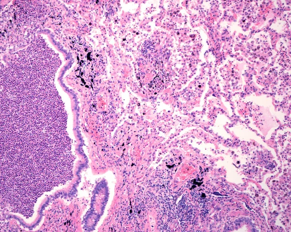
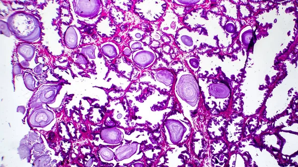
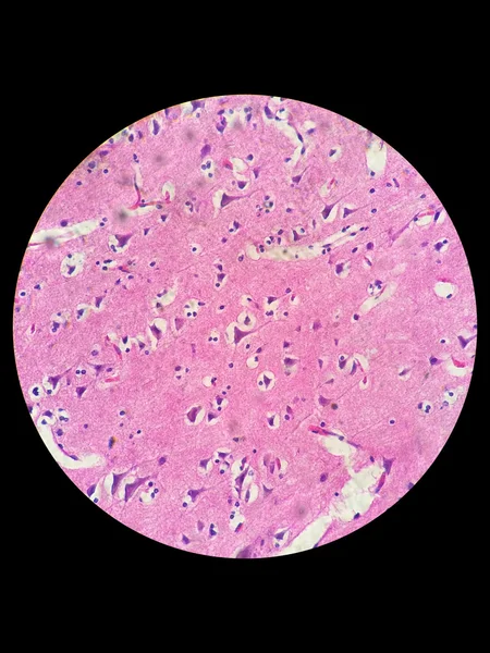
Human Lung Affected By An Acute Bronchopneumonia, Commonly A Hospital-acquired Bacterial Pneumonia. The Lumen Of Alveoli Is Occupied By Liquid Of Oedema Which Contains Acute Inflammatory Infiltrates (with Predominance Of Neutrophil Granulocytes). On

Image, 11.33MB, 3840 × 3072 jpg
stock image Portal triad

Portal Triad

Image, 15.47MB, 3840 × 3072 jpg
stock image Reticulosarcoma. Light micrograph from a lymph node in a case of reticulosarcoma, a malignant lymphoma type. The normal structure of the lymph node is totally occupied by sarcoma malignant cells.

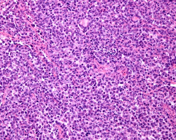
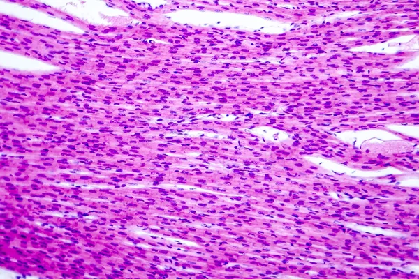
Reticulosarcoma. Light Micrograph From A Lymph Node In A Case Of Reticulosarcoma, A Malignant Lymphoma Type. The Normal Structure Of The Lymph Node Is Totally Occupied By Sarcoma Malignant Cells.

Image, 12.07MB, 3840 × 3072 jpg
stock image Pseudostratified epithelium is a type of epithelium that, though comprising only a single layer of cells.

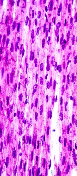
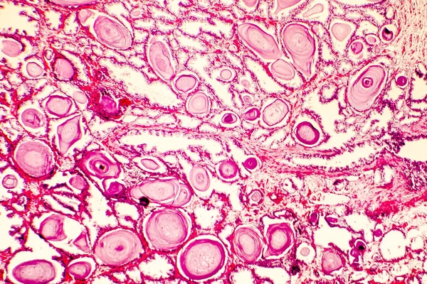
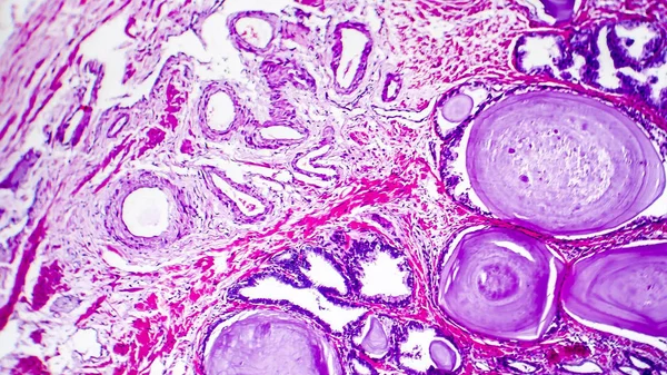
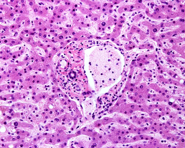
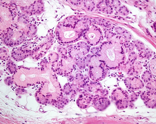
Pseudostratified Epithelium Is A Type Of Epithelium That, Though Comprising Only A Single Layer Of Cells.

Image, 11.24MB, 5840 × 3893 jpg
stock image Heart hypertrophy photomicrograph

Heart Hypertrophy Photomicrograph

Image, 5.33MB, 3876 × 2584 jpg
stock image Photomicrograph of prostate hyperplasia

Photomicrograph Of Prostate Hyperplasia

Image, 4.64MB, 3876 × 2584 jpg
stock image Onion epidermis with large cells under light microscope

Onion Epidermis With Large Cells Under Light Microscope

Image, 12.07MB, 4873 × 3228 jpg
stock image Nerve cells under light microscope

Nerve Cells Under Light Microscope

Image, 2.18MB, 2448 × 3264 jpg
stock image Mixed exocrine gland

Mixed Exocrine Gland

Image, 16.87MB, 3840 × 3072 jpg
stock image Human Cheek Epithelial Cells. The tissue that lines the inside of the mouth is known as the basal mucosa and is composed of squamous epithelial cells. Education pathology.

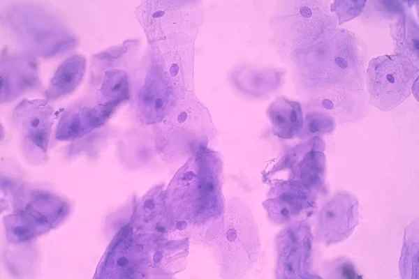
Human Cheek Epithelial Cells. The Tissue That Lines The Inside Of The Mouth Is Known As The Basal Mucosa And Is Composed Of Squamous Epithelial Cells. Education Pathology.

Image, 14.21MB, 6000 × 4000 jpg
stock image Education Spinal cord  and Motor Neuron under the microscope in Lab.

Education Spinal Cord And Motor Neuron Under The Microscope In Lab.

Image, 12.9MB, 6720 × 4480 jpg
stock image Education Spinal cord  and Motor Neuron under the microscope in Lab.

Education Spinal Cord And Motor Neuron Under The Microscope In Lab.

Image, 11.03MB, 6720 × 4480 jpg
Page 1 >> Next