
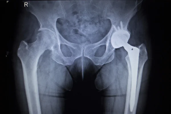
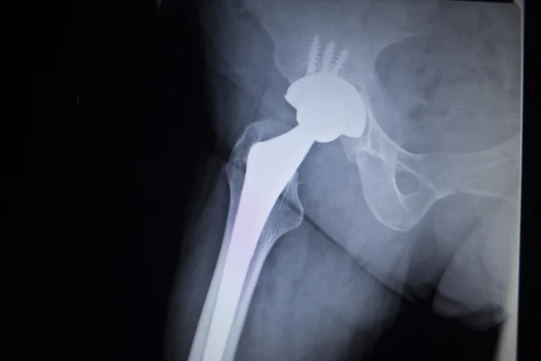
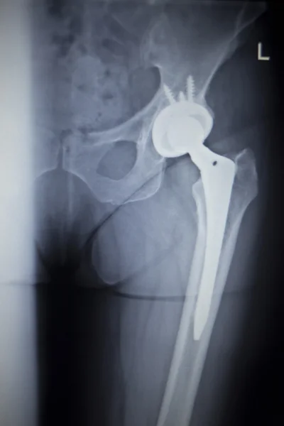
Stock image Lower Extremity
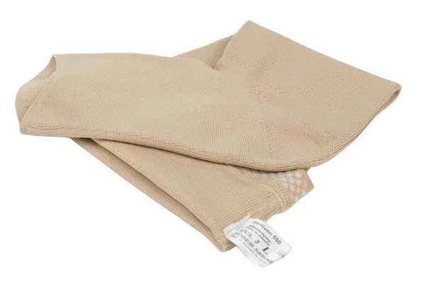
Burgas, Bulgaria - 06.03.2020: Close Up Of Mediven 550 Flat Knit Graduated Compression Garments For Leg Lymphedema, Edema And Lipedema. CCL 3. Illustrative Editorial.
Image, 5.86MB, 6000 × 4000 jpg
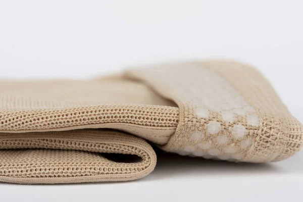
Close Up Of Flat Knit Graduated Compression Garments For Leg Lymphedema, Edema And Lipedema - Powerful Compression Stocking For Greater Edema Containment
Image, 7.35MB, 6000 × 4000 jpg
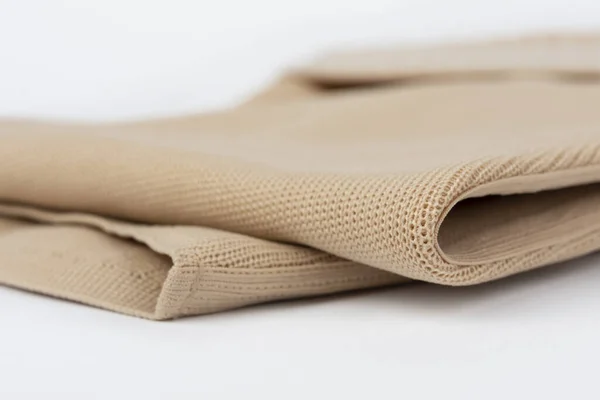
Close Up Of Flat Knit Graduated Compression Garments For Leg Lymphedema, Edema And Lipedema - Powerful Compression Stocking For Greater Edema Containment
Image, 6.31MB, 6000 × 4000 jpg
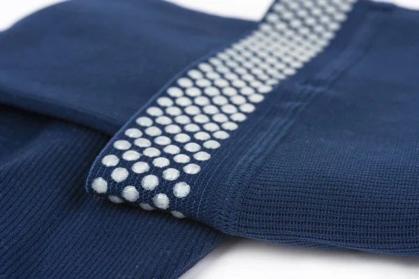
Close Up Of Flat Knit Graduated Compression Garments For Leg Lymphedema, Edema And Lipedema - Powerful Compression Stocking For Greater Edema Containment
Image, 11.05MB, 6000 × 4000 jpg
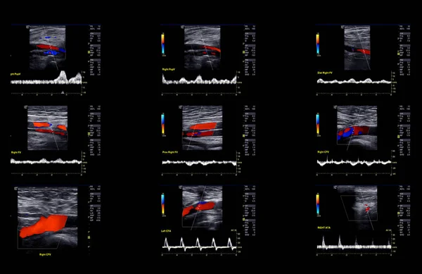
Ultrasound Doppler For Finding Deep Vein Thrombosis Of Lower Extremity.
Image, 1.35MB, 3280 × 2128 jpg
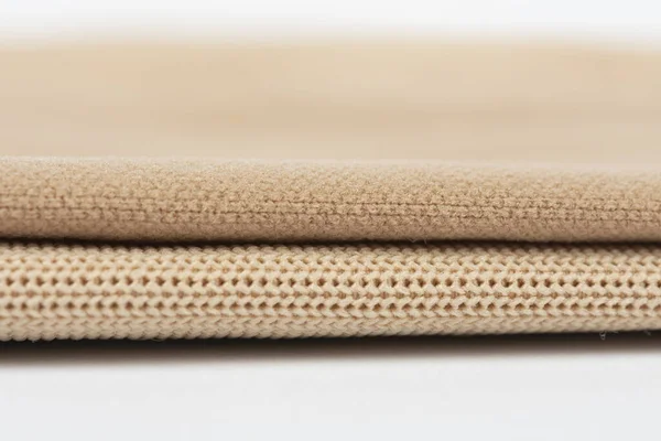
Close Up Of Compression Garments For Lymphedema, Edema And Lipedema - The Difference Between Flat Knit And Circular Knit
Image, 6.49MB, 5524 × 3682 jpg

Man Lying Supine While Radiologic Technologist Moving Examination Table Into CT Gantry Aperture With Hand-held Controller
Image, 13.39MB, 7008 × 4672 jpg


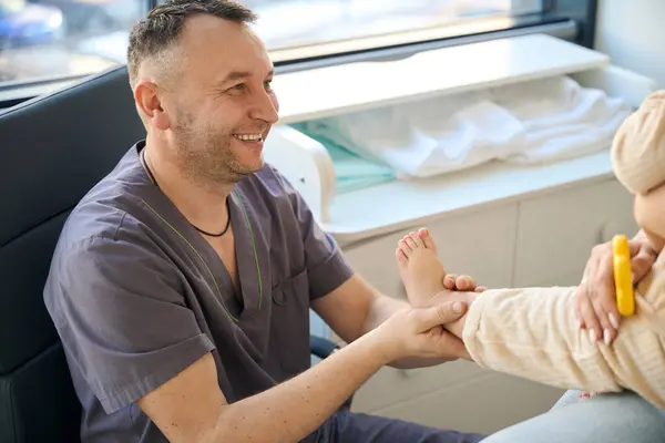
Child Seated On Mother Lap While Qualified Reflexologist Massaging Plantar Surface Of Its Foot
Image, 14.58MB, 6812 × 4541 jpg
Close Up Of Compression Garments For Lymphedema, Edema And Lipedema - The Difference Between Flat Knit And Circular Knit
Image, 7.07MB, 6000 × 3329 jpg

Cropped Photo Of Massotherapist Applying Pressure With Thumbs To Acupoints On Client Calf Muscle
Image, 14.75MB, 6847 × 4565 jpg

Masseur Performing Effleurage Strokes On Patient Calf Muscle While Pressing With His Forearm On Plantar Surface Of Her Foot
Image, 14.9MB, 7008 × 4672 jpg


Cutaneous Nerve Innervation Of The Lower Limbs, Highlighting The Various Nerve Branches And Regions Diagram Hand Drawn Schematic Vector Illustration. Medical Science Educational Illustration
Vector, 0.98MB, 5000 × 3750 eps

Cropped Photo Of Baby Seated On Parent Lap While Reflexologist Stimulating Acupressure Point On Top Of Its Foot
Image, 15.92MB, 6812 × 4541 jpg


Cutaneous Nerve Innervation Of The Lower Limbs, Highlighting The Various Nerve Branches And Regions Diagram Hand Drawn Schematic Raster Illustration. Medical Science Educational Illustration
Image, 3.17MB, 6000 × 4500 jpg


Closeup Of Doctor Hands In Nitrile Gloves Inserting Syringe Needle Into Patient Knee Under Ultrasound Guidance
Image, 14.27MB, 6812 × 4541 jpg

Cropped Photo Of Masseur Thumb Pressing On Acupoints On Patient Calf Muscle
Image, 15.34MB, 4565 × 6847 jpg

Side View Of Serious Massotherapist Performing Two-handed Kneading On Client Calf Muscles
Image, 15.5MB, 7008 × 4672 jpg


Smiling Massotherapist Looking At Baby Seated On Mother Lap While Massaging Its Calf Muscle
Image, 15.01MB, 6812 × 4541 jpg

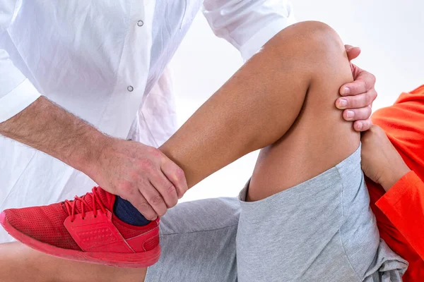
Woman Wearing Leg Brace After Surgery On Knee Fracture Trying To Walk With Walker. Rehabilitation After Injury. Closeup
Image, 11.3MB, 4016 × 6016 jpg

Page 1 >> Next