photo free
Home Images Vectors Videos

Stock image Lumber Vertebrae

stock image MRI scan of lumbar spines of a patient finding Spinal mass at Lt.side T12-L1 level Severe bulging disc L3-4 causing bilateral L4 nerve root compression  and spinal stenosis on arrow point.

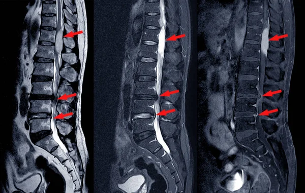
MRI Scan Of Lumbar Spines Of A Patient Finding Spinal Mass At Lt.side T12-L1 Level Severe Bulging Disc L3-4 Causing Bilateral L4 Nerve Root Compression And Spinal Stenosis On Arrow Point.

Image, 6.3MB, 6181 × 3928 jpg
stock image Cervical spine anatomy. Human skeleton. Medically accurate 3D illustration

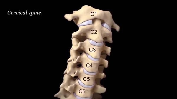
Cervical Spine Anatomy. Human Skeleton. Medically Accurate 3D Illustration

Image, 0.33MB, 3840 × 2160 jpg
stock vector Vertebral column

Vertebral Column

Vector, 3.08MB, 3801 × 5444 eps
stock image The cervical nerves are the spinal nerves from the cervical vertebrae in the cervical segment of the spinal cord

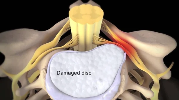
The Cervical Nerves Are The Spinal Nerves From The Cervical Vertebrae In The Cervical Segment Of The Spinal Cord

Image, 0.63MB, 3840 × 2160 jpg
stock vector Artistic style anatomical spine vector illustration with conceptual decorative elements. Cervical,thoracic,lumber sections scheme

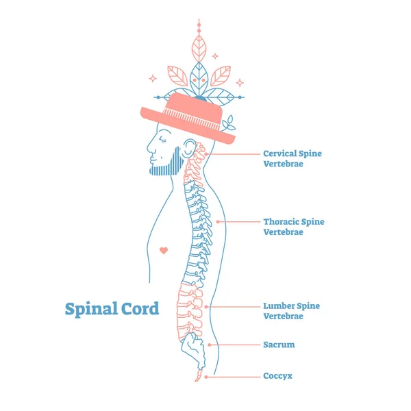
Artistic Style Anatomical Spine Vector Illustration With Conceptual Decorative Elements. Cervical,thoracic,lumber Sections Scheme

Vector, 1.12MB, 4000 × 4000 eps
stock image Businessman with lower back ache

Businessman With Lower Back Ache

Image, 3.56MB, 4592 × 3061 jpg
stock image Plain X ray lumbosacral spine revealed straightened, mild scoliotic deformity of  lumber spine, spondylotic changes, bilateral Sacroiliitis, mild narrowing of L4-L5, L5- S1 disc spaces

Plain X Ray Lumbosacral Spine Revealed Straightened, Mild Scoliotic Deformity Of Lumber Spine, Spondylotic Changes, Bilateral Sacroiliitis, Mild Narrowing Of L4-L5, L5- S1 Disc Spaces

Image, 14.74MB, 5765 × 3385 jpg
Page 1 >> Next