photo free
Home Images Vectors Videos

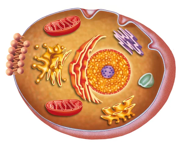
Stock image Membrane Receptors

stock image Cell membrane receptors

Cell Membrane Receptors

Image, 4.88MB, 3840 × 2160 jpg
stock vector Hormones, Receptors and Target Cells. each type of hormone is designed only certain cells. These cells will have receptors on them that are specific for a certain hormone. Vector illustration for medical, biological, and educational use

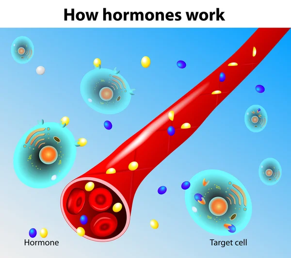
Hormones, Receptors And Target Cells. Each Type Of Hormone Is Designed Only Certain Cells. These Cells Will Have Receptors On Them That Are Specific For A Certain Hormone. Vector Illustration For Medical, Biological, And Educational Use

Vector, 2.77MB, 5013 × 5012 eps
stock vector medical illustration of elements of synapse

Medical Illustration Of Elements Of Synapse

Vector, 0MB, 4000 × 3866 zip
stock vector Medical illustration of elements of synapse

Medical Illustration Of Elements Of Synapse

Vector, 0.67MB, 5333 × 4000 eps
stock vector Mast Cell Activation Syndrome. White blood cell

Mast Cell Activation Syndrome. White Blood Cell

Vector, 1.84MB, 5000 × 4167 eps
stock vector Hormones work. Vector

Hormones Work. Vector

Vector, 10.94MB, 4958 × 4401 eps
stock vector Structure of the Plasma cell

Structure Of The Plasma Cell

Vector, 12.29MB, 4713 × 3438 eps
stock image Receiving cell

Receiving Cell

Image, 3.32MB, 3336 × 3077 jpg
stock image LDL particles binding to LDL receptors on the cell membrane. The binding of LDL particles to the LDL receptors mediates the endocytosis of the particles through clathrin coated vesicles. 3d rendering. Illustration

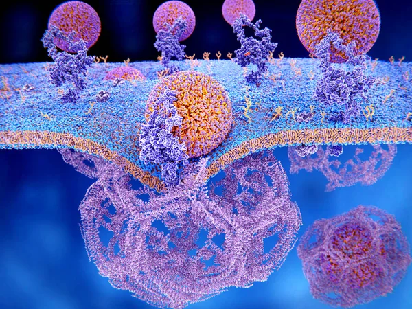
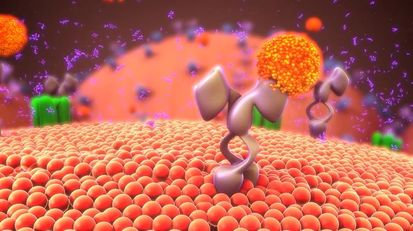
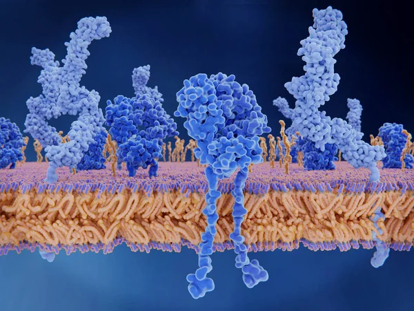
LDL Particles Binding To LDL Receptors On The Cell Membrane. The Binding Of LDL Particles To The LDL Receptors Mediates The Endocytosis Of The Particles Through Clathrin Coated Vesicles. 3d Rendering. Illustration

Image, 11.34MB, 8000 × 6000 jpg
stock image Anatomy and characteristics of the human cell

Anatomy And Characteristics Of The Human Cell

Image, 3.48MB, 3035 × 2400 jpg
stock image The T-cell receptor activates the immune response to antigens in T-lymphocytes. T-cell receptors (dark blue), CD4 molecules (light blue), glycolipids (orange). 3d rendering. Illustration

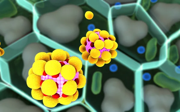
The T-cell Receptor Activates The Immune Response To Antigens In T-lymphocytes. T-cell Receptors (dark Blue), CD4 Molecules (light Blue), Glycolipids (orange). 3d Rendering. Illustration

Image, 3.11MB, 8000 × 6000 jpg
stock image Molecule structure

Molecule Structure

Image, 18.78MB, 8000 × 5000 jpg
stock image Virus pathogen or virus particle interacting with cell - 3d illustration

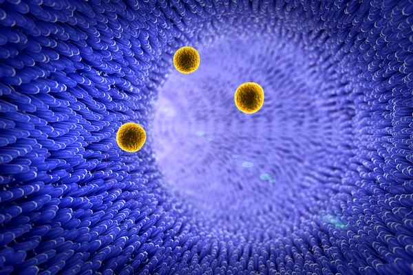
Virus Pathogen Or Virus Particle Interacting With Cell - 3d Illustration

Image, 9.73MB, 5800 × 3013 jpg
stock image Artery, cholesterol

Artery, Cholesterol

Image, 8.55MB, 6000 × 4000 jpg
stock image Capillary and cells

Capillary And Cells

Image, 16.55MB, 8000 × 6000 jpg
stock image Ebola glycoprotein molecule isolated on black

Ebola Glycoprotein Molecule Isolated On Black

Image, 3.22MB, 3607 × 3404 jpg
stock image Ebola glycoprotein molecule isolated on blue

Ebola Glycoprotein Molecule Isolated On Blue

Image, 3.4MB, 3757 × 3546 jpg
stock image Steroids hormones background concept glowing

Steroids Hormones Background Concept Glowing

Image, 2.68MB, 6000 × 3963 jpg
stock image an activated platelet, disc-like form transforms upon activation, developing spiky protrusions called pseudopodia. These extend outward, resembling thorny rays, and help platelets adhere to each other and injured blood vessel walls.

An Activated Platelet, Disc-like Form Transforms Upon Activation, Developing Spiky Protrusions Called Pseudopodia. These Extend Outward, Resembling Thorny Rays, And Help Platelets Adhere To Each Other And Injured Blood Vessel Walls.

Image, 5.74MB, 5000 × 5000 jpg
Page 1 >> Next