photo free
Home Images Vectors Videos

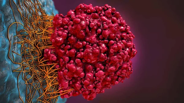
Stock image Metastasis page 4

stock image CT colonography , This imaging technique is often employed for colorectal cancer screening, providing detailed images of the colon's interior 3D rendering.

CT Colonography , This Imaging Technique Is Often Employed For Colorectal Cancer Screening, Providing Detailed Images Of The Colon's Interior 3D Rendering.

Image, 0.5MB, 3840 × 2160 jpg
stock image Prostate cancer symptoms, a small walnut-shaped gland in men that produces the seminal fluid that nourishes and transports sperm

Prostate Cancer Symptoms, A Small Walnut-shaped Gland In Men That Produces The Seminal Fluid That Nourishes And Transports Sperm

Image, 8.06MB, 5472 × 3648 jpg
stock image cancer cells and Lymphocytes

Cancer Cells And Lymphocytes

Image, 9.36MB, 8000 × 6000 jpg
stock image Primary myelofibrosis (PMF) cells in blood flow - isometric view 3d illustration

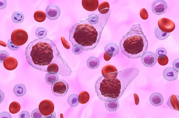
Primary Myelofibrosis (PMF) Cells In Blood Flow - Isometric View 3d Illustration

Image, 7.02MB, 10000 × 6600 jpg
stock image Hepatocellular cancer of liver of a human

Hepatocellular Cancer Of Liver Of A Human

Image, 8.46MB, 2592 × 1944 jpg
stock image T-Cells of the immune System attacking growing Cancer cells

T-Cells Of The Immune System Attacking Growing Cancer Cells

Image, 15.92MB, 6000 × 4000 jpg
stock vector breast cancer

Breast Cancer

Vector, 0.18MB, 4167 × 4167 eps
stock image Acute myeloid leukemia (AML) cells in blood flow - section view 3d illustration

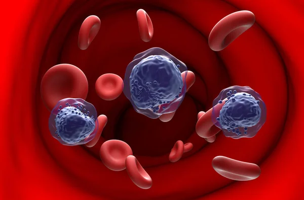
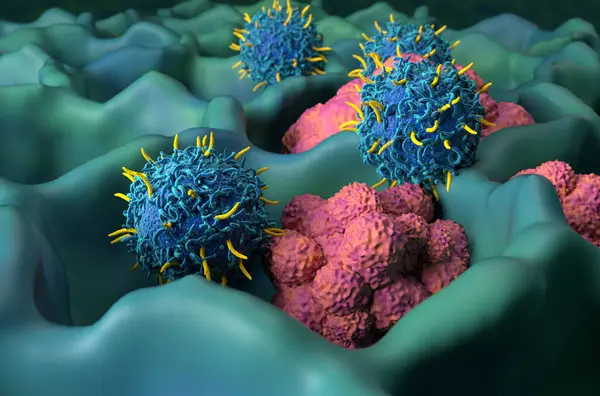
Acute Myeloid Leukemia (AML) Cells In Blood Flow - Section View 3d Illustration

Image, 7.29MB, 10000 × 6600 jpg
stock image 3d rendered illustration of the female pancreas

3d Rendered Illustration Of The Female Pancreas

Image, 10.82MB, 5337 × 6359 jpg
stock vector lymph node

Lymph Node

Vector, 12.72MB, 5642 × 4290 eps
stock vector medical Illustration of the symptoms of testicular cancer

Medical Illustration Of The Symptoms Of Testicular Cancer

Vector, 0MB, 4000 × 3820 zip
stock image tumor cell in the moment that divides, 3d illustration

Tumor Cell In The Moment That Divides, 3d Illustration

Image, 15.59MB, 5001 × 3865 jpg
stock vector Mesothelioma tumor cells poster. Lung cancer concept. Respiratory system illness. Asbestos related diseases. Shortness of breath, pain in chest, breathing problem, medical flat vector illustration.

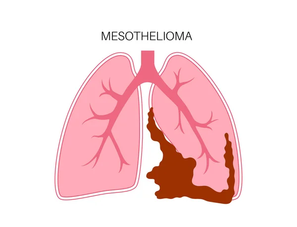
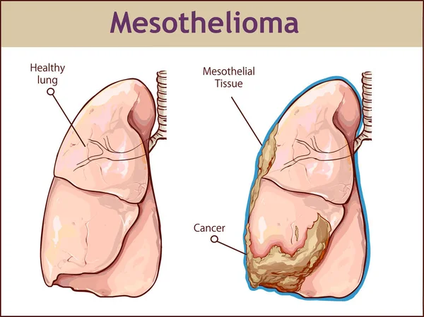
Mesothelioma Tumor Cells Poster. Lung Cancer Concept. Respiratory System Illness. Asbestos Related Diseases. Shortness Of Breath, Pain In Chest, Breathing Problem, Medical Flat Vector Illustration.

Vector, 0.35MB, 4764 × 3647 eps
stock vector  vector illustration of a Pleural mesothelioma.

Vector Illustration Of A Pleural Mesothelioma.

Vector, 7.07MB, 9576 × 7171 eps
stock image A growing Cancer Tumor spreading on healthy Tissue

A Growing Cancer Tumor Spreading On Healthy Tissue

Image, 6.99MB, 5000 × 3333 jpg
stock image Doctor attentively examines the MRI scan of the patient

Doctor Attentively Examines The MRI Scan Of The Patient

Image, 3.81MB, 4541 × 3000 jpg
stock image Human Skin cancer. Illustration

Human Skin Cancer. Illustration

Image, 2.94MB, 4000 × 3000 jpg
stock image 3d illustration of a brain cancer cell

3d Illustration Of A Brain Cancer Cell

Image, 23.6MB, 9200 × 5175 jpg
stock image CAR T cell therapy in Multiple myeloma (MM) - isometric view 3d illustration

CAR T Cell Therapy In Multiple Myeloma (MM) - Isometric View 3d Illustration

Image, 7.7MB, 10000 × 6600 jpg
stock image A PET-CT scan image is a diagnostic visualization combining Positron Emission Tomography (PET) and Computed Tomography (CT) for Helps in finding cancer recurrence.

A PET-CT Scan Image Is A Diagnostic Visualization Combining Positron Emission Tomography (PET) And Computed Tomography (CT) For Helps In Finding Cancer Recurrence.

Image, 2.45MB, 5056 × 3424 jpg
stock image Breast MRI revealing BI-RADS 4 in women indicates suspicious findings warranting further investigation for potential malignancy and  biopsy to confirm the presence of cancerous lesions.

Breast MRI Revealing BI-RADS 4 In Women Indicates Suspicious Findings Warranting Further Investigation For Potential Malignancy And Biopsy To Confirm The Presence Of Cancerous Lesions.

Image, 2.84MB, 6096 × 3215 jpg
stock image A PET-CT scan image is a diagnostic visualization combining Positron Emission Tomography (PET) and Computed Tomography (CT) for Helps in finding cancer recurrence.

A PET-CT Scan Image Is A Diagnostic Visualization Combining Positron Emission Tomography (PET) And Computed Tomography (CT) For Helps In Finding Cancer Recurrence.

Image, 1.87MB, 6096 × 3215 jpg
stock image Breast MRI revealing BI-RADS 4 in women indicates suspicious findings warranting further investigation for potential malignancy and  biopsy to confirm the presence of cancerous lesions.

Breast MRI Revealing BI-RADS 4 In Women Indicates Suspicious Findings Warranting Further Investigation For Potential Malignancy And Biopsy To Confirm The Presence Of Cancerous Lesions.

Image, 2.39MB, 5096 × 2687 jpg
stock image A PET-CT scan image is a diagnostic visualization combining Positron Emission Tomography (PET) and Computed Tomography (CT) for Helps in finding cancer recurrence.

A PET-CT Scan Image Is A Diagnostic Visualization Combining Positron Emission Tomography (PET) And Computed Tomography (CT) For Helps In Finding Cancer Recurrence.

Image, 0.61MB, 4692 × 3583 jpg
stock image Breast MRI revealing BI-RADS 4 in women indicates suspicious findings warranting further investigation for potential malignancy and  biopsy to confirm the presence of cancerous lesions.

Breast MRI Revealing BI-RADS 4 In Women Indicates Suspicious Findings Warranting Further Investigation For Potential Malignancy And Biopsy To Confirm The Presence Of Cancerous Lesions.

Image, 3.07MB, 5096 × 2687 jpg
stock image Breast MRI revealing BI-RADS 4 in women indicates suspicious findings warranting further investigation for potential malignancy and  biopsy to confirm the presence of cancerous lesions.

Breast MRI Revealing BI-RADS 4 In Women Indicates Suspicious Findings Warranting Further Investigation For Potential Malignancy And Biopsy To Confirm The Presence Of Cancerous Lesions.

Image, 2.45MB, 6096 × 3215 jpg
stock image Breast MRI revealing BI-RADS 4 in women indicates suspicious findings warranting further investigation for potential malignancy and  biopsy to confirm the presence of cancerous lesions.

Breast MRI Revealing BI-RADS 4 In Women Indicates Suspicious Findings Warranting Further Investigation For Potential Malignancy And Biopsy To Confirm The Presence Of Cancerous Lesions.

Image, 2.09MB, 4096 × 2916 jpg
stock image Breast MRI revealing BI-RADS 4 in women indicates suspicious findings warranting further investigation for potential malignancy and  biopsy to confirm the presence of cancerous lesions.

Breast MRI Revealing BI-RADS 4 In Women Indicates Suspicious Findings Warranting Further Investigation For Potential Malignancy And Biopsy To Confirm The Presence Of Cancerous Lesions.

Image, 2.71MB, 5096 × 2687 jpg
stock image Breast MRI revealing BI-RADS 4 in women indicates suspicious findings warranting further investigation for potential malignancy and  biopsy to confirm the presence of cancerous lesions.

Breast MRI Revealing BI-RADS 4 In Women Indicates Suspicious Findings Warranting Further Investigation For Potential Malignancy And Biopsy To Confirm The Presence Of Cancerous Lesions.

Image, 0.73MB, 4096 × 2160 jpg
stock vector Urothelial carcinoma portrait poster. Urinary bladder cancer banner. Medical print. Bladder tumor. Adenocarcinoma. Hematuria. Vertical print. Editable vector illustration on colorful background

Urothelial Carcinoma Portrait Poster. Urinary Bladder Cancer Banner. Medical Print. Bladder Tumor. Adenocarcinoma. Hematuria. Vertical Print. Editable Vector Illustration On Colorful Background

Vector, 25.06MB, 4572 × 5468 eps
stock image 3d rendering of close up view of blood vessel and cancer cell

3d Rendering Of Close Up View Of Blood Vessel And Cancer Cell

Image, 0.52MB, 3840 × 2160 jpg
stock image Diffuse large B-cell lymphoma (DLBCL) - closeup view 3d illustration

Diffuse Large B-cell Lymphoma (DLBCL) - Closeup View 3d Illustration

Image, 7.6MB, 10000 × 6600 jpg
stock image Plasma cell leukemia (PCL) - isometric view 3d illustration

Plasma Cell Leukemia (PCL) - Isometric View 3d Illustration

Image, 5.45MB, 10000 × 6600 jpg
stock image Monoclonal antibody treatment in Plasma cell leukemia (PCL) - closeup view 3d illustration

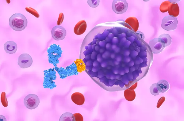
Monoclonal Antibody Treatment In Plasma Cell Leukemia (PCL) - Closeup View 3d Illustration

Image, 4.81MB, 10000 × 6600 jpg
stock vector Yellow and black color with line striped label banner with word kidney cancer awareness

Yellow And Black Color With Line Striped Label Banner With Word Kidney Cancer Awareness

Vector, 5.31MB, 8000 × 3200 eps
stock vector Childhood cancer, tumor in kids international month. Landscape poster, banner with oncological sign. Medical line concept. Pediatric oncology. Editable vector illustration isolated on white background

Childhood Cancer, Tumor In Kids International Month. Landscape Poster, Banner With Oncological Sign. Medical Line Concept. Pediatric Oncology. Editable Vector Illustration Isolated On White Background

Vector, 6.46MB, 6811 × 3671 eps
stock image New targeted therapy, breast cancer. microRNA (miR-182-3p) inhibits the telomeric protein TRF2, responsible for tumor. 3D rendering

New Targeted Therapy, Breast Cancer. MicroRNA (miR-182-3p) Inhibits The Telomeric Protein TRF2, Responsible For Tumor. 3D Rendering

Image, 12.57MB, 4631 × 4800 jpg
stock image Dendritic cells (DC) recognize Acute Lymphocytic Leukemia (ALL) - isometric view 3d illustration

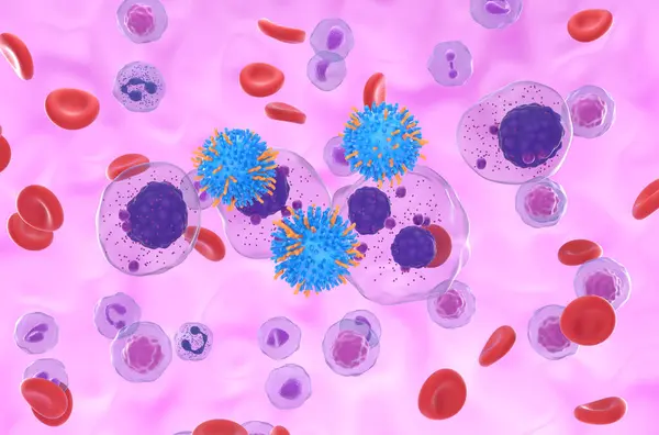
Dendritic Cells (DC) Recognize Acute Lymphocytic Leukemia (ALL) - Isometric View 3d Illustration

Image, 6.91MB, 10000 × 6600 jpg
stock vector Gallbladder, bild duct cancer icon. Outline medical sign. Digestive system disease. Gastroenterology, general surgery, oncology concept. Editable vector illustration isolated on a white background

Gallbladder, Bild Duct Cancer Icon. Outline Medical Sign. Digestive System Disease. Gastroenterology, General Surgery, Oncology Concept. Editable Vector Illustration Isolated On A White Background

Vector, 5.72MB, 5000 × 5000 eps
stock image Dendritic cell (DC) recognize Acute myeloid leukemia (AML) - closeup view 3d illustration

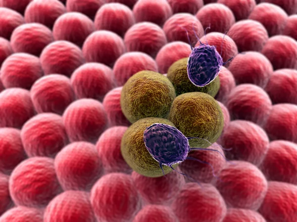
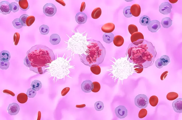
Dendritic Cell (DC) Recognize Acute Myeloid Leukemia (AML) - Closeup View 3d Illustration

Image, 6.09MB, 10000 × 6600 jpg
stock image CAR T cell therapy in Thyroid cancer - isometric view 3d illustration

CAR T Cell Therapy In Thyroid Cancer - Isometric View 3d Illustration

Image, 10.79MB, 10000 × 6600 jpg
stock image CAR T cell therapy in Chronic Myelogenous Leukemia (CML) - isometric view 3d illustration

CAR T Cell Therapy In Chronic Myelogenous Leukemia (CML) - Isometric View 3d Illustration

Image, 8.33MB, 10000 × 6600 jpg
stock image CAR T cell therapy in lung cancer (LC) - closeup view 3d illustration

CAR T Cell Therapy In Lung Cancer (LC) - Closeup View 3d Illustration

Image, 8.33MB, 10000 × 6600 jpg
stock image Examining woman with cancer

Examining Woman With Cancer

Image, 10.03MB, 5616 × 3744 jpg
stock image Woman with cancer during mole examining

Woman With Cancer During Mole Examining

Image, 10.95MB, 3744 × 5616 jpg
stock image Woman with cancer during findings consulting

Woman With Cancer During Findings Consulting

Image, 11.16MB, 3744 × 5616 jpg
stock image 3d  human character fighting with cancer

3d Human Character Fighting With Cancer

Image, 1.86MB, 5000 × 3750 jpg
stock image 3D illustration of MENINGIOMA  title on a medical document

3D Illustration Of MENINGIOMA Title On A Medical Document

Image, 2.55MB, 4500 × 3000 jpg
stock image Hepatocellular carcinoma - liver cancer in rat, 60x zoom

Hepatocellular Carcinoma - Liver Cancer In Rat, 60x Zoom

Image, 7.66MB, 4000 × 2667 jpg
Previous << Page 4 >> Next