
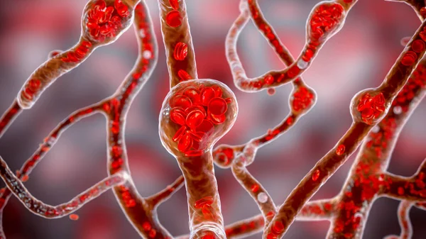
Stock image Microaneurysms

Non-proliferative Diabetic Retinopathy, 3D Illustration Showing Multiple Microaneurysms On The Eye Retina And Closeup View Of Microaneurysms, Microscopic Buldges In The Artery Walls Filled With Blood
Image, 11.11MB, 10431 × 6954 jpg
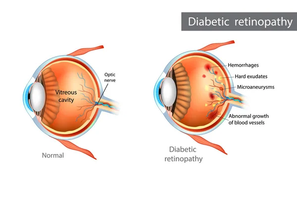
Diabetic Retinopathy. Difference Between Normal Retina And Diabetic Retinopathy
Vector, 2.35MB, 5100 × 3396 eps

Non-proliferative Diabetic Retinopathy, Illustration Showing Normal Eye Retina And Retina With Hard Exudates, Microaneurysms, Dot Haemorrhages, Flame-shaped And Splinter Retinal Haemorrhages
Image, 6.89MB, 11738 × 6603 jpg
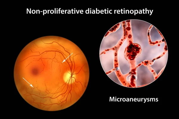
Non-proliferative Diabetic Retinopathy, 3D Illustration Showing Multiple Microaneurysms On The Eye Retina And Closeup View Of Microaneurysms, Microscopic Buldges In The Artery Walls Filled With Blood
Image, 10.46MB, 10431 × 6954 jpg

Non-proliferative Diabetic Retinopathy, 3D Illustration Showing Hard Exudates, Microaneurysms, Dot Haemorrhages, Flame-shaped And Splinter Retinal Haemorrhages, Ophthalmoscope View
Image, 13.11MB, 5352 × 5352 jpg
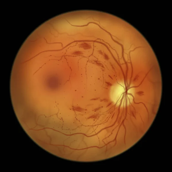
Non-proliferative Diabetic Retinopathy, Illustration Showing Microaneurysms, Dot Haemorrhages, Flame-shaped And Splinter Retinal Haemorrhages, Ophthalmoscope View
Image, 2.86MB, 5000 × 5000 jpg
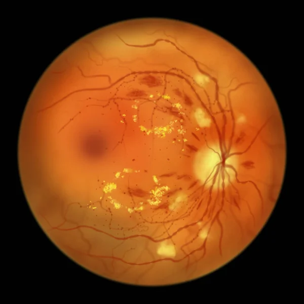
Diabetic Retinopathy Non-proliferative, Illustration Showing Hard Exudates, Cotton Wool Spots, Microaneurysms, Dot Haemorrhages, Flame-shaped And Splinter Retinal Haemorrhages, IRMAs, Venous Beading
Image, 3.07MB, 5000 × 5000 jpg
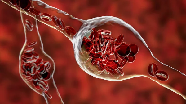
Microaneurysms, Microscopic Buldges In The Artery Walls Filled With Blood, 3D Illustration. Found In The Eye Retina In Diabetic Retinopathy, And Also In Brain (Charcot-Bouchard Aneurysms)
Image, 7.58MB, 7200 × 4050 jpg

Non-proliferative Diabetic Retinopathy, Illustration Showing Normal Eye Retina And Retina With Hard Exudates, Microaneurysms, Dot Haemorrhages, Flame-shaped And Splinter Retinal Haemorrhages
Image, 7.35MB, 11738 × 6603 jpg

Diabetic Retinopathy Non-proliferative, Illustration Showing Hard Exudates, Cotton Wool Spots, Microaneurysms, Dot Haemorrhages, Flame-shaped And Splinter Retinal Haemorrhages, IRMAs, Venous Beading
Image, 7.22MB, 11738 × 6603 jpg

Non-proliferative Diabetic Retinopathy, Illustration Showing IRMAs (intraretinal Microvascular Abnormalities), Venous Beading, And Microaneurysms
Image, 2.85MB, 5000 × 5000 jpg
Microaneurysms, Microscopic Buldges In The Artery Walls Filled With Blood, 3D Illustration. Found In The Eye Retina In Diabetic Retinopathy, And Also In Brain (Charcot-Bouchard Aneurysms)
Image, 6.95MB, 7200 × 4050 jpg
Page 1 >> Next