
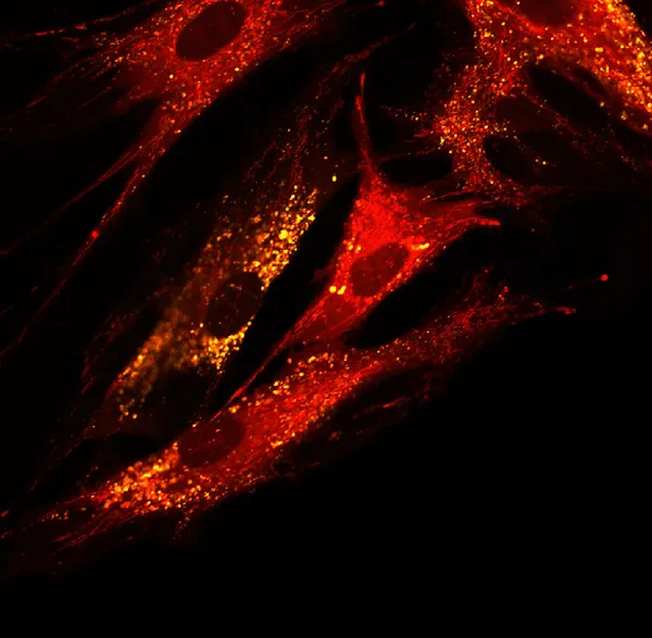
Stock image Microfilaments
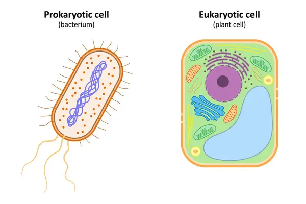

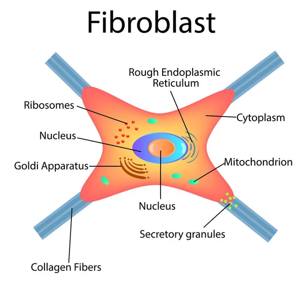
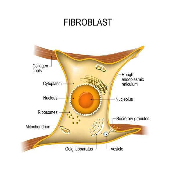
Fibroblast Anatomy. Structure Of Cell. Diagram With Golgi Apparatus, Nucleus, Mitochondrion And Ribosomes. Vector Illustration. Poster
Vector, 14.88MB, 4444 × 4444 eps

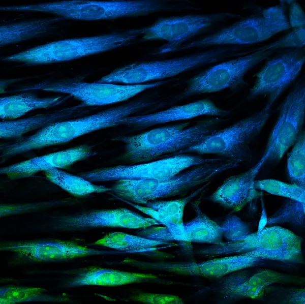
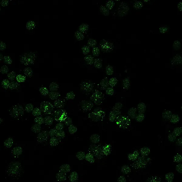
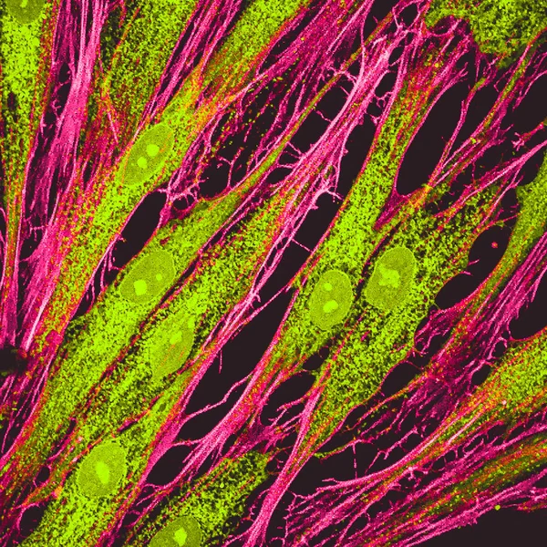
Real Fluorescence Microscopic View Of Human Skin Cells In Culture. Actin Filaments Are In Pink, Tubulin Was Labeled With Green
Image, 11.97MB, 4000 × 4000 jpg

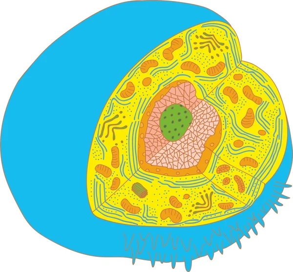

Fibroblast Structure. Fibroblast Cell. Vector Illustration Isolated
Vector, 19.48MB, 5000 × 5000 eps
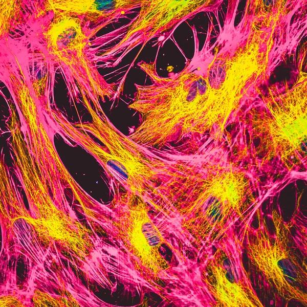
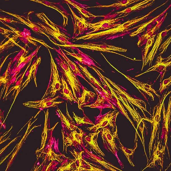
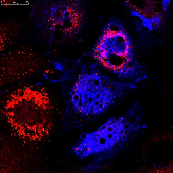
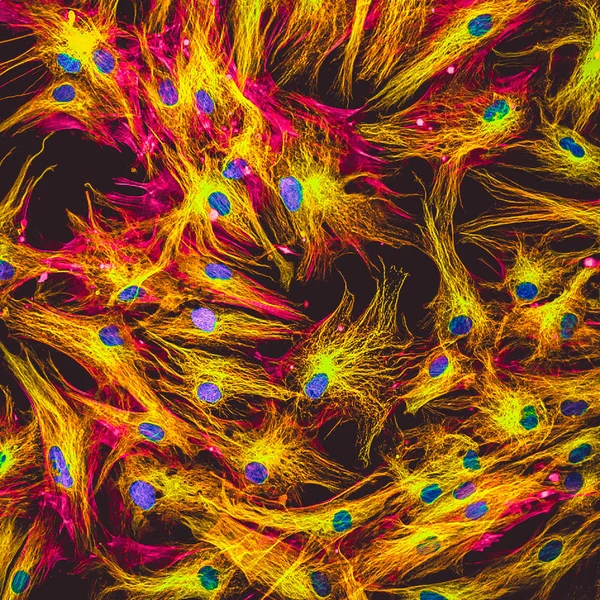
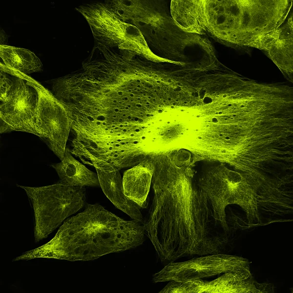
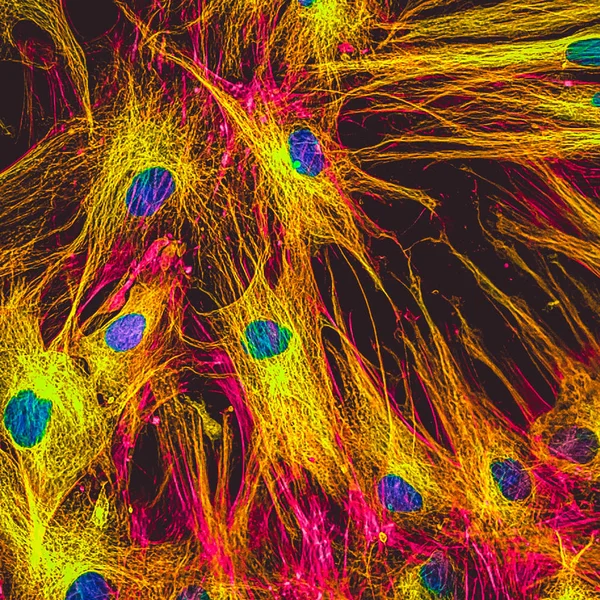
Real Fluorescence Microscopic View Of Human Skin Cells In Culture. Nucleus Are In Blue, Actin Filaments Are In Pink, Tubulin Was Labeled With Yellow
Image, 17.2MB, 4000 × 4000 jpg


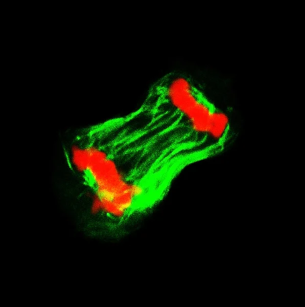
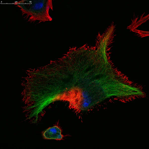
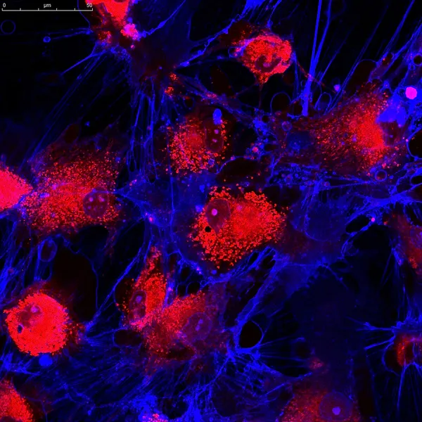
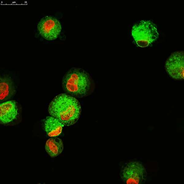
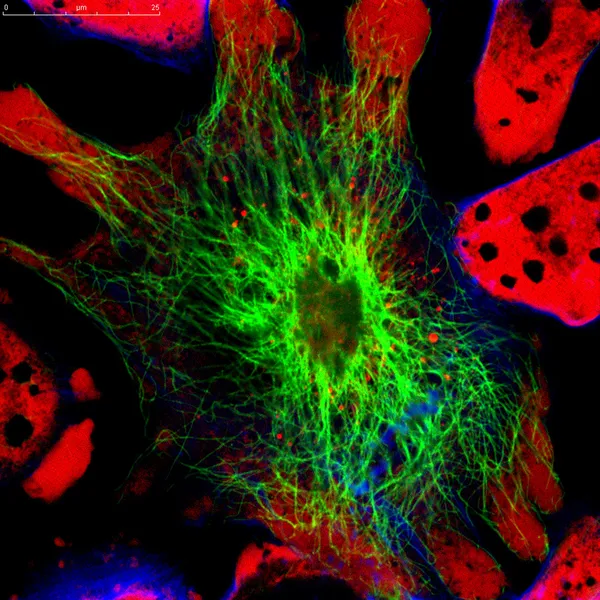
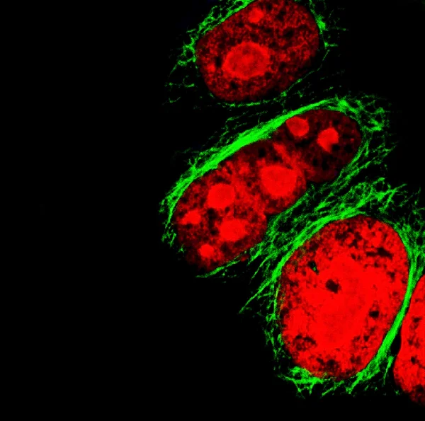
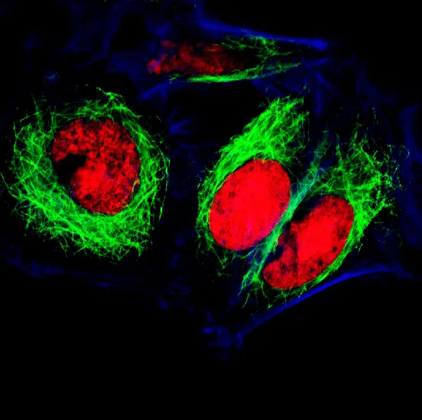
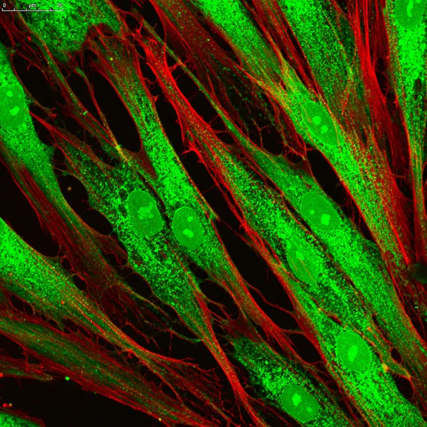
Real Fluorescence Microscopic View Of Human Skin Cells In Culture. Actin Filaments Are In Red, Tubulin Was Labeled With Green
Image, 16.93MB, 4000 × 4000 jpg
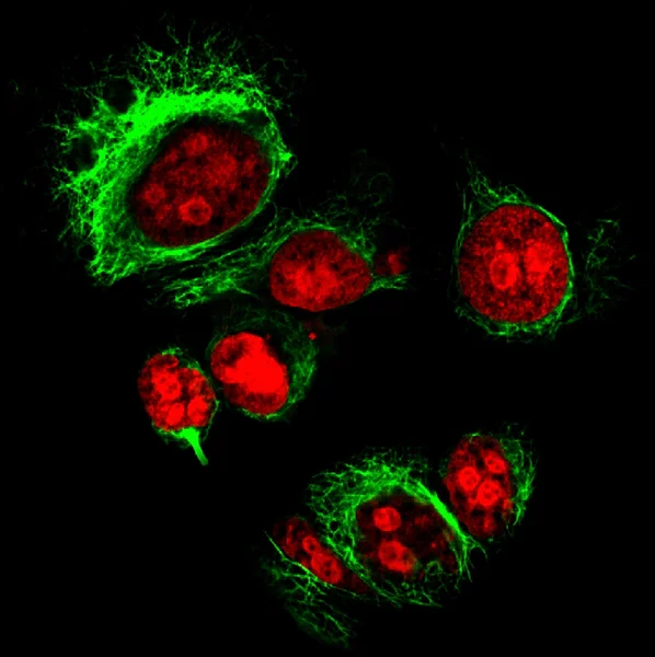

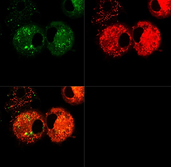
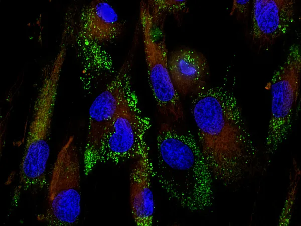
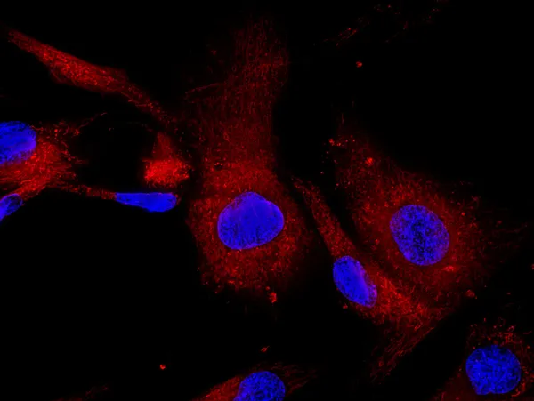
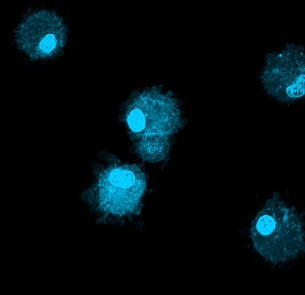
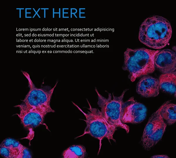
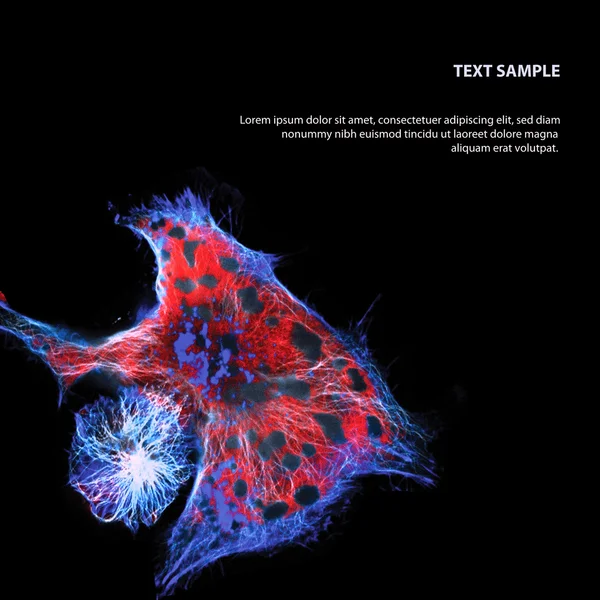
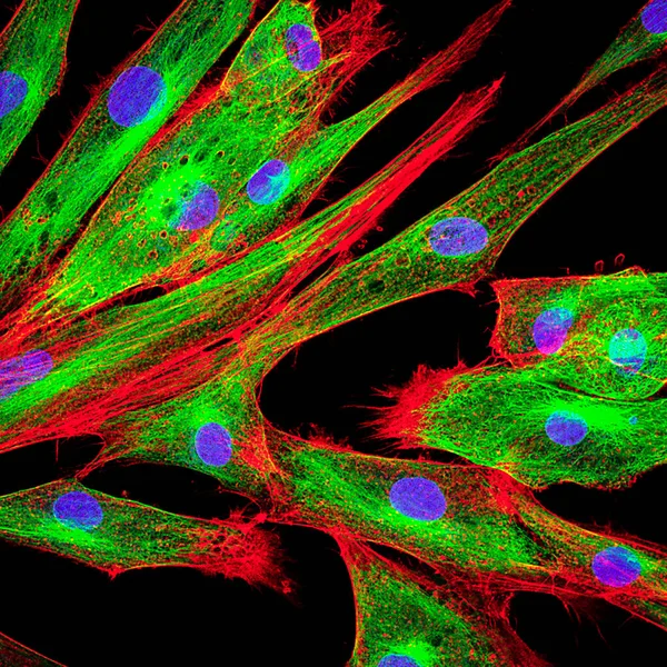
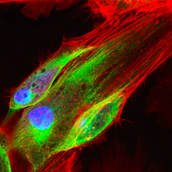
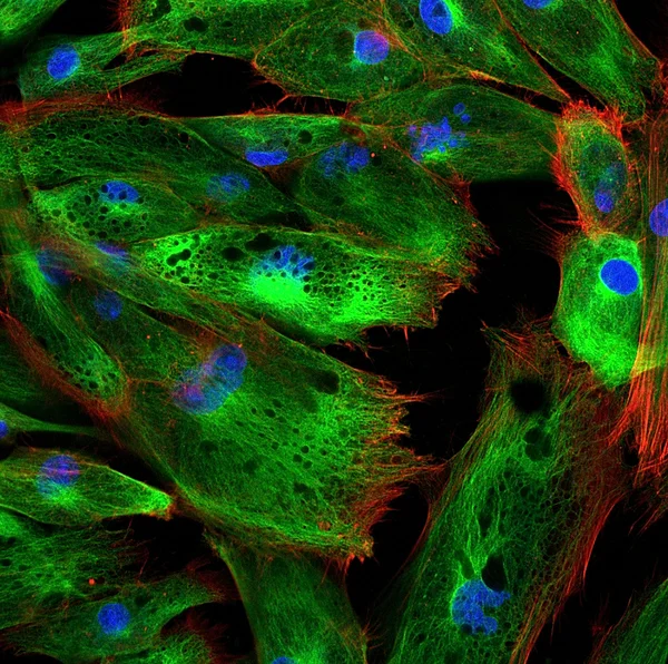
Real Fluorescence Microscopic View Of Human Skin Cells In Culture. Nucleus Are In Blue, Actin Filaments Are In Pink, Tubulin Was Labeled With Green
Image, 16.31MB, 4000 × 4000 jpg


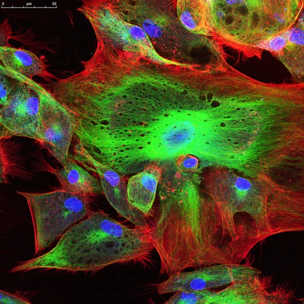
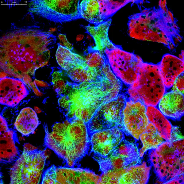
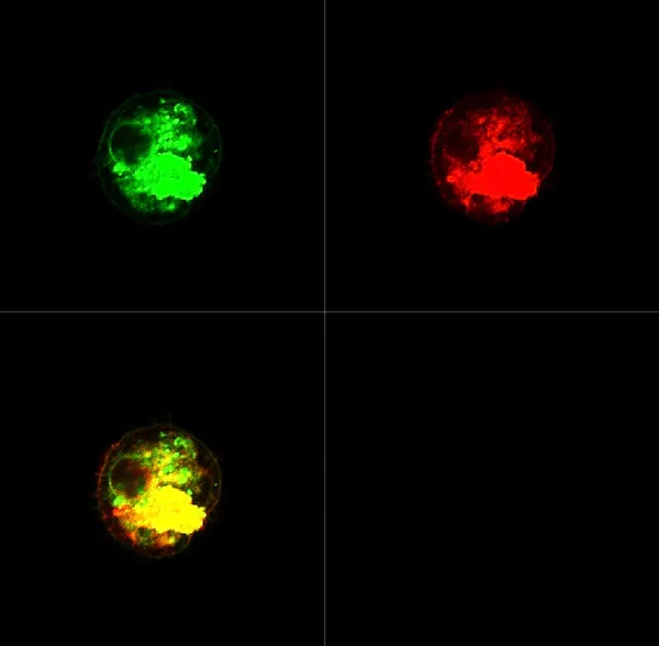
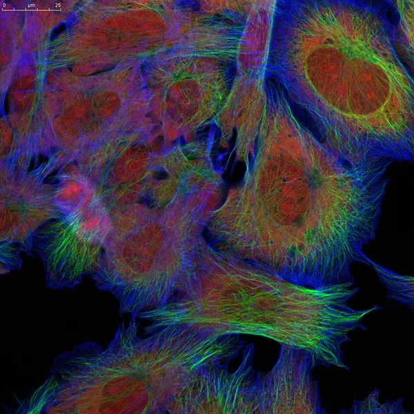
Real Fluorescence Microscopic View Of Human Skin Cells In Culture. Nucleus Are In Blue, Actin Filaments Are In Red, Tubulin Was Labeled With Green
Image, 11.6MB, 4000 × 4000 jpg
Real Fluorescence Microscopic View Of Human Skin Cells In Culture. Nucleus Are In Blue, Actin Filaments Are In Pink, Tubulin Was Labeled With Yellow
Image, 15.09MB, 4000 × 4000 jpg

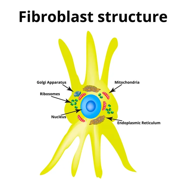
Fibroblast Structure. Fibroblast Cell. Vector Illustration On Isolated Background
Vector, 19.29MB, 5000 × 5000 eps
Page 1 >> Next