
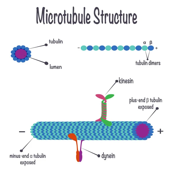
Stock image Microtubule

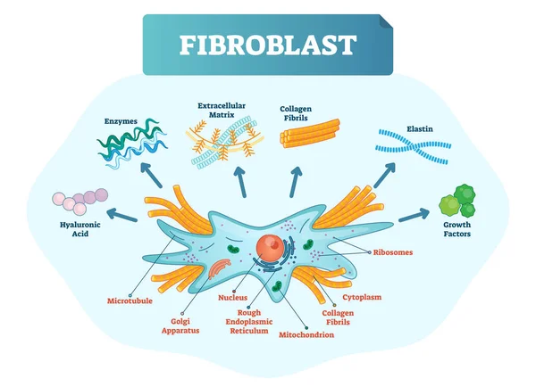
Fibroblast Vector Illustration. Scheme With Extracellular, Collagen Fibrils, Elastin, Hyaluronic Acid, Microtubule, Golgi Apparatus, Nucleus And Ribosomes.
Vector, 8.13MB, 5877 × 4252 eps
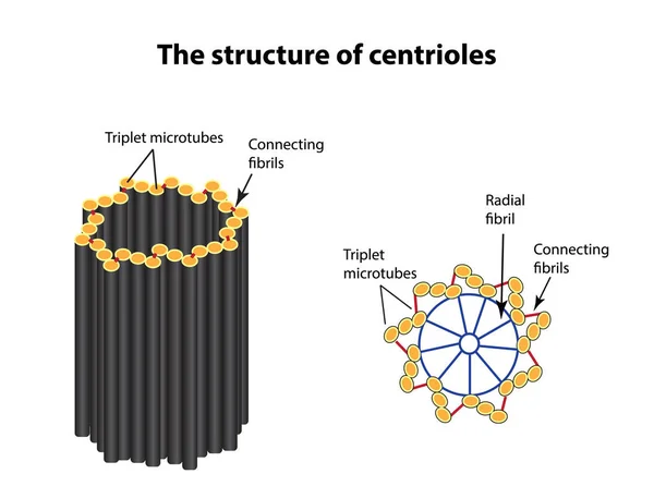
The Structure Of Centrioles. Infographics. Vector Illustration On Isolated Background
Vector, 0.97MB, 5000 × 3722 eps


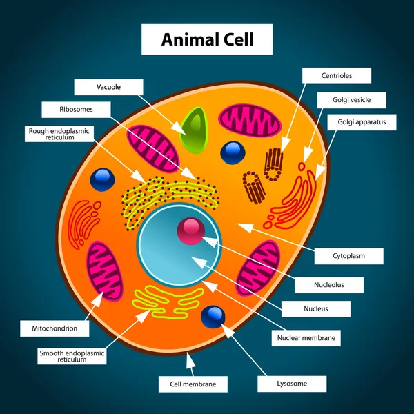
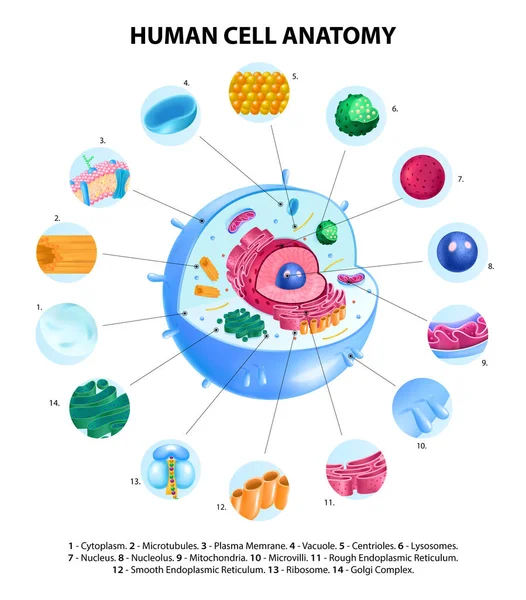
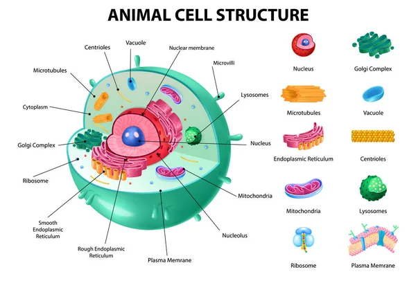
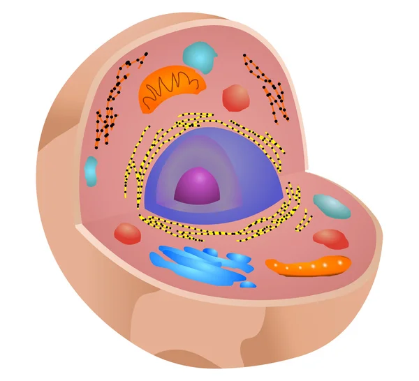
Animal Cell Anatomy Infographics With Detailed Educative Diagram And Labelled Elements Realistic Vector Illustration.
Vector, 3.51MB, 5500 × 4620 eps
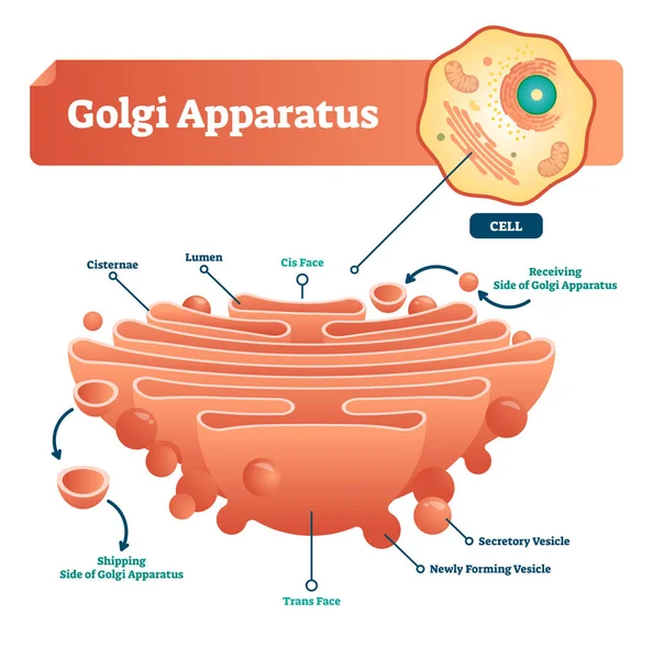
Golgi Apparatus Vector Illustration. Labeled Microscopic Scheme With Cisternae, Lumen, Secretory And Newly Forming Vesicle. Diagram With Receiving And Shipping Side.
Vector, 5.83MB, 4000 × 4039 eps


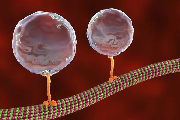
Intracellular Transport, Kinesin Proteins Transport Molecules Moving Across Microtubules
Image, 1.92MB, 4500 × 3000 jpg

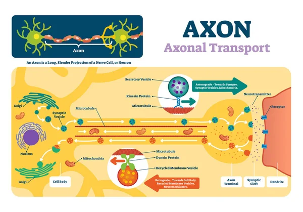
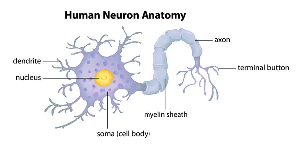
Axon Vector Illustration. Labeled Diagram With Explanation And Structure.
Vector, 5.97MB, 5000 × 3498 eps
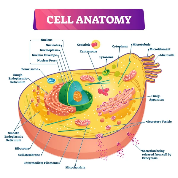
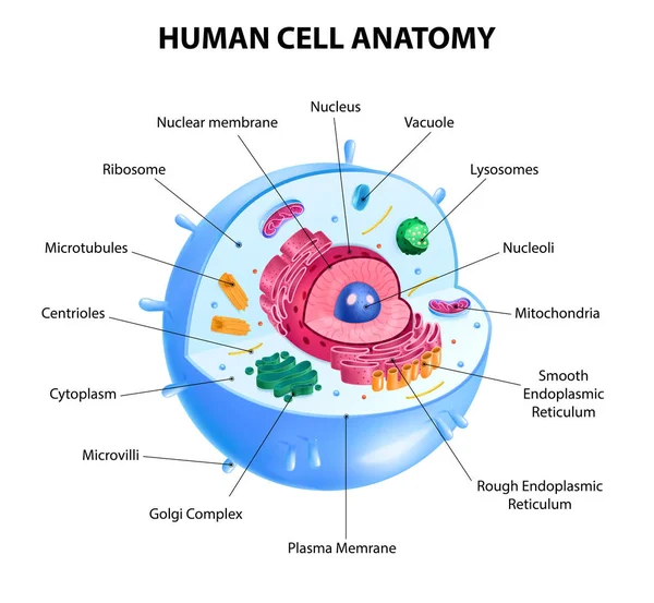
Cell Anatomy Vector Illustration. Labeled Educational Structure Diagram.
Vector, 12.61MB, 4000 × 4000 eps
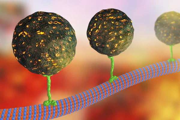
Intracellular Transport, Kinesin Proteins Transport Molecules Moving Across Microtubules
Image, 2.61MB, 4500 × 3000 jpg

Intracellular Transport, Kinesin Proteins Transport Molecules Moving Across Microtubules
Image, 4.16MB, 4500 × 4500 jpg
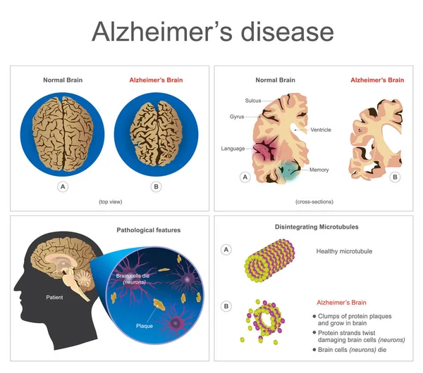
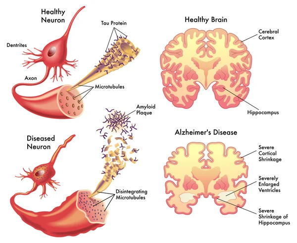
Alzheimers Disease. Brain Cells Die, Neuron Diseased, Certain Areas Of Brain Shrink Memory Loss Or Changes In Memory For People At Risk Could Affect Younger People. Info Graphic Vector.
Vector, 4.02MB, 6000 × 5306 eps
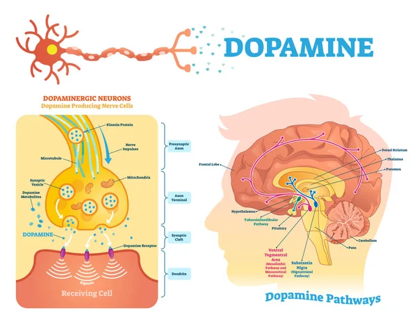
Dopamine Vector Illustration. Labeled Diagram With Its Action And Pathways.
Vector, 7.99MB, 5000 × 3890 eps
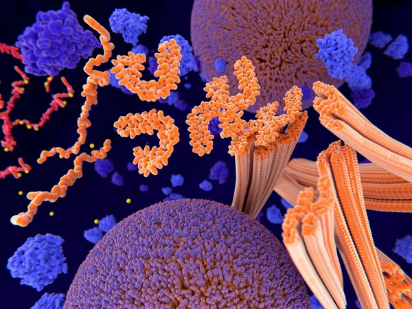
Pathological Phosphorylation (yellow) Of Tau Proteins (red-orange) Leads To Disintegration Of Microtubuli In The Neuron Axon An Aggregation Of The Tau Proteins. The Transport Of Synaptic Vesicles (orange-violet Spheres) Is Interrupted.
Image, 9.48MB, 8000 × 6000 jpg
Intracellular Transport, Kinesin Proteins Transport Molecules Moving Across Microtubules
Image, 1.92MB, 4500 × 3000 jpg

Intracellular Transport, Kinesin Proteins Transport Molecules Moving Across Microtubules
Image, 4.28MB, 4500 × 4500 jpg
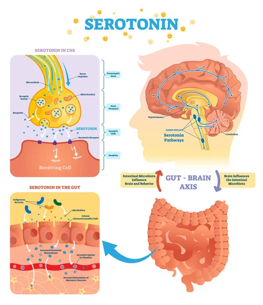
Serotonin Vector Illustration. Labeled Diagram With Gut Brain Axis And CNS.
Vector, 8.95MB, 4000 × 4640 eps

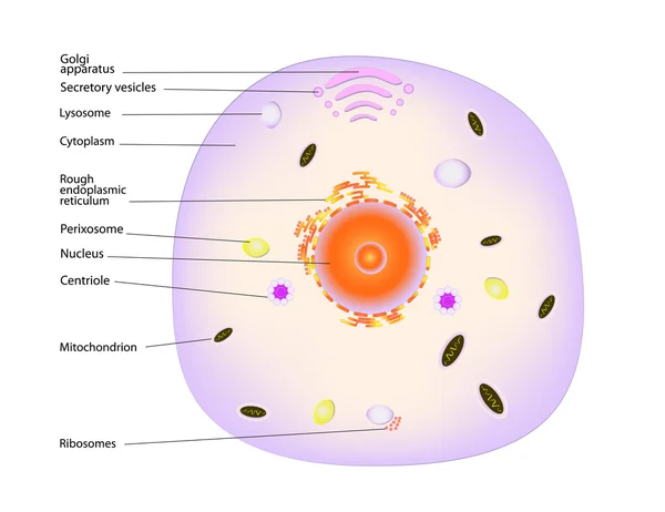
Animal Cell Anatomy Infographics With Detailed Educative Diagram And Labelled Elements Realistic Vector Illustration.
Vector, 2.71MB, 5280 × 4840 eps
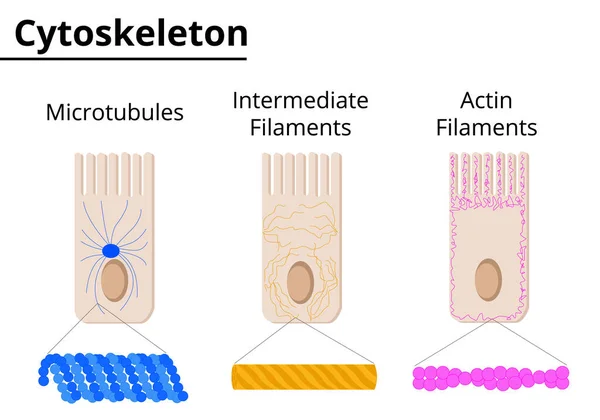
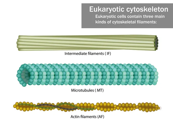
Different Structures Of Cytoskeleton. Microtubules, Intermediate Filaments And Actin Filaments. Vector Illustration. Didatic Illustration.
Vector, 0.59MB, 5000 × 3500 ai
Page 1 >> Next