
Stock image Mitral Stenosis
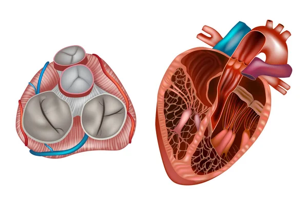
Heart Valves Anatomy. Mitral Valve, Pulmonary Valve, Aortic Valve And The Tricuspid Valve.
Vector, 11.68MB, 5000 × 3334 eps
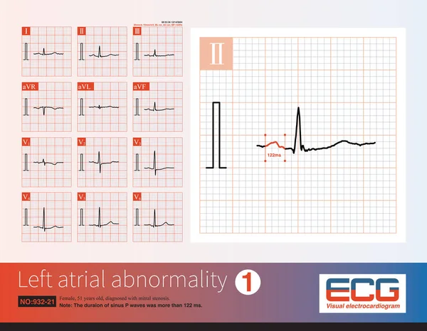
Female, 51 Years Old, Diagnosed With Mitral Stenosis. When This ECG Was Taken, The Patient Still Maintained Sinus Rhythm.Note That The P Wave Duration Was Widened.
Image, 14.21MB, 10000 × 7772 jpg


Mitral Stenosis. A Narrowing Of The Mitral Orifice, Obstructing The Flow Of Blood From The Left Atrium To The Left Ventricle. Simple Black And White Vector Illustration.
Vector, 0.4MB, 5650 × 2985 eps

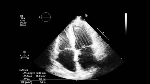
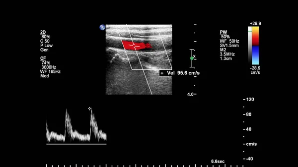
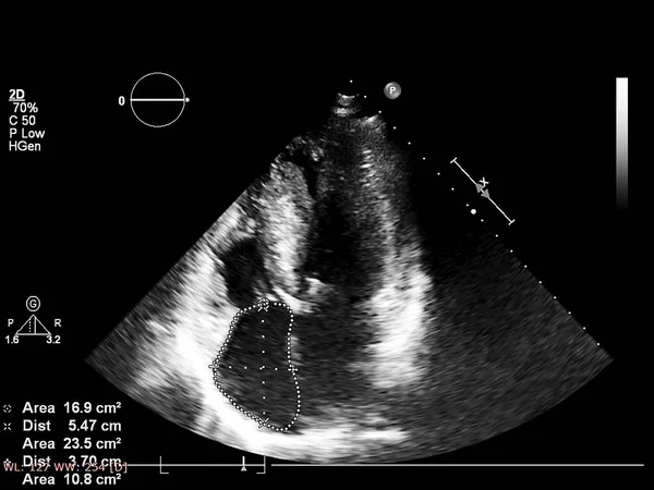
Image Of The Heart In Gray-scale Mode During Transesophageal Ultrasound.
Image, 0.82MB, 4000 × 2250 jpg

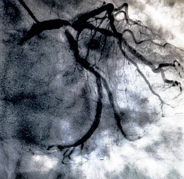
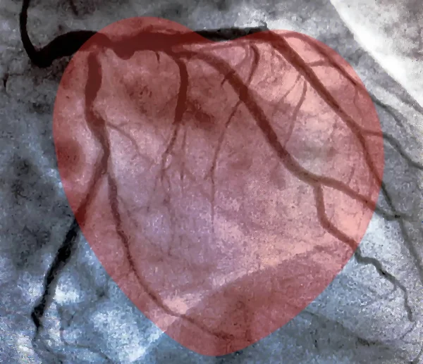
Coronary Artery Angiogram Of Left Coronary Artery During Cardiac Catheterization . Catheterization. Cardiac Ventriculography Is A Medical Imaging Test Used To Determine A Patient Cardiac Function
Image, 3.81MB, 2574 × 2500 jpg

Coronary Artery Angiogram Of Left Coronary Artery During Cardiac Catheterization . Catheterization. Cardiac Ventriculography Is A Medical Imaging Test Used To Determine A Patient Cardiac Function
Image, 5.72MB, 3000 × 2252 jpg
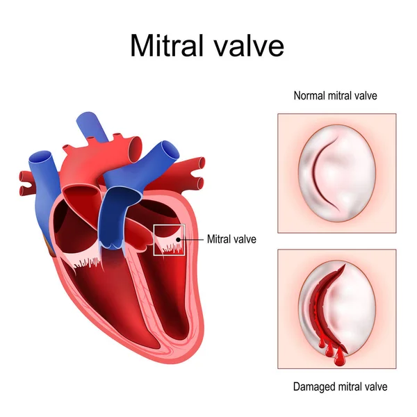
Heart Anatomy. Close-up Of Normal Mitral Valve And Damaged Mitral Valve. Cross Section Of Human Heart. Detailed Diagram. Vector Poster
Vector, 4.25MB, 4444 × 4445 eps
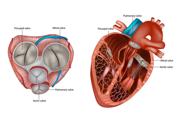
Structure Of The Heart Valves Anatomy. Mitral Valve, Pulmonary Valve, Aortic Valve And The Tricuspid Valve.
Vector, 10.89MB, 6000 × 4000 eps
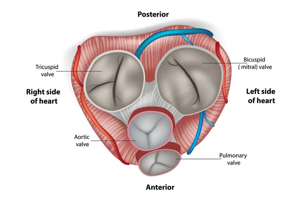
Structure Of The Heart Valves. Mitral Valve, Pulmonary Valve, Aortic Valve And The Tricuspid Valve.
Vector, 9.69MB, 5000 × 3334 eps
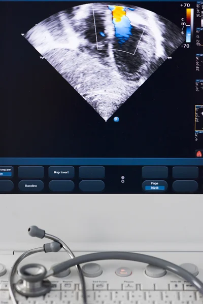


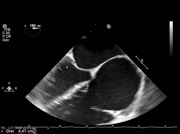
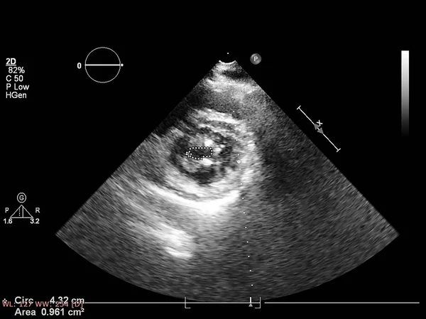
Image Of The Heart In Gray-scale Mode During Transesophageal Ultrasound.
Image, 1.13MB, 3500 × 1969 jpg
Catheterization And Small Red Heart. Cardiac Ventriculography Is A Medical Imaging Test Used To Determine A Patient Cardiac Function In The Right Or Left Ventricle
Image, 1.12MB, 3013 × 2600 jpg
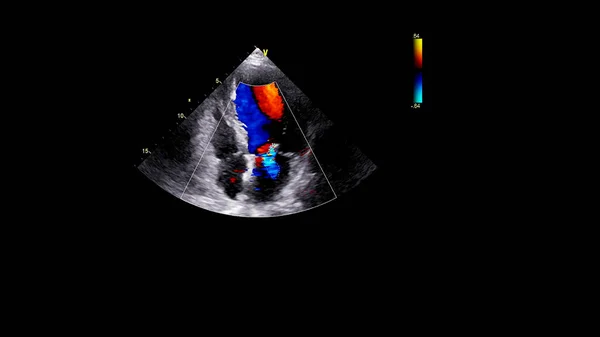
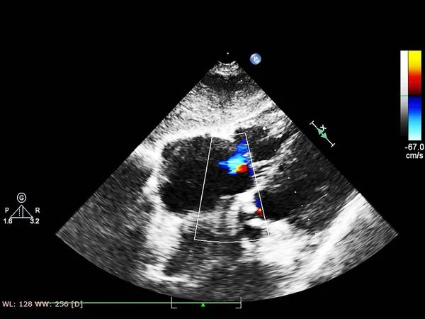
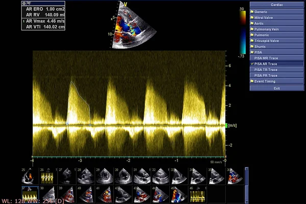
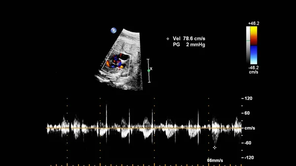
Image Of The Heart During Transesophageal Ultrasound With Doppler Mode.
Image, 0.51MB, 4000 × 2250 jpg

Catheterization. Cardiac Ventriculography Is A Medical Imaging Test Used To Determine A Patient Cardiac Function In The Right Or Left Ventricle
Image, 2.72MB, 2510 × 2500 jpg

Catheterization And Small Red Heart. Cardiac Ventriculography Is A Medical Imaging Test Used To Determine A Patient Cardiac Function In The Right Or Left Ventricle
Image, 0.94MB, 3150 × 2000 jpg
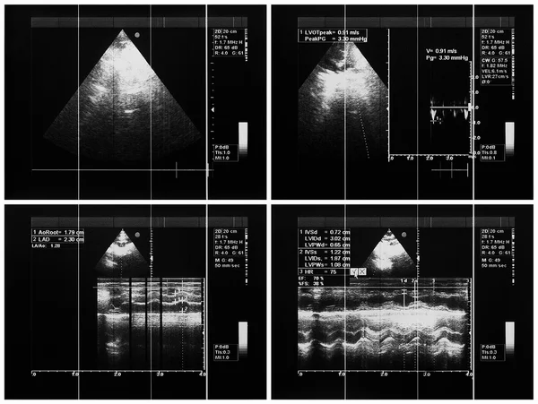

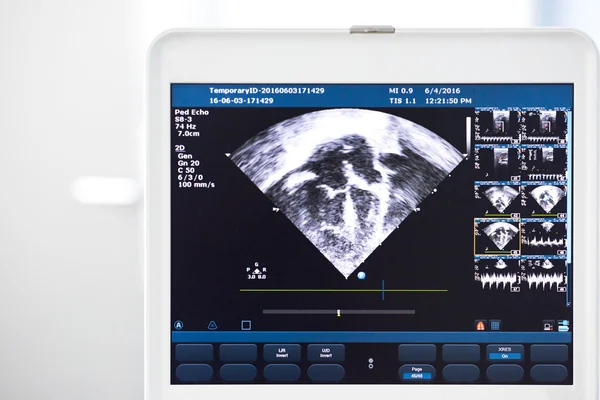


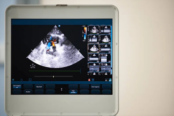
On The Screen Of The Ultrasound Apparatus, The Scan Of The Left Ventricle Of The Heart In The Position For Measuring The Ejection Fraction By Teyolz. Black And White Image.
Image, 8.04MB, 5000 × 3337 jpg


Coronary Artery Angiogram Of Left Coronary Artery During Cardiac Catheterization . Catheterization. Cardiac Ventriculography Is A Medical Imaging Test Used To Determine A Patient Cardiac Function
Image, 1.12MB, 3001 × 2590 jpg


Cardiomyopathy Is Inflammation In The Heart Muscle, Resulting In Its Enlargement And Weakening That Impairs The Blood's Pumping Ability. 3D Illustration
Image, 1.17MB, 3508 × 2480 jpg
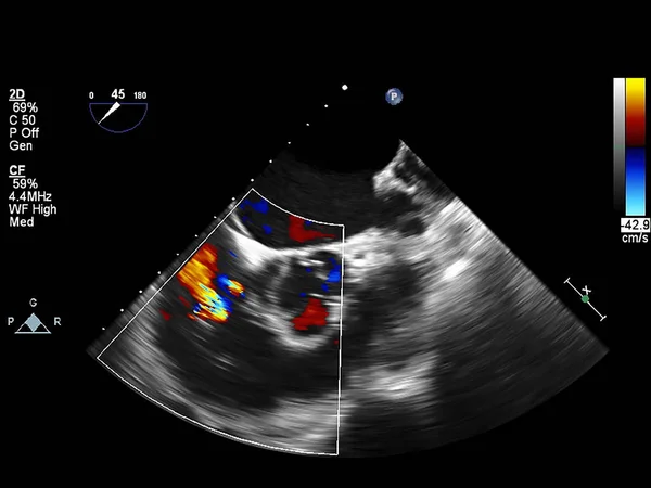


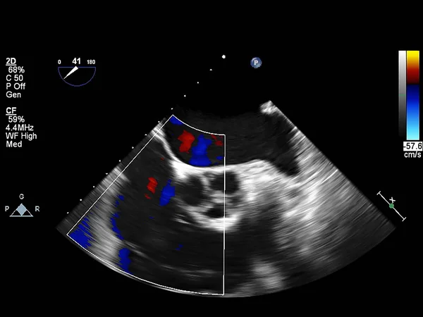
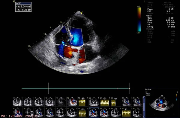
On The Screen Of A Modern Ultrasound Scanner, An Image Of The Heart Cavities With Red And Blue Streams Of Blood Regurgitation Depicted By The Doppler Method.
Image, 12.96MB, 6000 × 4004 jpg

Catheterization. Cardiac Ventriculography Is A Medical Imaging Test Used To Determine A Patient Cardiac Function In The Right Or Left Ventricle
Image, 2.42MB, 3007 × 2930 jpg
Page 1 >> Next