
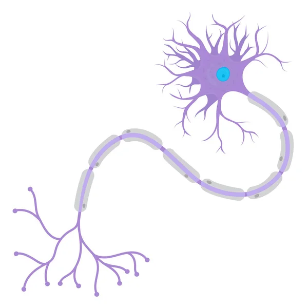
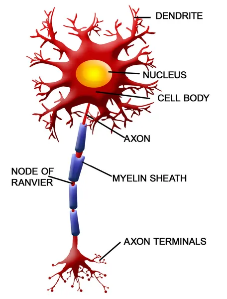
Stock image Motor Neuron
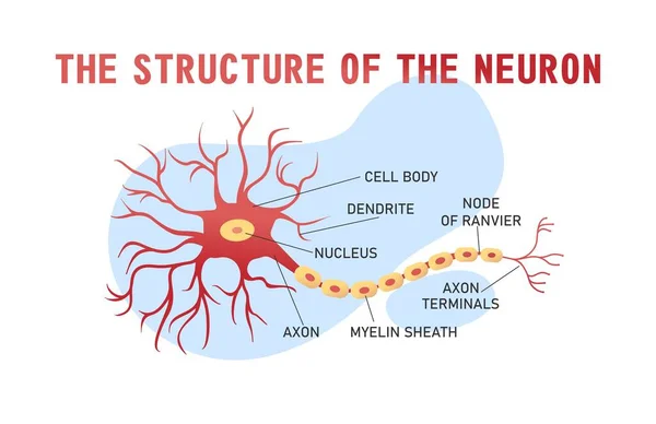
Educational Horizontal Banner Of Brain Neuron Structure On White Background, Vector Illustration
Vector, 0.39MB, 5000 × 3238 eps
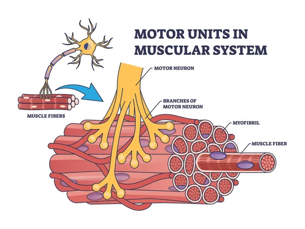
Motor Units In Muscular System With Fibers Neuron Anatomy Outline Diagram. Labeled Educational Medical Scheme With Myofibril And Muscle Fiber Closeup Vector Illustration. Nerve Functional Contraction
Vector, 6.89MB, 5000 × 3750 eps
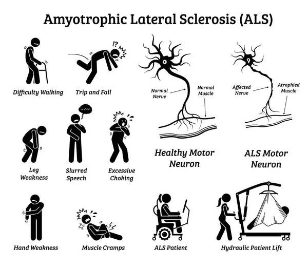
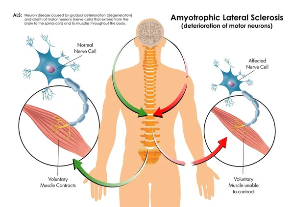
Amyotrophic Lateral Sclerosis ALS Disease Signs And Symptoms. Illustrations Depict Nervous System Or Neurological Disease In ALS Patient.
Vector, 4.71MB, 7500 × 6500 eps
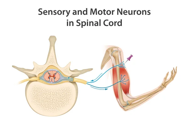
Somatic Motor Reflex, Somatic Nervous System, Peripheral Nervous System, Voluntary Control Of Body Movements Via Skeletal Muscles, Afferent And Efferent Nerves
Image, 2.5MB, 4093 × 3508 jpg

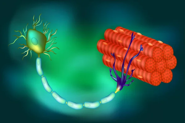
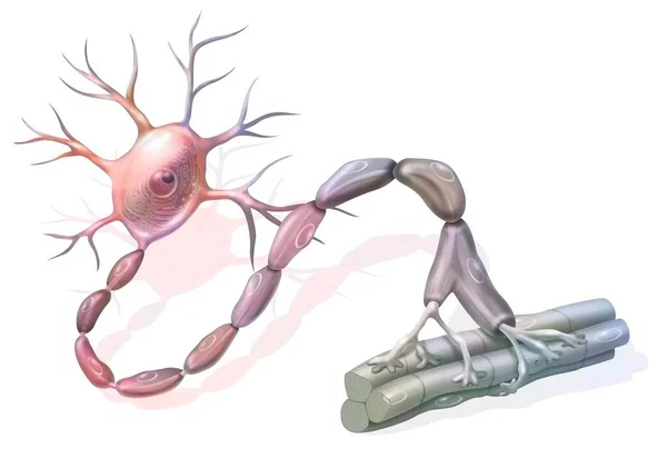
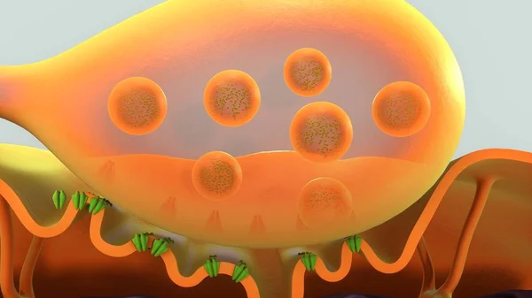
Motor Neuron Connecting To Muscle Fiber, 3D Illustration. A Neuromuscular Junction Allows The Motor Neuron To Transmit A Signal To The Muscle Causing Contraction. It Is Affected By Toxins And Diseases
Image, 11.3MB, 7200 × 4050 jpg
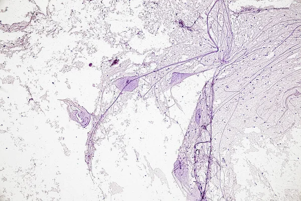
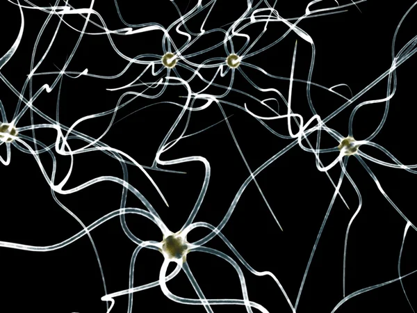
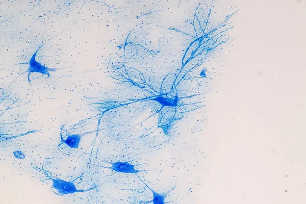
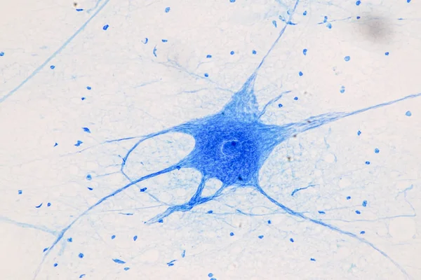
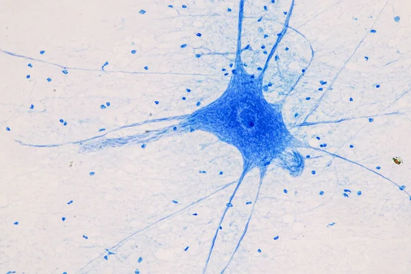
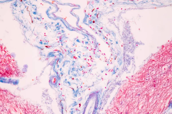
Motor Neuron, Spinal Cord, Nerve Fibres And Nerve Cells Under The Microscope In Lab.
Image, 13.05MB, 6000 × 4000 jpg
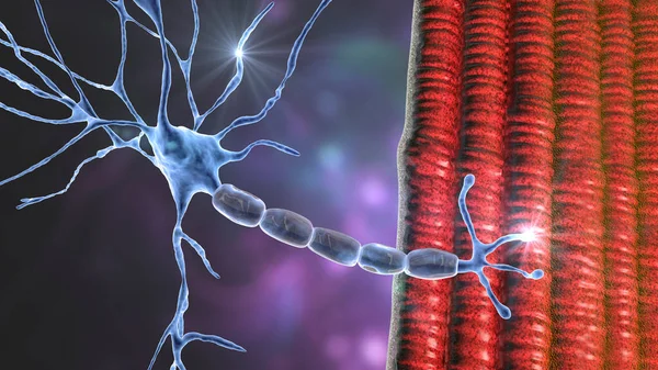
Motor Neuron Connecting To Muscle Fiber, 3D Illustration. A Neuromuscular Junction Allows The Motor Neuron To Transmit A Signal To The Muscle Causing Contraction. It Is Affected By Toxins And Diseases
Image, 18.96MB, 7200 × 4050 jpg
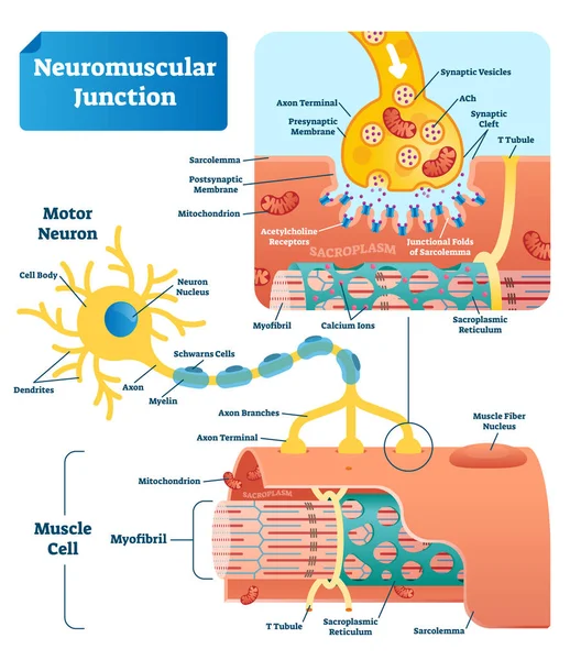
Neuromuscular Junction Vector Illustration Scheme. Labeled Cell Infographic
Vector, 7.8MB, 4000 × 4630 eps
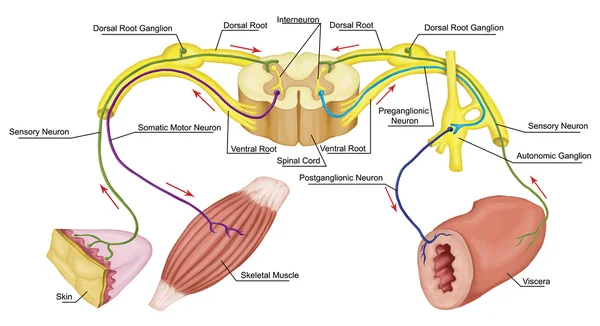
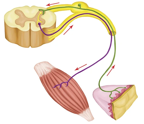
Somatic And Automatic Motor Reflex, Somatic And Automatic Nervous System, Peripheral And Visceral Nervous System, Voluntary And Involuntary Control Of Body And Visceral Functions
Image, 6.39MB, 8268 × 4503 jpg
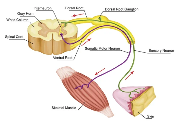
Somatic Motor Reflex, Somatic Nervous System, Peripheral Nervous System, Voluntary Control Of Body Movements Via Skeletal Muscles, Afferent And Efferent Nerves
Image, 3.86MB, 5906 × 4322 jpg
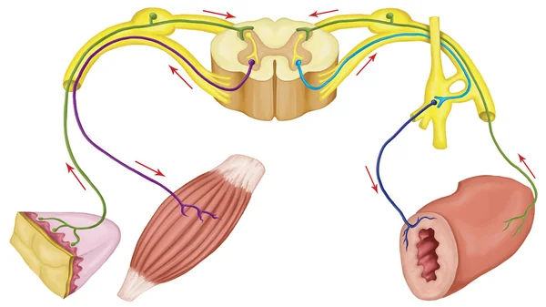
Somatic And Automatic Motor Reflex, Somatic And Automatic Nervous System, Peripheral And Visceral Nervous System, Voluntary And Involuntary Control Of Body And Visceral Functions
Image, 3.86MB, 5906 × 3321 jpg

Educational Horizontal Banner Of Brain Neuron Structure On White Background, Vector Illustration
Vector, 0.48MB, 5000 × 3238 eps

Motor Neuron Connecting To Muscle Fiber, 3D Illustration. A Neuromuscular Junction Allows The Motor Neuron To Transmit A Signal To The Muscle Causing Contraction. It Is Affected By Toxins And Diseases
Image, 16.63MB, 7200 × 4050 jpg
Neuromuscular Junction. A Synaptic Connection Between The Terminal End Of A Motor Nerve And A Muscle. Presynaptic (nerve Terminal), Postsynaptic Part, Synaptic Cleft.
Vector, 5.77MB, 6250 × 6250 eps

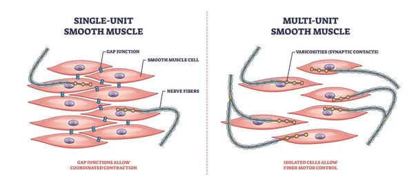
Single Unit Vs Multi Unit Smooth Muscle Structure Differences Outline Diagram
Vector, 6.26MB, 6000 × 2727 eps
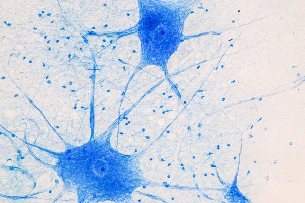
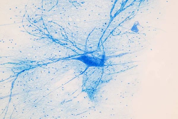

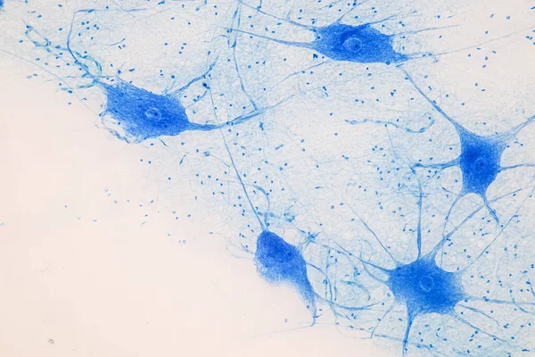


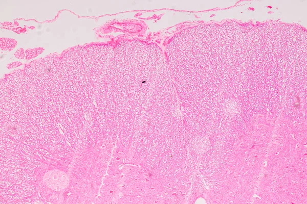
Education Spinal Cord, Nerve, Cerebellum, Cortex And Motor Neuron Human Under The Microscope In Lab.
Image, 19.22MB, 6720 × 4480 jpg

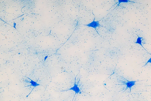
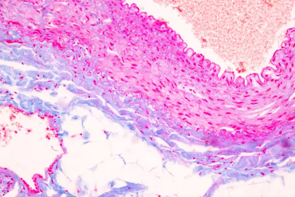
Cerebellum, Thalamus, Medulla Oblongata, Spinal Cord And Motor Neuron Human Under The Microscope In Lab.
Image, 13.45MB, 6000 × 4000 jpg


Cerebellum, Thalamus, Medulla Oblongata, Spinal Cord And Motor Neuron Human Under The Microscope In Lab.
Image, 12.22MB, 6000 × 4000 jpg

Neurological Physical Examination Of The Hands Reflexes. Doctor Neurologist Checks The Status Of The Patient's Reflexes In Office In Hospital. Selective Focus, Space For Text.
Image, 8.47MB, 5767 × 3850 jpg
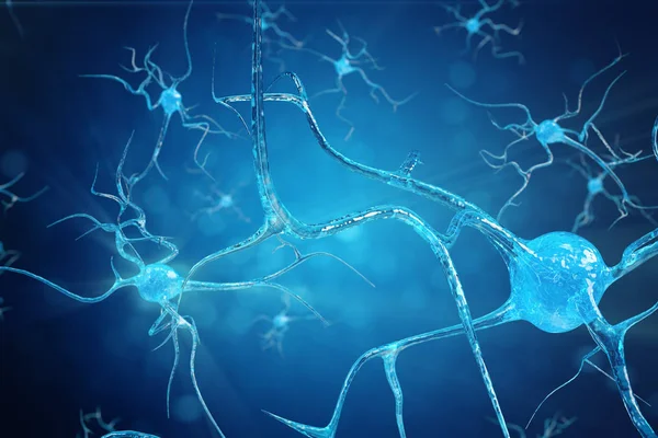
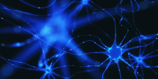
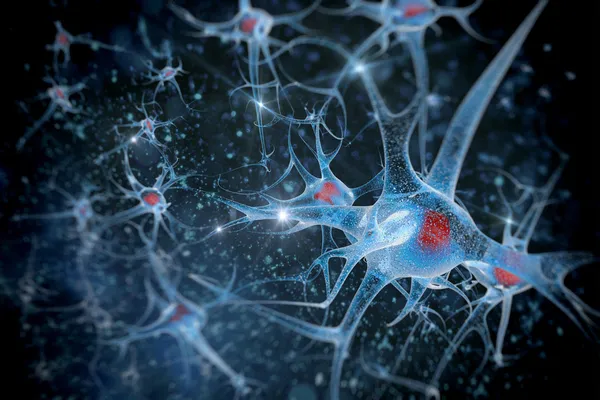
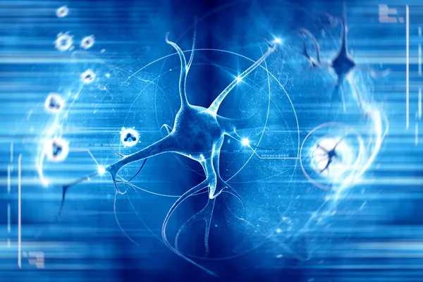
Conceptual Illustration Of Neuron Cells With Glowing Link Knots. Synapse And Neuron Cells Sending Electrical Chemical Signals. Neuron Of Interconnected Neurons With Electrical Pulses, 3D Illustration
Image, 2.69MB, 6000 × 4000 jpg
Page 1 >> Next