
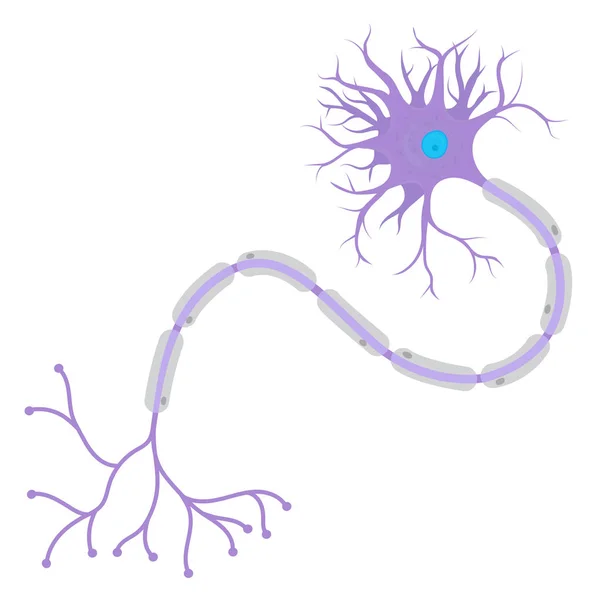
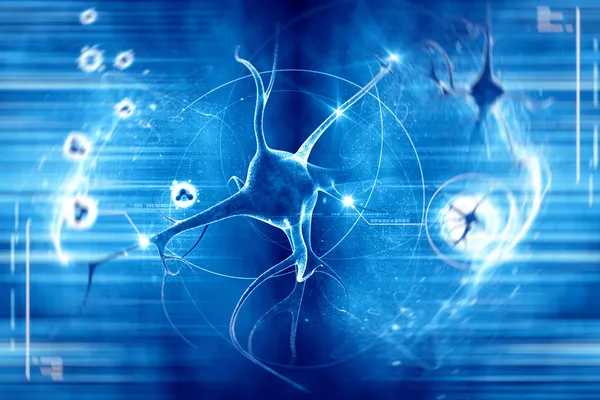
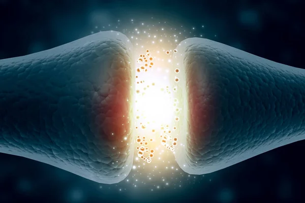
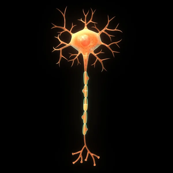
Stock image Motor Neuron Human
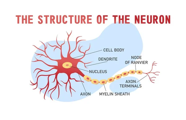
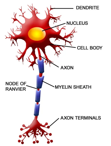
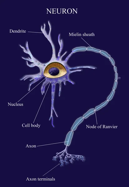
Educational Horizontal Banner Of Brain Neuron Structure On White Background, Vector Illustration
Vector, 0.39MB, 5000 × 3238 eps
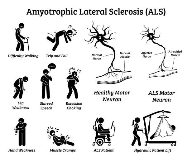
Amyotrophic Lateral Sclerosis ALS Disease Signs And Symptoms. Illustrations Depict Nervous System Or Neurological Disease In ALS Patient.
Vector, 4.71MB, 7500 × 6500 eps
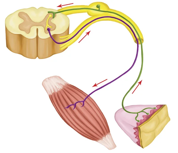
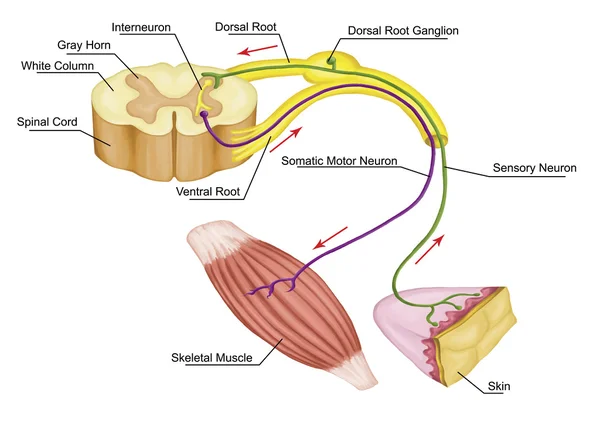
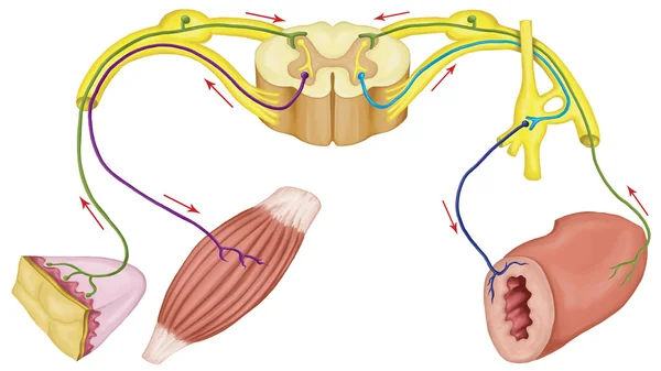
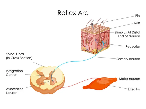
Somatic Motor Reflex, Somatic Nervous System, Peripheral Nervous System, Voluntary Control Of Body Movements Via Skeletal Muscles, Afferent And Efferent Nerves
Image, 2.5MB, 4093 × 3508 jpg
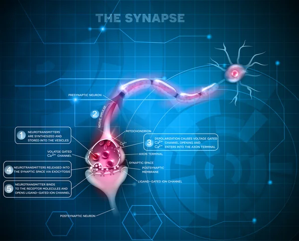
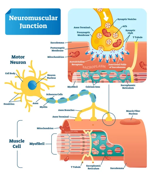
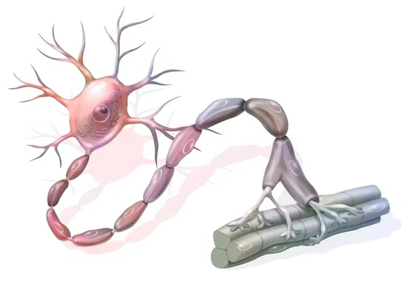
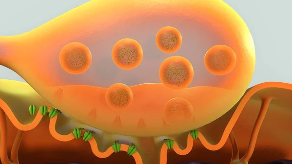
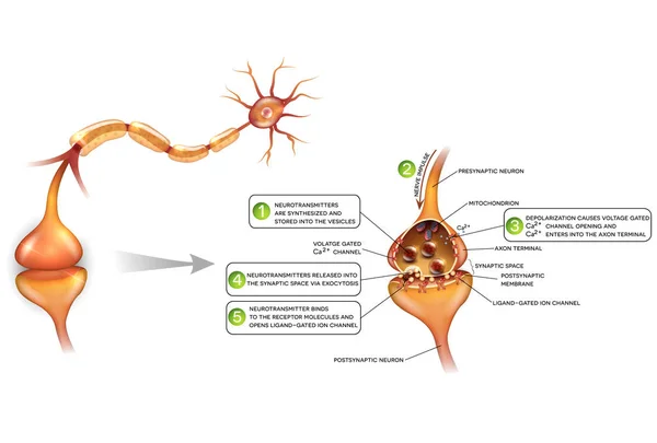
Neuromuscular Junction Vector Illustration Scheme. Labeled Cell Infographic
Vector, 7.8MB, 4000 × 4630 eps
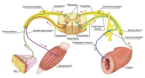
Somatic And Automatic Motor Reflex, Somatic And Automatic Nervous System, Peripheral And Visceral Nervous System, Voluntary And Involuntary Control Of Body And Visceral Functions
Image, 6.39MB, 8268 × 4503 jpg
Somatic Motor Reflex, Somatic Nervous System, Peripheral Nervous System, Voluntary Control Of Body Movements Via Skeletal Muscles, Afferent And Efferent Nerves
Image, 3.86MB, 5906 × 4322 jpg
Somatic And Automatic Motor Reflex, Somatic And Automatic Nervous System, Peripheral And Visceral Nervous System, Voluntary And Involuntary Control Of Body And Visceral Functions
Image, 3.86MB, 5906 × 3321 jpg

Educational Horizontal Banner Of Brain Neuron Structure On White Background, Vector Illustration
Vector, 0.48MB, 5000 × 3238 eps


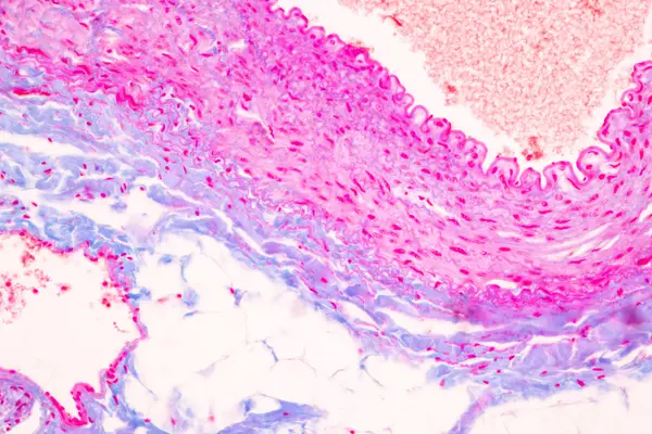
Cerebellum, Thalamus, Medulla Oblongata, Spinal Cord And Motor Neuron Human Under The Microscope In Lab.
Image, 13.45MB, 6000 × 4000 jpg
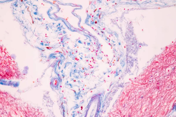
Cerebellum, Thalamus, Medulla Oblongata, Spinal Cord And Motor Neuron Human Under The Microscope In Lab.
Image, 12.22MB, 6000 × 4000 jpg

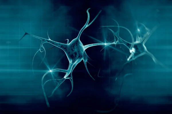
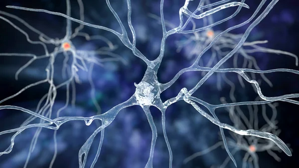
Neurons, 3D Illustration Showing Brain Cells Located In The Precentral Gyrus Of The Frontal Cortex Of The Human Brain. They Control Movements Of The Contralateral Side Of The Body
Image, 4MB, 7200 × 4050 jpg

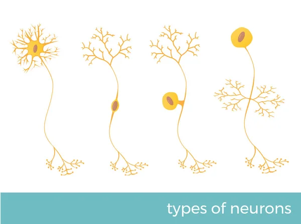
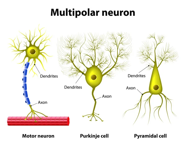
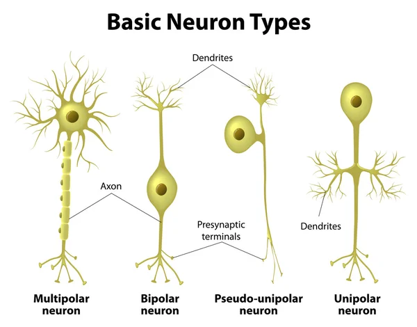
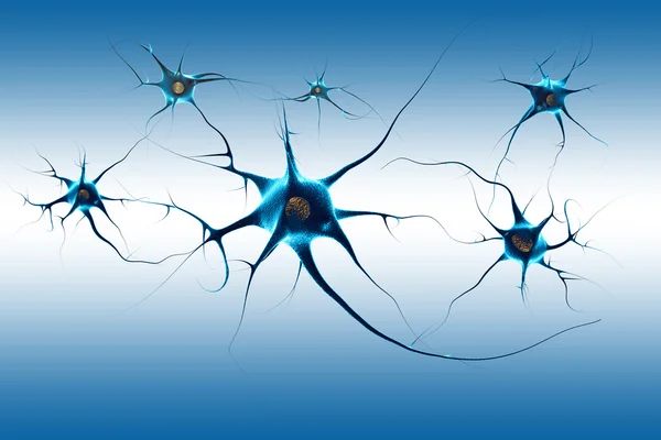
Types Of Neurons - Part Of Human's Central Nervous System. Vector Format Illustration.
Vector, 5.2MB, 8041 × 6000 eps
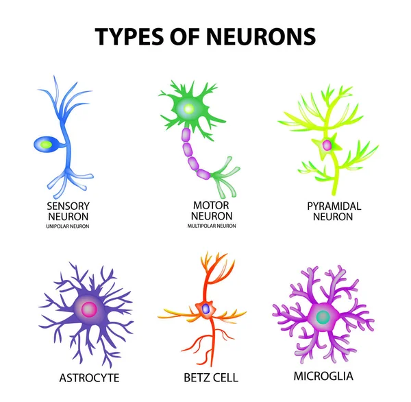
Types Of Neurons. Structure Sensory, Motor Neuron, Astrocyte, Pyromidal, Betz Cell, Microglia. Set. Infographics. Vector Illustration On Isolated Background
Vector, 3.45MB, 5000 × 4890 eps

Types Of Neurons. Structure Sensory, Motor Neuron, Astrocyte, Pyromidal, Betz Cell, Microglia. Set. Infographics. Vector
Vector, 3.45MB, 5000 × 4890 eps

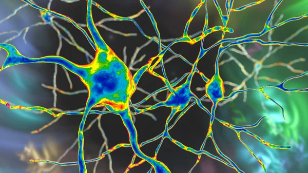
Neurons, 3D Illustration Showing Brain Cells Located In The Precentral Gyrus Of The Frontal Cortex Of The Human Brain. They Control Movements Of The Contralateral Side Of The Body
Image, 8.69MB, 7200 × 4050 jpg
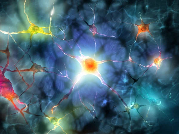
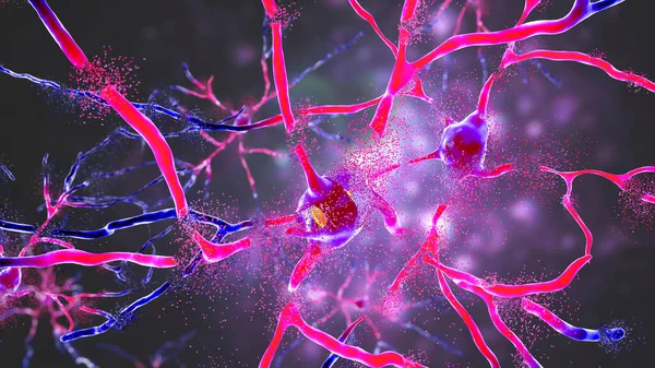
Neurons Of Dorsal Striatum, 3D Illustration. The Dorsal Striatum Is A Nucleus In The Basal Ganglia, Degrading Of Its Neurons Plays A Crucial Role In The Development Of Huntington's Disease
Image, 8.02MB, 7200 × 4050 jpg
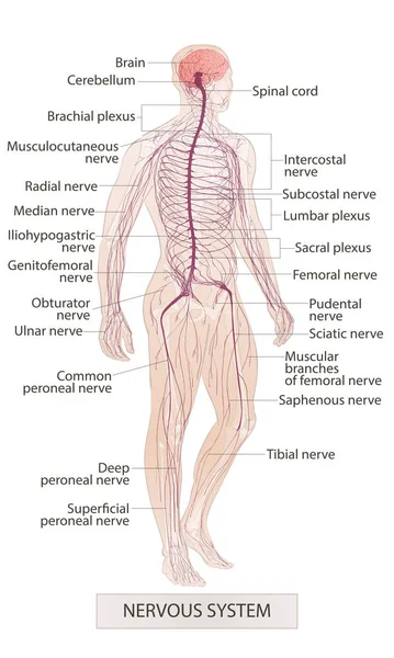
Nervous System. Human Body Parts. Man Anatomy. Hand Drown Vector Sketch Illustration Isolated
Vector, 7.04MB, 3543 × 5906 eps

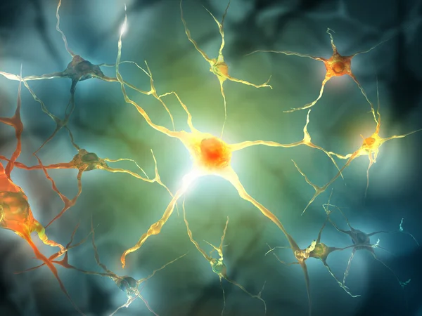
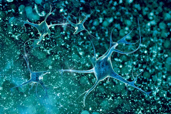
Neurons Of Dorsal Striatum, 3D Illustration. Dorsal Striatum Is A Nucleus In The Basal Ganglia, Degrading Of Its Neurons Plays Crucial Role In Development Of Huntington's Disease
Image, 8.32MB, 7200 × 4050 jpg

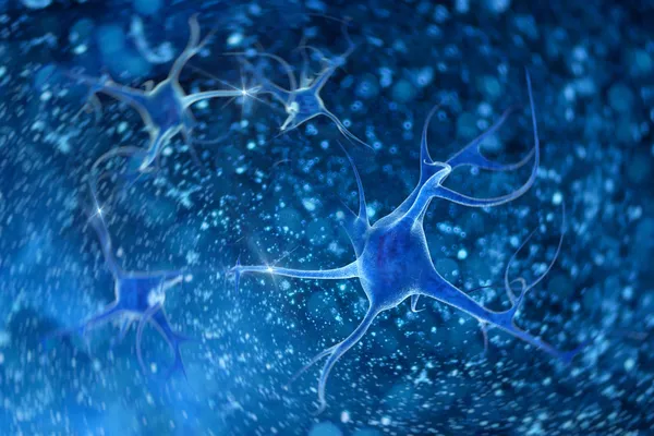
Neurons Of Dorsal Striatum, 3D Illustration. Dorsal Striatum Is A Nucleus In The Basal Ganglia, Degrading Of Its Neurons Plays Crucial Role In Development Of Huntington's Disease
Image, 12.86MB, 7200 × 4050 jpg
Page 1 >> Next