photo free
Home Images Vectors Videos

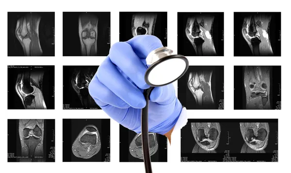
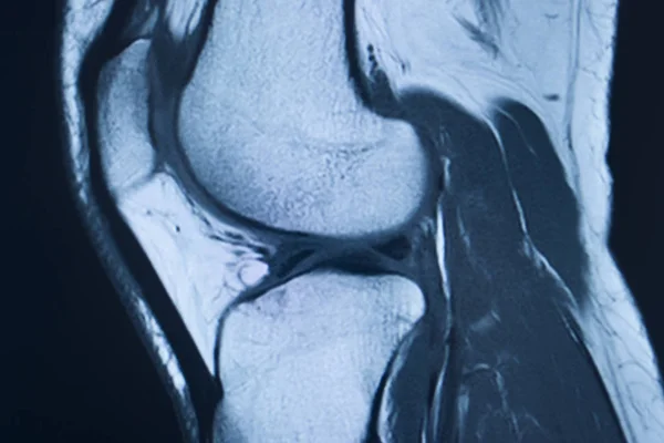
Stock image Mri Knee Joint

stock image Mri knee

Mri Knee

Image, 3.67MB, 3872 × 2592 jpg
stock image Magnetic Resonance images of  The Knee joint Sagittal Proton density Images  (MRI Knee joint) showing the anatomy of the knee

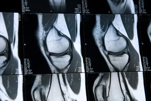
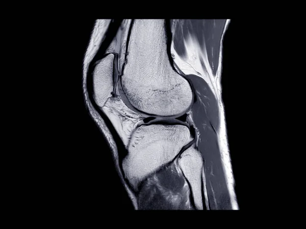
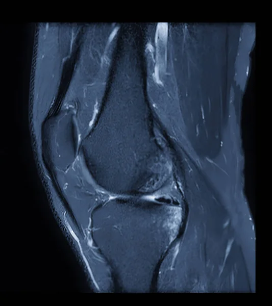
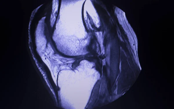
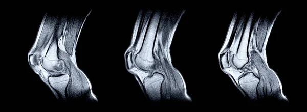
Magnetic Resonance Images Of The Knee Joint Sagittal Proton Density Images (MRI Knee Joint) Showing The Anatomy Of The Knee

Image, 5.23MB, 5760 × 3240 jpg
stock image MRI knee or Magnetic resonance imaging of knee joint stir technique of  coronal view for fat suppression.

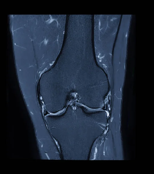
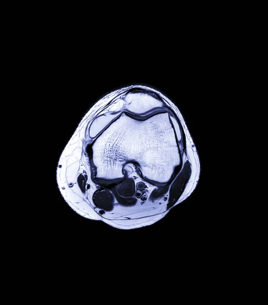
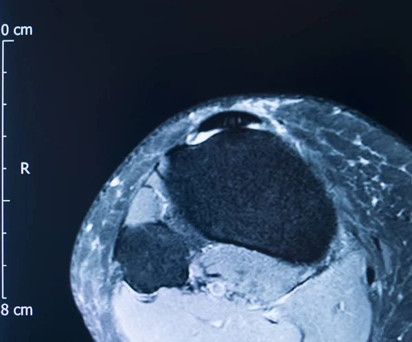
MRI Knee Or Magnetic Resonance Imaging Of Knee Joint Stir Technique Of Coronal View For Fat Suppression.

Image, 1.77MB, 2348 × 2664 jpg
stock image Magnetic resonance image of the knee joint  (MRI knee)in sagittal plan showing complete Anterior cruciate ligament tear (ACL tear)

Magnetic Resonance Image Of The Knee Joint (MRI Knee)in Sagittal Plan Showing Complete Anterior Cruciate Ligament Tear (ACL Tear)

Image, 13.94MB, 5100 × 4982 jpg
stock image MRI images

MRI Images

Image, 2.96MB, 4689 × 2796 jpg
stock image A male doctor makes an MRI of the knee joint. A young patient on an automatic table comes out of a closed-type MRI machine. Modern equipment, a coil on the patient's knee.

A Male Doctor Makes An MRI Of The Knee Joint. A Young Patient On An Automatic Table Comes Out Of A Closed-type MRI Machine. Modern Equipment, A Coil On The Patient's Knee.

Image, 8.18MB, 8044 × 5365 jpg
stock image Infographic diagram of human skeleton lower limb anatomy bone system or leg bone anterior view- 3D- Human Anatomy- Medical Diagram- educational and Human Body concept- Isolated on white background

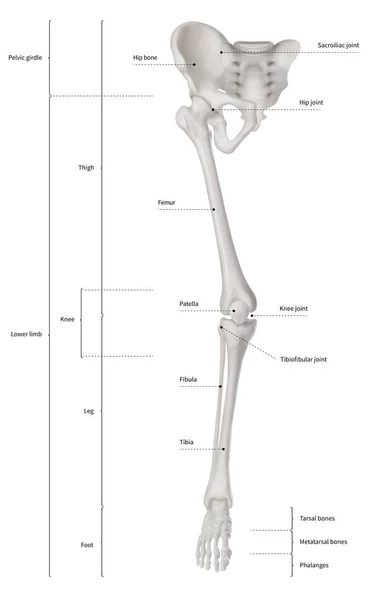
Infographic Diagram Of Human Skeleton Lower Limb Anatomy Bone System Or Leg Bone Anterior View- 3D- Human Anatomy- Medical Diagram- Educational And Human Body Concept- Isolated On White Background

Image, 6.28MB, 9405 × 15000 jpg
stock image MRI image of knee joint leg

MRI Image Of Knee Joint Leg

Image, 8.79MB, 4300 × 2867 jpg
stock image Compare of MRI Knee joint or Magnetic resonance imaging  sagital view for detect tear or sprain of the anterior cruciate  ligament (ACL).

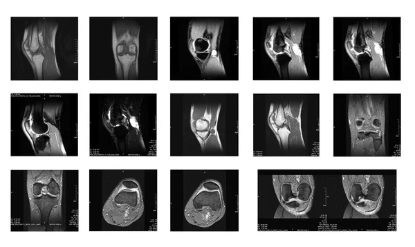
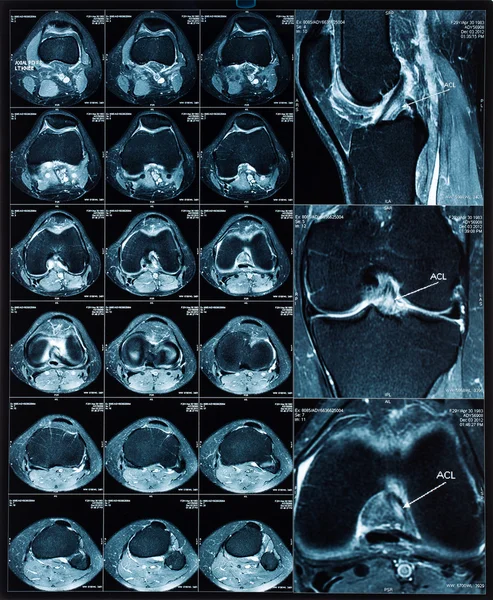
Compare Of MRI Knee Joint Or Magnetic Resonance Imaging Sagital View For Detect Tear Or Sprain Of The Anterior Cruciate Ligament (ACL).

Image, 2.76MB, 4448 × 2768 jpg
stock image Magnetic resonance imaging or MRI  knee joint  comparison coronal and sagittal view for detect tear or sprain of the anterior cruciate  ligament (ACL)

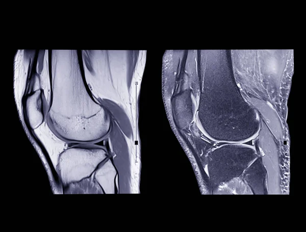
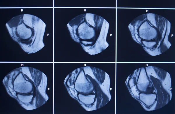
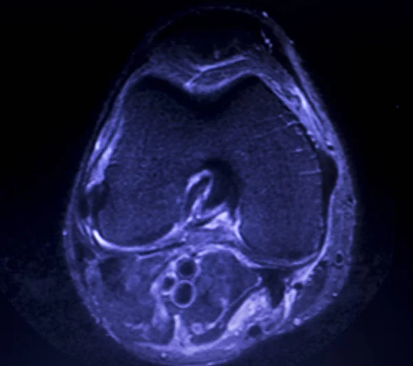
Magnetic Resonance Imaging Or MRI Knee Joint Comparison Coronal And Sagittal View For Detect Tear Or Sprain Of The Anterior Cruciate Ligament (ACL)

Image, 1.92MB, 3149 × 2173 jpg
stock image MRI Knee joint or Magnetic resonance imaging  sagittal view for detect tear or sprain of the anterior cruciate  ligament (ACL).

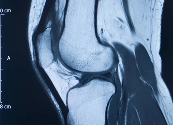
MRI Knee Joint Or Magnetic Resonance Imaging Sagittal View For Detect Tear Or Sprain Of The Anterior Cruciate Ligament (ACL).

Image, 1.44MB, 3195 × 2400 jpg
stock image MRI knee or Magnetic resonance imaging of knee joint stir technique of sagittal view for fat suppression.

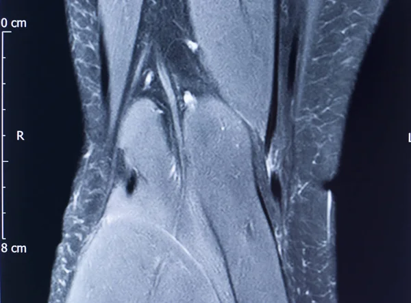
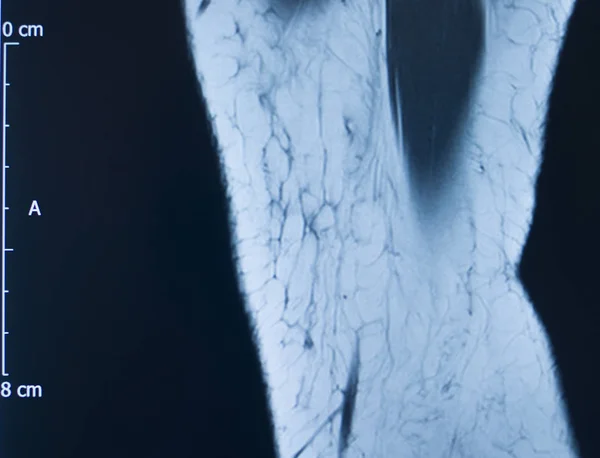
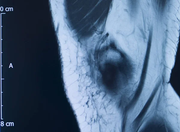
MRI Knee Or Magnetic Resonance Imaging Of Knee Joint Stir Technique Of Sagittal View For Fat Suppression.

Image, 1.96MB, 2370 × 2664 jpg
stock image Female basketball player visiting doctor after injury

Female Basketball Player Visiting Doctor After Injury

Image, 8.81MB, 6016 × 4016 jpg
stock image MRI Knee joint  3view

MRI Knee Joint 3view

Image, 9.29MB, 8238 × 9360 jpg
stock image Magnetic resonance imaging or MRI knee comparison sagittal PDW and TIW view for detect tear or sprain of the anterior cruciate  ligament (ACL).clipping path.

Magnetic Resonance Imaging Or MRI Knee Comparison Sagittal PDW And TIW View For Detect Tear Or Sprain Of The Anterior Cruciate Ligament (ACL).clipping Path.

Image, 2.65MB, 3968 × 3014 jpg
stock image Vertical photo of a close up x-ray scan of a knee joint. Doctor room, examining for diagnosis.

Vertical Photo Of A Close Up X-ray Scan Of A Knee Joint. Doctor Room, Examining For Diagnosis.

Image, 10.07MB, 3333 × 5000 jpg
stock image Close up hand doctor holding a pen and explain the results patient to know Magnetic resonance (MRI) Knee joint.

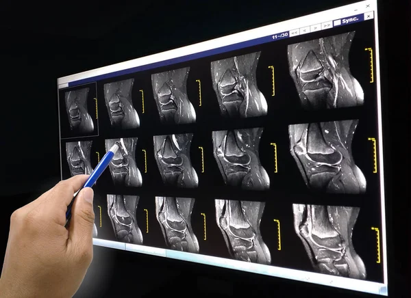
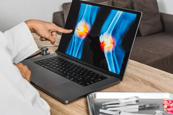
Close Up Hand Doctor Holding A Pen And Explain The Results Patient To Know Magnetic Resonance (MRI) Knee Joint.

Image, 4.3MB, 4128 × 3004 jpg
stock image Close up x-ray scan of a knee joint. Doctor room, examining for diagnosis.

Close Up X-ray Scan Of A Knee Joint. Doctor Room, Examining For Diagnosis.

Image, 10.29MB, 5000 × 3333 jpg
stock image Close up hand doctor holding a pen and explain the results  patient to know MRI of right knee.

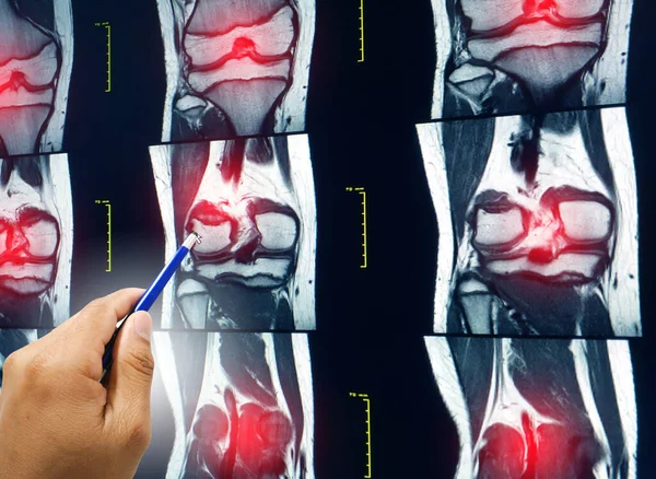
Close Up Hand Doctor Holding A Pen And Explain The Results Patient To Know MRI Of Right Knee.

Image, 7.17MB, 4044 × 2957 jpg
stock image A male doctor makes an MRI of the knee joint. A young patient on an automatic table comes out of a closed-type MRI machine. Modern equipment, a coil on the patient's knee.

A Male Doctor Makes An MRI Of The Knee Joint. A Young Patient On An Automatic Table Comes Out Of A Closed-type MRI Machine. Modern Equipment, A Coil On The Patient's Knee.

Image, 1.81MB, 3543 × 2362 jpg
stock image Radiologist woman doctor explaining good results of CT scan to young man showing xray with images, observing and analyzing CT scan in modern clinic next to modern CT scanner

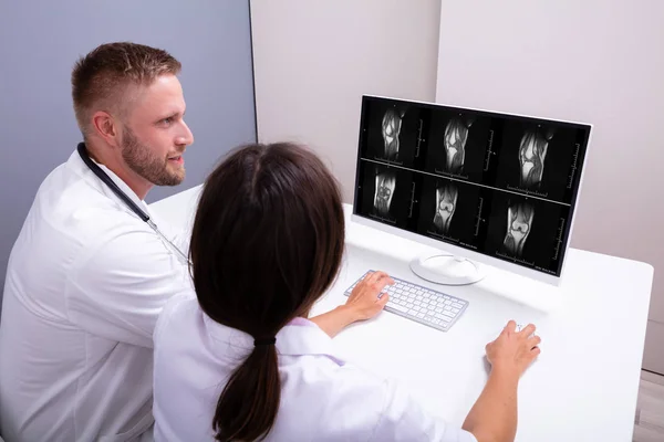
Radiologist Woman Doctor Explaining Good Results Of CT Scan To Young Man Showing Xray With Images, Observing And Analyzing CT Scan In Modern Clinic Next To Modern CT Scanner

Image, 10.54MB, 7289 × 4862 jpg
stock image Radiography of Human Bones

Radiography Of Human Bones

Image, 13.28MB, 4500 × 4494 jpg
stock image Doctors Looking At An MRI Scan At Computer In Clinic

Doctors Looking At An MRI Scan At Computer In Clinic

Image, 16.79MB, 8688 × 5792 jpg
stock image Meniscal root tears are less common than meniscal body tears and frequently go undetected.3D rendering

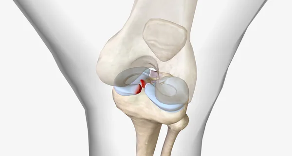
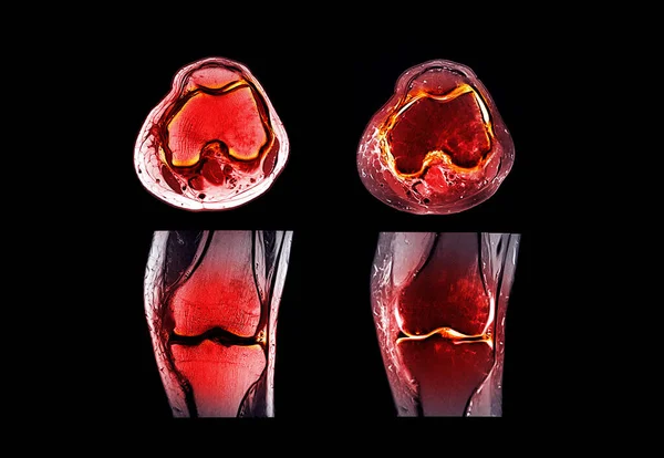
Meniscal Root Tears Are Less Common Than Meniscal Body Tears And Frequently Go Undetected.3D Rendering

Image, 1.96MB, 7304 × 3920 jpg
stock image Knee injury mri mcl tear

Knee Injury Mri Mcl Tear

Image, 16.99MB, 5304 × 3920 jpg
stock image MRI images

MRI Images

Image, 3.51MB, 4689 × 2796 jpg
stock image MRI scan test results knee meniscus injury

MRI Scan Test Results Knee Meniscus Injury

Image, 7.24MB, 3876 × 3456 jpg
stock image Collection MRI Knee: at distal lateral femoral metaphysis, representing the pre-existing location of the tumor, surrounding with a well-defined bony destruction with multiple cystic.

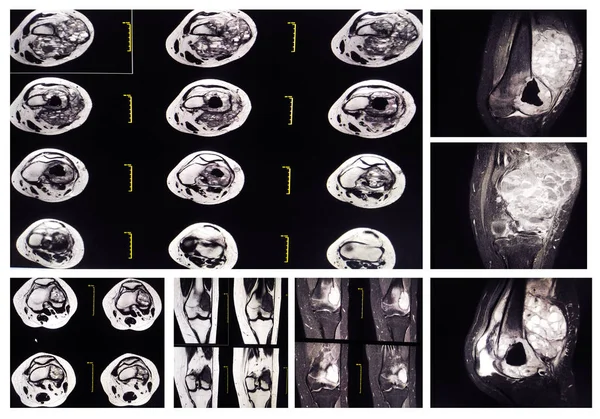
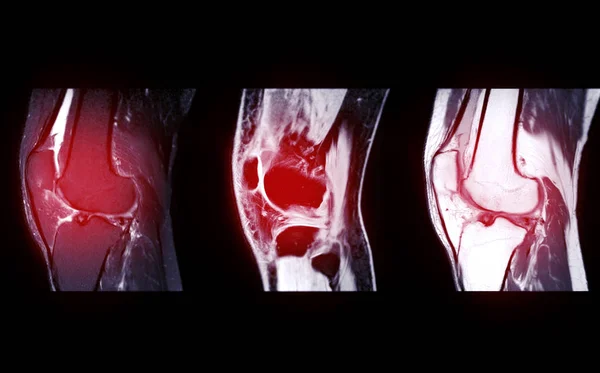
Collection MRI Knee: At Distal Lateral Femoral Metaphysis, Representing The Pre-existing Location Of The Tumor, Surrounding With A Well-defined Bony Destruction With Multiple Cystic.

Image, 8.76MB, 5672 × 3967 jpg
stock image A snapshot of the diagnosis of an MRI of the knee in which arthrosis and arthritis. The concept of joint diagnosis using x-rays, treatment of diseases of the knee joint, bursitis, periarticular bag

A Snapshot Of The Diagnosis Of An MRI Of The Knee In Which Arthrosis And Arthritis. The Concept Of Joint Diagnosis Using X-rays, Treatment Of Diseases Of The Knee Joint, Bursitis, Periarticular Bag

Image, 9.4MB, 4077 × 3648 jpg
stock image MRT images of knee

MRT Images Of Knee

Image, 8.32MB, 2634 × 3205 jpg
stock vector Orthopedics joints and rheumatic disorder medical health care. Vector human skeleton parts hand, foot and pelvis, spine. Knee and shoulder joints orthopedy clinic mri tomography images medicine poster

Orthopedics Joints And Rheumatic Disorder Medical Health Care. Vector Human Skeleton Parts Hand, Foot And Pelvis, Spine. Knee And Shoulder Joints Orthopedy Clinic Mri Tomography Images Medicine Poster

Vector, 1.05MB, 6927 × 4908 eps
stock image Knee injury mri mcl tear

Knee Injury Mri Mcl Tear

Image, 12.69MB, 5301 × 3820 jpg
stock image Doctor explaining x-ray scan during consultation

Doctor Explaining X-ray Scan During Consultation

Image, 8.45MB, 5744 × 3835 jpg
stock image Knee medical exam: X-ray and MRI scan

Knee Medical Exam: X-ray And MRI Scan

Image, 14.92MB, 4500 × 4494 jpg
stock vector Rheumatology and orthopedic medical clinic vector banners with joints and bones anatomy. MRI and CT scans of arthritis pain diagnostics with leg, hand, knee and spine, foot, pelvis, shoulder and elbow

Rheumatology And Orthopedic Medical Clinic Vector Banners With Joints And Bones Anatomy. MRI And CT Scans Of Arthritis Pain Diagnostics With Leg, Hand, Knee And Spine, Foot, Pelvis, Shoulder And Elbow

Vector, 1.09MB, 6033 × 5967 eps
stock image Magnetic resonance imaging MRI knee posterior horn medial meniscus tear scantest results.

Magnetic Resonance Imaging MRI Knee Posterior Horn Medial Meniscus Tear Scantest Results.

Image, 14.46MB, 6000 × 3792 jpg
stock vector X-rays of human joint anatomy with pain parts. X-ray examination of bones. Medical and science illustration.

X-rays Of Human Joint Anatomy With Pain Parts. X-ray Examination Of Bones. Medical And Science Illustration.

Vector, 0.3MB, 9500 × 4000 eps
stock image Knee medical exam: X-ray and MRI scan

Knee Medical Exam: X-ray And MRI Scan

Image, 8.35MB, 4500 × 4494 jpg
stock image MRI scan test results knee meniscus injury

MRI Scan Test Results Knee Meniscus Injury

Image, 10.19MB, 4994 × 3255 jpg
stock image Knee injury mri mcl tear

Knee Injury Mri Mcl Tear

Image, 10.29MB, 5045 × 3854 jpg
stock image Close up of doctor showing a x-ray of pain in the knee. High quality photo

Close Up Of Doctor Showing A X-ray Of Pain In The Knee. High Quality Photo

Image, 15.81MB, 7008 × 4672 jpg
stock image Magnetic resonance imaging (MRI) of right knee.

Magnetic Resonance Imaging (MRI) Of Right Knee.

Image, 0.91MB, 3386 × 1242 jpg
stock image Knee injury mri mcl tear

Knee Injury Mri Mcl Tear

Image, 13.03MB, 5072 × 3723 jpg
stock image Knee injury mri mcl tear

Knee Injury Mri Mcl Tear

Image, 11.53MB, 4618 × 3836 jpg
stock image Sports player visiting doctor after injury

Sports Player Visiting Doctor After Injury

Image, 3.56MB, 6016 × 4016 jpg
stock image Knee sports injury mri mcl grade 2 tear magnetic resonance imaging orthopedic traumatology scan.

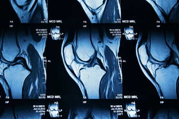
Knee Sports Injury Mri Mcl Grade 2 Tear Magnetic Resonance Imaging Orthopedic Traumatology Scan.

Image, 15.89MB, 5523 × 3935 jpg
stock image Magnetic resonance imaging MRI knee posterior horn medial meniscus tear scantest results.

Magnetic Resonance Imaging MRI Knee Posterior Horn Medial Meniscus Tear Scantest Results.

Image, 11.56MB, 4512 × 4000 jpg
stock image Magnetic resonance imaging MRI knee posterior horn medial meniscus tear scantest results.

Magnetic Resonance Imaging MRI Knee Posterior Horn Medial Meniscus Tear Scantest Results.

Image, 12.43MB, 4640 × 3888 jpg
stock image Knee injury mri mcl tear

Knee Injury Mri Mcl Tear

Image, 18.28MB, 6000 × 4000 jpg
stock vector Rheumatology, joints and rheumatic disorder medical health care. Vector human skeleton parts hand, foot and pelvis, spine, knee and shoulder joints mri or computed tomography images, medicine poster

Rheumatology, Joints And Rheumatic Disorder Medical Health Care. Vector Human Skeleton Parts Hand, Foot And Pelvis, Spine, Knee And Shoulder Joints Mri Or Computed Tomography Images, Medicine Poster

Vector, 0.73MB, 4745 × 7166 eps
Page 1 >> Next