
Stock image Mri Of Shoulder Joint

MRI Shoulder History Of Rotator Cuff Tear With Suspected Lipoma Of Left Shoulder Findings Tear Of Supraspinatous Tendon.Medical Image Concept.
Image, 5.61MB, 7230 × 4000 jpg
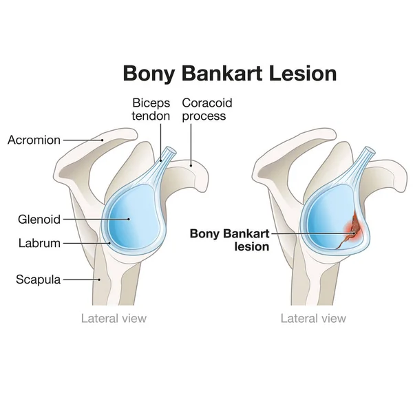
A Bony Bankart Lesion In The Shoulder Involves A Fracture Of The Anterior Glenoid Rim, Often Resulting From Dislocation, Leading To Instability And Requiring Surgical Repair For Stability Restoration.
Image, 2.88MB, 5000 × 5000 jpg
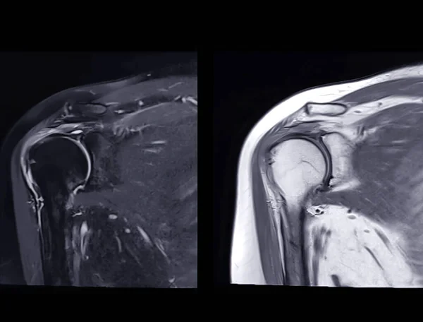
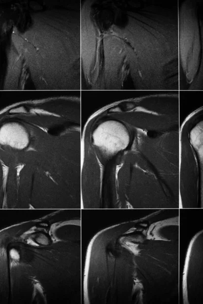
Magnetic Resonance Imaging Or MRI Of Shoulder Joint Coronal T2 FS And PDW For Diagnostic Shoulder Pain.
Image, 3.42MB, 4035 × 3071 jpg
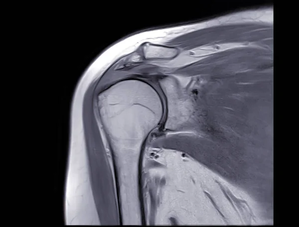
Magnetic Resonance Imaging Or MRI Of Shoulder Joint Coronal PDW For Diagnostic Shoulder Pain.
Image, 3.34MB, 4035 × 3071 jpg
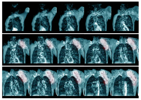
Magnetic Resonance Imaging(MRI) Left.shoulder History:Case Lt. Shoulder Mass . Impression:-The Mass Soft Tissue Sarcoma And Metastasis.Medical And Healthcare Concept.
Image, 16.6MB, 8552 × 6068 jpg
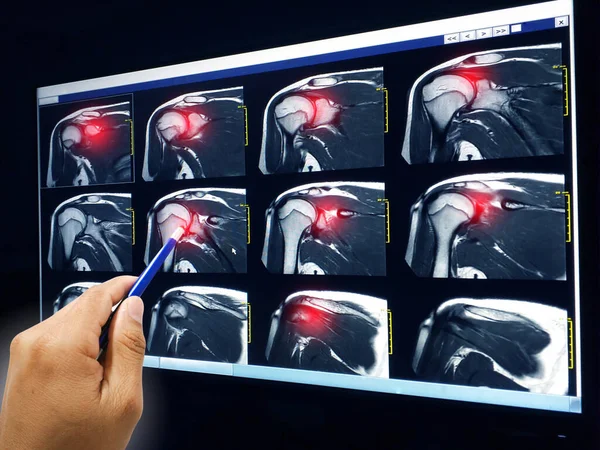
Hand Doctor Point MRI Of Shoulder Explain The Results To The Patient To Know Rotator Cuff Tendon Tear Red Highlight Focus
Image, 5.8MB, 4128 × 3096 jpg
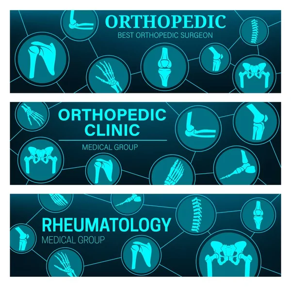
Rheumatology And Orthopedic Medical Clinic Vector Banners With Joints And Bones Anatomy. MRI And CT Scans Of Arthritis Pain Diagnostics With Leg, Hand, Knee And Spine, Foot, Pelvis, Shoulder And Elbow
Vector, 1.09MB, 6033 × 5967 eps
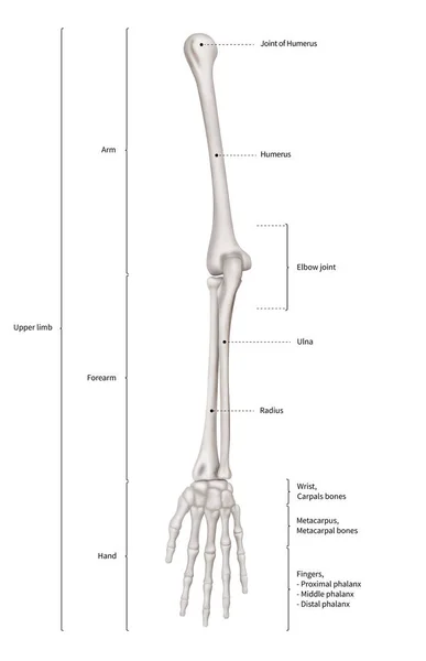
Infographic Diagram Of Human Skeleton Upper Limb Bone Anatomy System Or Arm Bone Anterior View- 3D- Human Anatomy- Medical Diagram- Educational And Human Body Concept- Isolated On White Background
Image, 7.21MB, 9692 × 15000 jpg
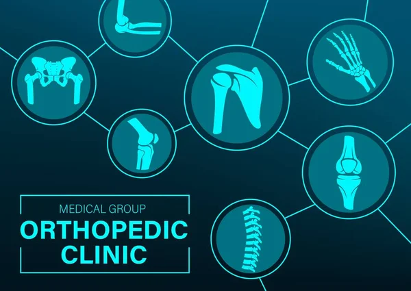
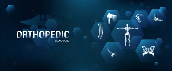
Orthopedics Joints And Rheumatic Disorder Medical Health Care. Vector Human Skeleton Parts Hand, Foot And Pelvis, Spine. Knee And Shoulder Joints Orthopedy Clinic Mri Tomography Images Medicine Poster
Vector, 1.05MB, 6927 × 4908 eps
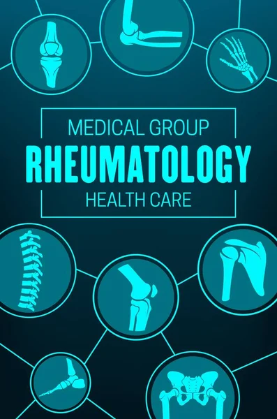
Rheumatology, Joints And Rheumatic Disorder Medical Health Care. Vector Human Skeleton Parts Hand, Foot And Pelvis, Spine, Knee And Shoulder Joints Mri Or Computed Tomography Images, Medicine Poster
Vector, 0.73MB, 4745 × 7166 eps
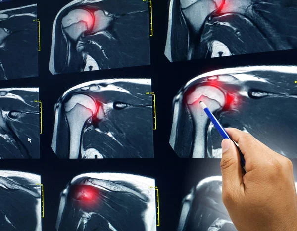
Hand Doctor Point MRI Of Shoulder Explain The Results To The Patient To Know Rotator Cuff Tendon Tear Red Highlight Focus.
Image, 5.86MB, 3840 × 2993 jpg

Orthopedy Medicine, Joints Treatment, Xray, Rheumatic Disorder Medical Health Care. Vector Human Skeleton Parts Elbow, Foot And Pelvis, Spine, Knee And Shoulder Joints Mri, Computed Tomography Poster
Vector, 0.89MB, 5831 × 5831 eps

Doctor, Senior And Patient In Bed For Consulting With Xray On Screen For Evaluation Of Joint Condition And Arthritis. Old Man, Wife And Support In Hospital For Mri Or Scan On Shoulder And Bursitis
Image, 6.94MB, 6934 × 4912 jpg

AC Joint Separation Is A Shoulder Injury Involving Ligament Damage At The Acromioclavicular Joint. It Causes Pain, Swelling, And Possible Deformity, With Treatment Options Ranging From Conservative Approaches To Surgical Intervention Based On The Sev
Image, 3.69MB, 5000 × 5000 jpg
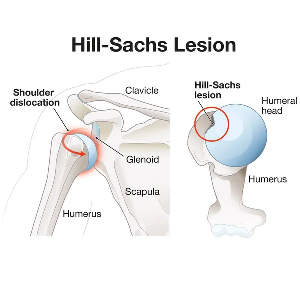
A Hill-Sachs Lesion Is A Divot-like Defect On The Humeral Head, Often Resulting From Shoulder Dislocation. It Can Contribute To Instability And Limited Range Of Motion In The Joint.
Image, 2.83MB, 5000 × 5000 jpg
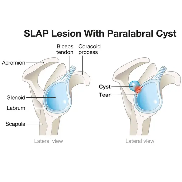
A SLAP Lesion Paralabral Cyst In The Shoulder Is A Tear In The Labrum Accompanied By A Cyst, Often Causing Pain, Instability, And Functional Limitations, Typically Requiring Surgical Intervention For Treatment.
Image, 2.9MB, 5000 × 5000 jpg
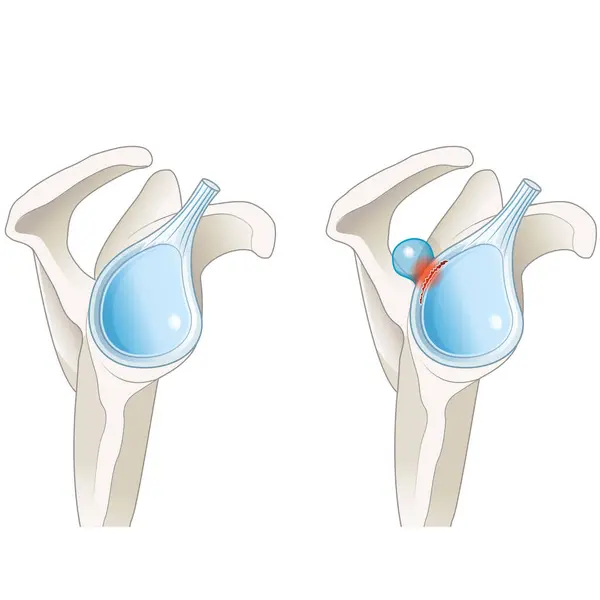
A SLAP Lesion Paralabral Cyst In The Shoulder Is A Tear In The Labrum Accompanied By A Cyst, Often Causing Pain, Instability, And Functional Limitations, Typically Requiring Surgical Intervention For Treatment.
Image, 2.86MB, 5000 × 5000 jpg

AC Joint Separation Is A Shoulder Injury Involving Ligament Damage At The Acromioclavicular Joint. It Causes Pain, Swelling, And Possible Deformity, With Treatment Options Ranging From Conservative Approaches To Surgical Intervention Based On The Sev
Image, 3.86MB, 5000 × 5000 jpg

Doctor, Senior And Patient With Xray On Tablet For Consultation With Screen For Evaluation Of Joint Condition And Arthritis. Old Man, Wife And Support In Hospital For Mri On Shoulder And Bursitis
Image, 7.51MB, 6448 × 4912 jpg
Page 1 >> Next