
Stock image Mycology


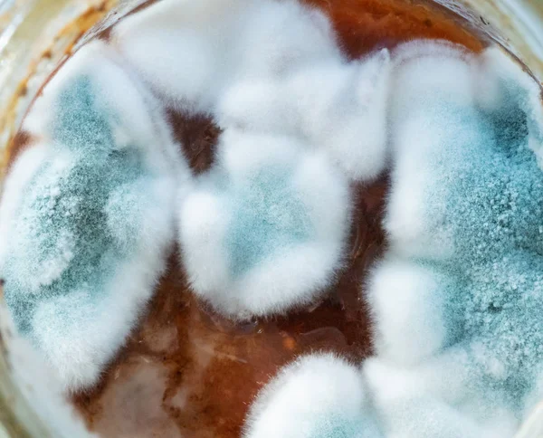
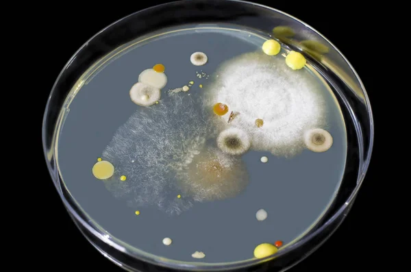
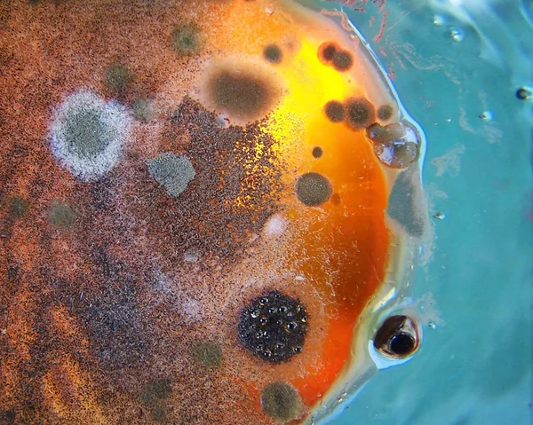
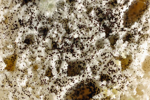
Microbiology. Growth Of Aspergillosis Aspergillus Flavus On Jam Surface, Macro, Top View
Image, 10.04MB, 4011 × 3240 jpg


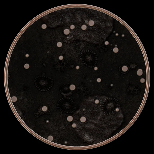
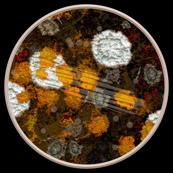
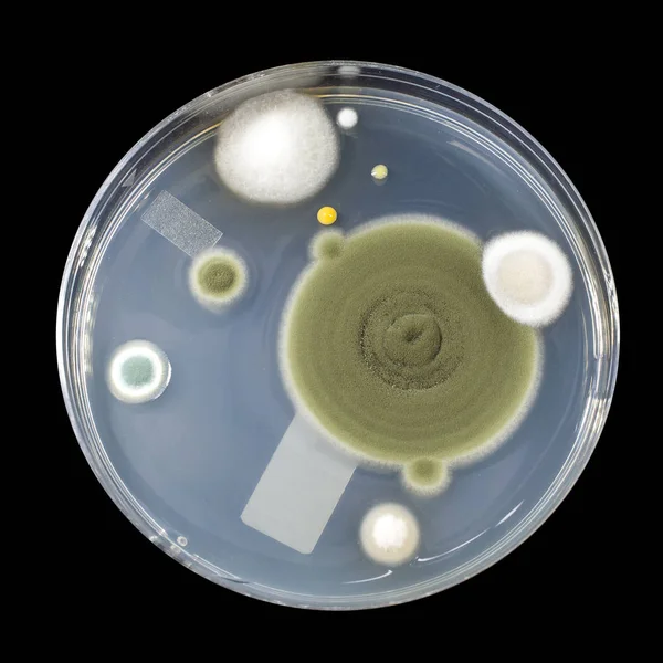
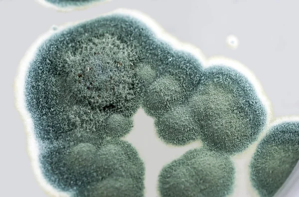
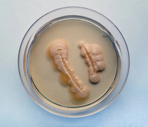
Fungal Mycelium Petri Dish. White Background. Mushroom Mycelium On Agar. Reishi Mushroom Mycelium On Potato Dextrose Agar. Laboratory Accessories. Mycology Growing In A Petri Dishes.
Image, 7.68MB, 6000 × 4000 jpg

Laboratory Accessories. White Background. Fungal Mycelium Petri Dish. Mycology Growing In A Petri Dishes. Mushroom Mycelium On Agar. Reishi Mushroom Mycelium On Potato Dextrose Agar.
Image, 10.8MB, 6000 × 4000 jpg
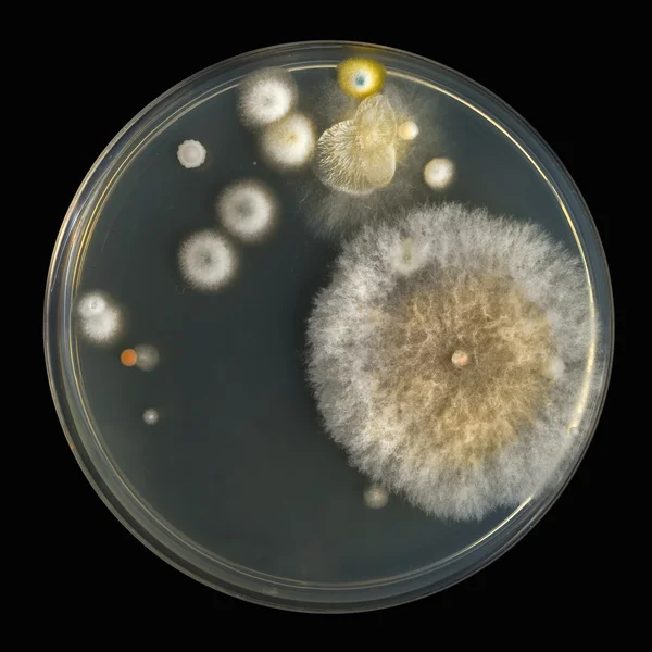
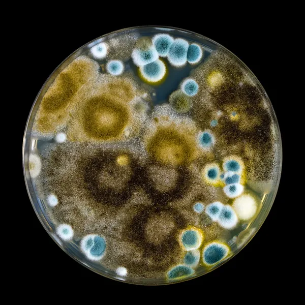
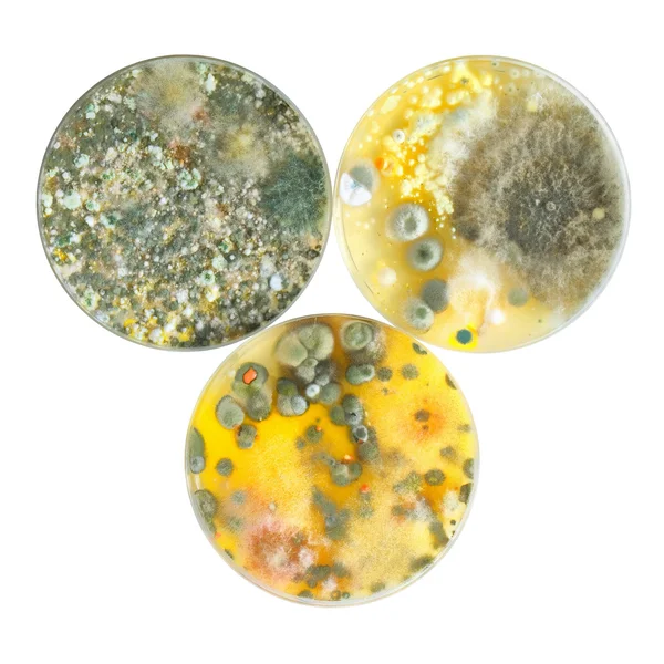
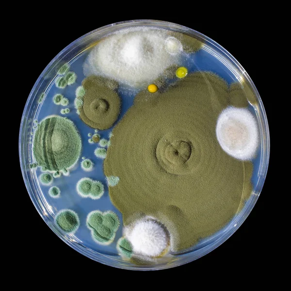
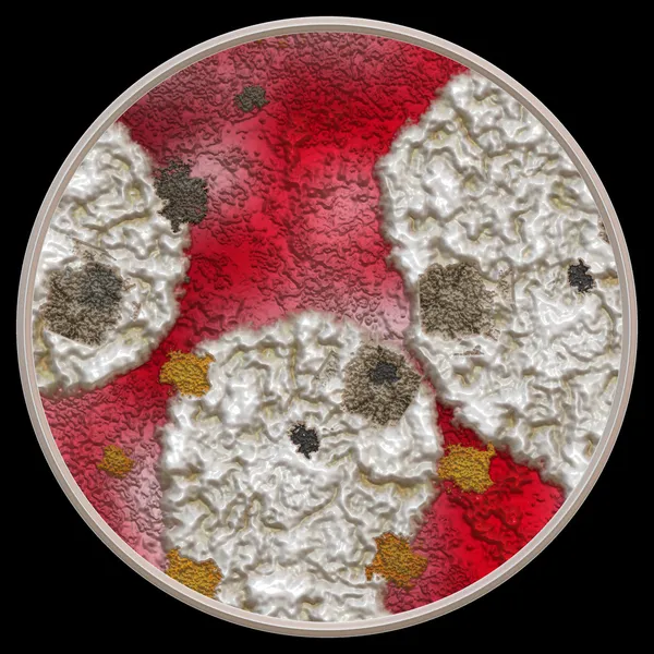
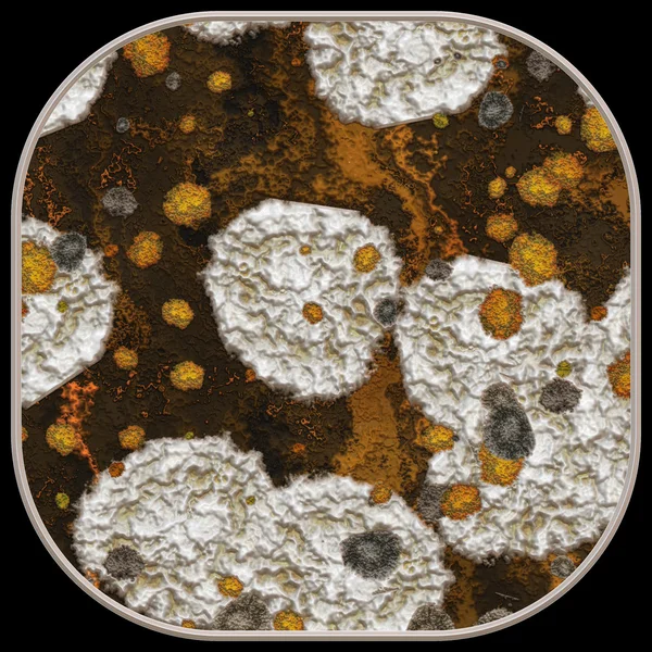
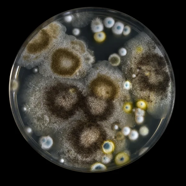
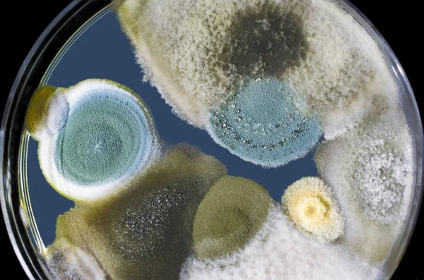
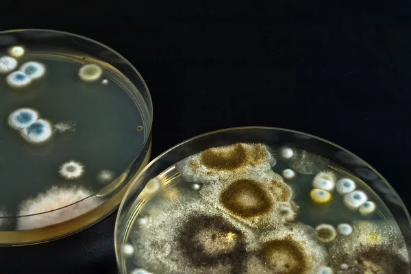
Mold Beautiful, Colony Of Characteristics Of Fungus (Mold) In Culture Medium Plate From Laboratory Microbiology.
Image, 11.64MB, 6612 × 4408 jpg
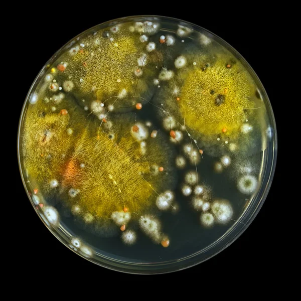


Microbiology. Growth Of Aspergillosis. Aspergillus Flavus On Jam Surface, Macro
Image, 8.71MB, 4163 × 3122 jpg


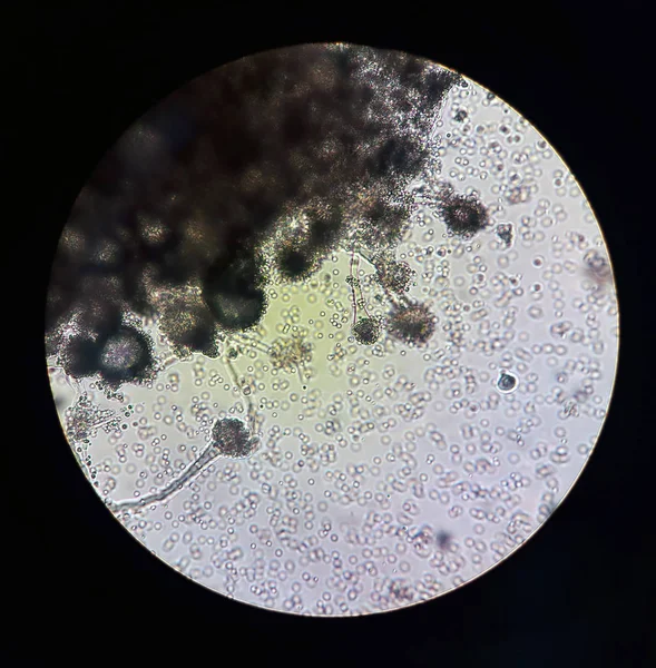
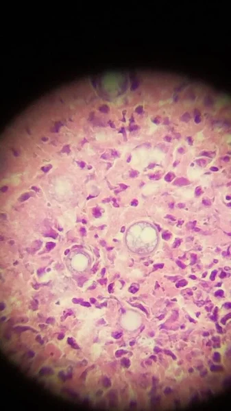
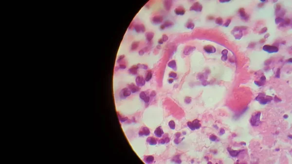
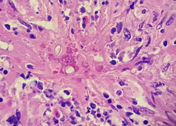
Zygomycota, Or Zygote Fungi ,Downy Mildew Of Cruzifers Host Tissue With Conidia Living In Decaying Plant On Slide Under The Microscope For Education.
Image, 21MB, 6720 × 4480 jpg


Zygomycota, Or Zygote Fungi ,Downy Mildew Of Cruzifers Host Tissue With Conidia Living In Decaying Plant On Slide Under The Microscope For Education.
Image, 21.35MB, 6720 × 4480 jpg


Page 1 >> Next