
Stock image Myelogram
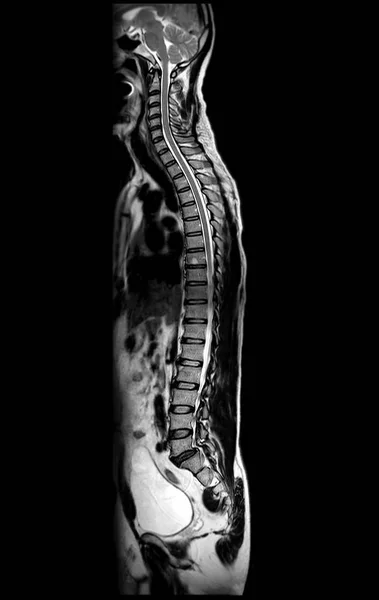
MRI Of Whole Spine T2W Sagittal Plane For Diagnostic Spinal Cord Compression.
Image, 4.7MB, 4624 × 7314 jpg


MRI Of Whole Spine T2W Sagittal Plane For Diagnostic Spinal Cord Compression.
Image, 1.94MB, 3088 × 4136 jpg

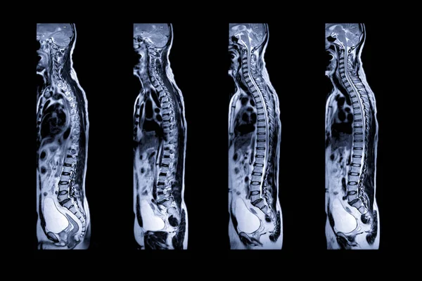
MRI Of The Whole Spine Reveals Detailed Images Of The Spinal Cord For Comprehensive Evaluation.
Image, 4.48MB, 3536 × 7369 jpg


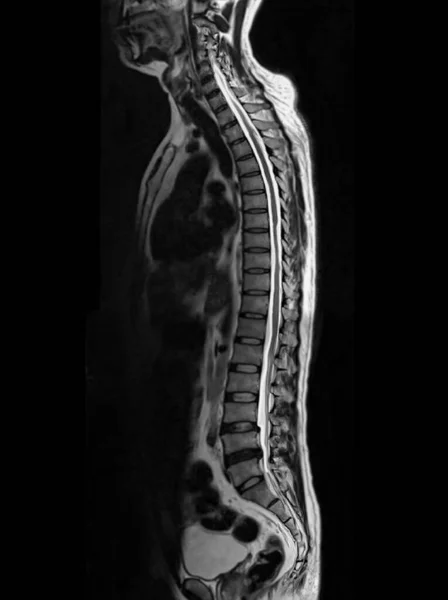
MRI Of The Whole Spine Reveals Detailed Images Of The Spinal Cord For Comprehensive Evaluation.
Image, 2.58MB, 3695 × 7246 jpg

MRI Of The Whole Spine Reveals Detailed Images Of The Spinal Cord For Comprehensive Evaluation.
Image, 2.45MB, 3689 × 7666 jpg
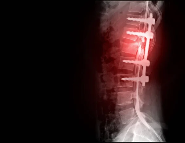
Myelography Is Particularly Sensitive At Detecting Small Disk Herniations Compressing Nerves Of The Spine And Spinal Cord Injury.
Image, 3.14MB, 4816 × 3719 jpg

MRI Of The Whole Spine Reveals Detailed Images Of The Spinal Cord For Comprehensive Evaluation.
Image, 4.7MB, 3578 × 7571 jpg
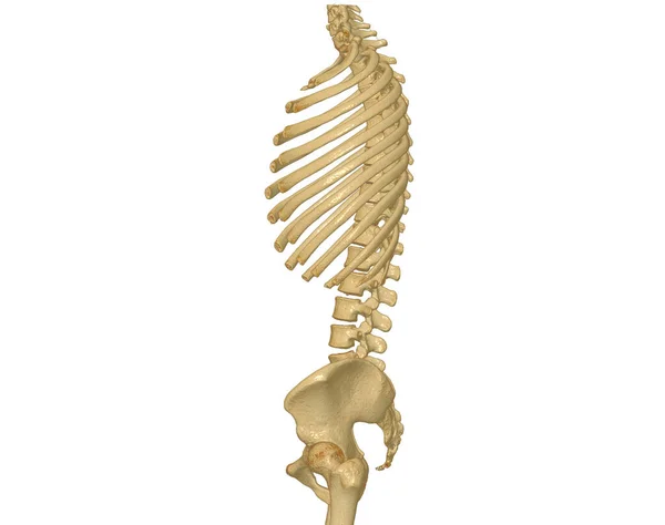
CT Scan Of Whole Spine 3D Rendering Showing Profile Human Spine. Musculoskeletal System Human Body. Structure Spine. Studying Problem Disease And Treatment Methods. Isolated On White Background. Clipping Path.
Image, 1.59MB, 3840 × 3036 jpg

MRI Cervical Spine Myelogram Seqeunce For Diagnostic Spinal Cord Compression.
Image, 1.8MB, 3536 × 3536 jpg

MRI Cervical Spine Myelogram Seqeunce For Diagnostic Spinal Cord Compression.
Image, 1.78MB, 3536 × 3536 jpg

MRI Of The Whole Spine Reveals Detailed Images Of The Spinal Cord For Comprehensive Evaluation.
Image, 1.85MB, 3536 × 3536 jpg

MRI Of The Whole Spine Reveals Detailed Images Of The Spinal Cord For Comprehensive Evaluation.
Image, 8.06MB, 6149 × 7571 jpg


Page 1 >> Next