
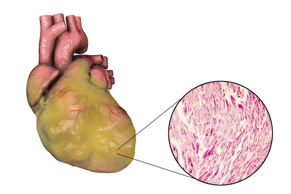
Stock image Myocardial Hypertrophy
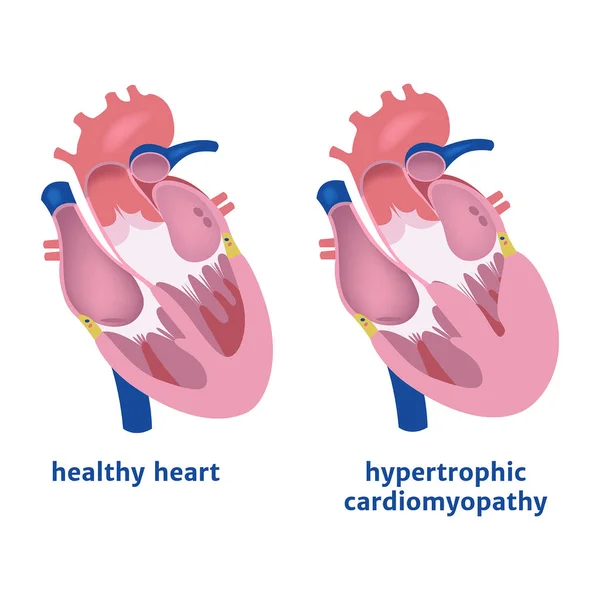
Hypertrophic Cardiomyopathy. Expansion Of The Ventricle Of The Heart. Medical Poster Vector Illustration
Vector, 6.01MB, 2000 × 2000 eps
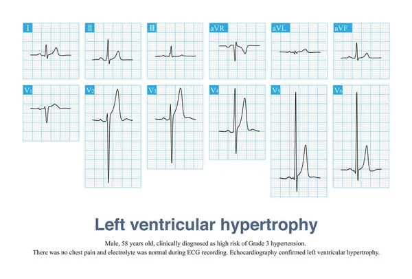
Sometimes, Left Ventricular Hypertrophy With Tall T Waves Is Easily Misdiagnosed As Hyperkalemia And Hyperacute T Waves, And ECG Needs To Be Carefully Identified In Combination With Clinic.
Image, 13.77MB, 10000 × 6782 jpg
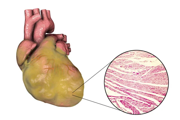


Cardiomyopathy Is Inflammation In The Heart Muscle, Resulting In Its Enlargement And Weakening That Impairs The Blood's Pumping Ability. 3D Illustration
Image, 1.17MB, 3508 × 2480 jpg


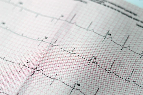
ECG ElectroCardioGraph Paper That Shows Sinus Rhythm Abnormality Of Right Ventricular Hypertrophy, Inferior T Wave Due To Hypertrophy And Ischemia, Abnormal ECG Study, Unconfirmed Diagnosis
Image, 17.21MB, 6016 × 4000 jpg

Heart Failure ( Film X-ray Chest PA Upright : Show Cardiomegaly And Interstitial Infiltrate Both Lung )
Image, 6.71MB, 3915 × 3456 jpg
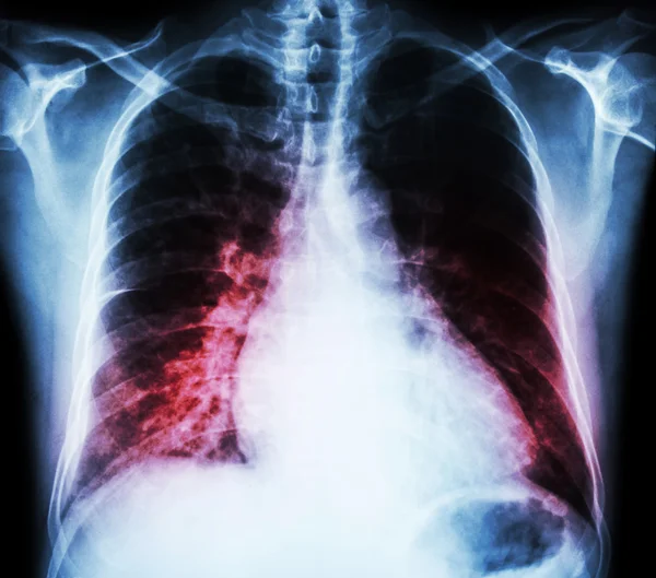
Heart Failure ( Film X-ray Chest PA Upright : Show Cardiomegaly And Interstitial Infiltrate Both Lung )
Image, 6.45MB, 3915 × 3456 jpg
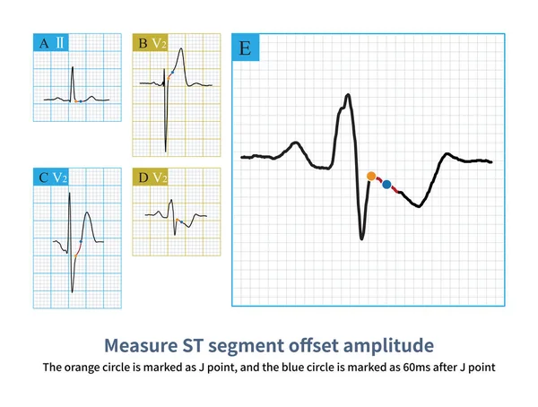
Firstly, Select Point J As The Reference Point, And Then Select 60ms After Point J As The Measurement Point To Evaluate The ST Segment Offset Morphology And Amplitude.
Image, 10.33MB, 10000 × 7579 jpg
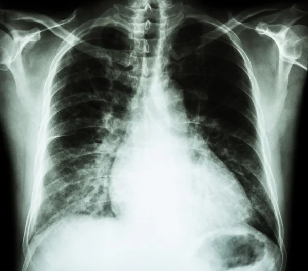
Heart Failure ( Film X-ray Chest PA Upright : Show Cardiomegaly And Interstitial Infiltrate Both Lung )
Image, 4.93MB, 3915 × 3456 jpg
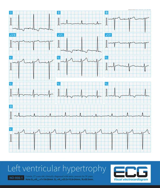
Sometimes, Because The QRS Axis Is In The Upper Left Quadrant, The High-amplitude R Wave Of Left Ventricular Hypertrophy Occurs In The Limb Leads, And Left Chest Leads Is Normal.
Image, 31.4MB, 10000 × 11694 jpg
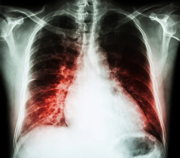
Heart Failure ( Film X-ray Chest PA Upright : Show Cardiomegaly And Interstitial Infiltrate Both Lung )
Image, 5.88MB, 3915 × 3456 jpg
Page 1 >> Next