
Stock image Nerve Root Compression page 2


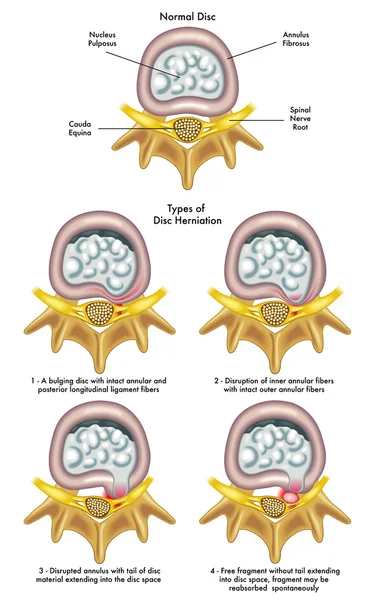
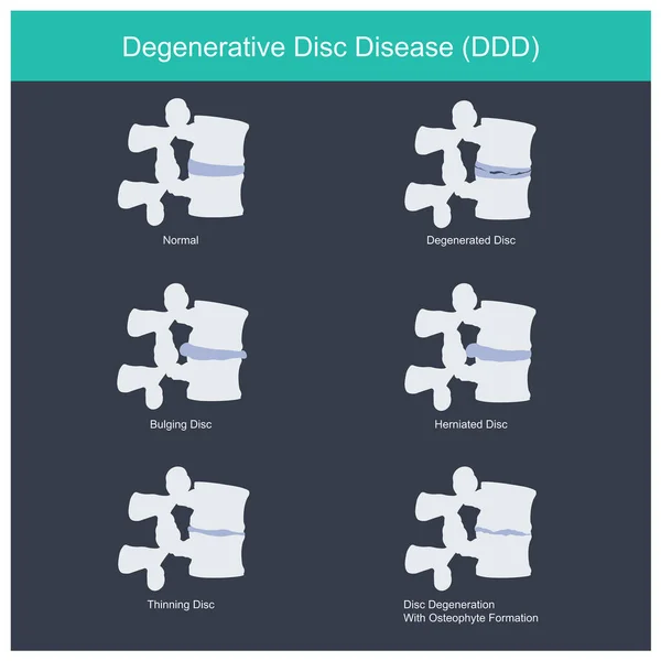
Spinal Disc Herniation. Difference Between Bulging Disc And Herniated Disc. Vector Illustration
Vector, 9.64MB, 5907 × 3000 eps

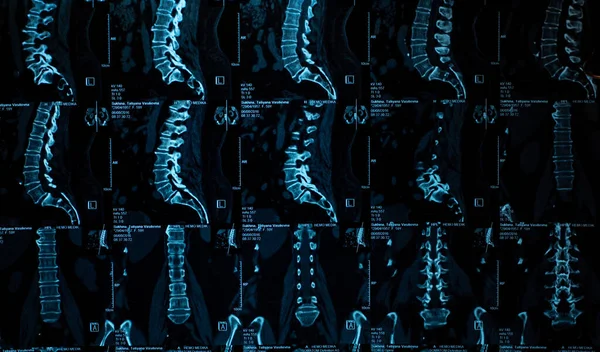
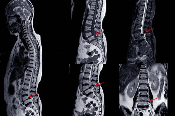
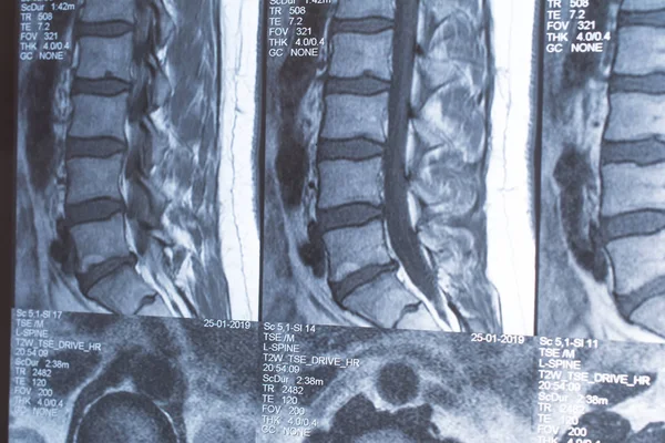
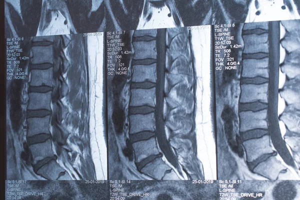
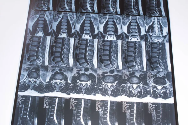
MRI OF THORACOLUMBAR SPINE:IMPRESSION: Moderate Pathological Compression Of T11 And L2 Levels With Enhancing Multiple Marrow Lesions At T1, T10 ToT12, L2, L3 To L5 Levels.multiple Bone Metastases Should Be Considered,hematologic Malignancy .
Image, 7.76MB, 5760 × 4348 jpg
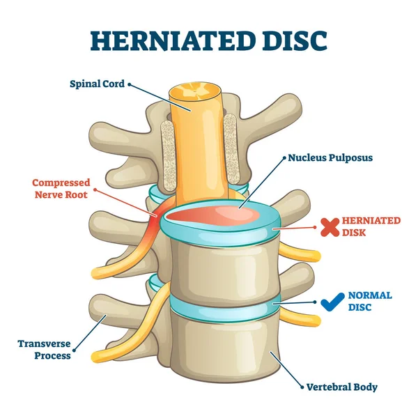
Herniated Disc Injury 3D Side View On Spine Bone Skeleton Vector Illustration
Vector, 10.23MB, 4000 × 4000 eps
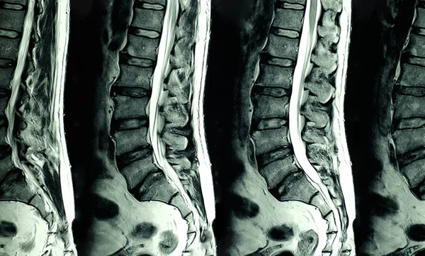
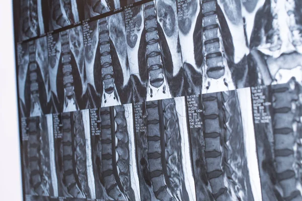
MRI Scans Of The Lumbosacral Spine.. MRI Shows Degenerative Changes In The Spine, Hernia Of The Lumbar Discs And Compression Of The Nerve Roots.
Image, 3.03MB, 3173 × 1861 jpg
MRI Scan Of Lumbar Spines Of A Patient With Chronic Back Pain Showing Degenerative Change Of The L Spines With Right Lateral Protrution Of L4-5 Disc And L4 Nerve Root Compression
Image, 11.99MB, 5967 × 3596 jpg
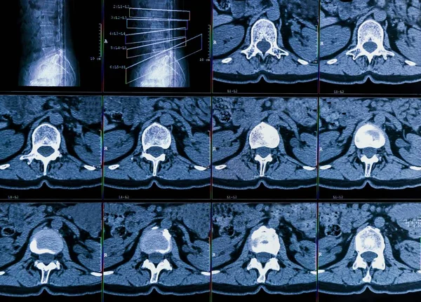
Results Of Computer Tomography Or CT Imaging Of Human Spine Of A Patient With Chronic Back Pain, Shows Degenerative Changes Of Spines, Lumbar Discs Herniation And Nerve Roots Compression
Image, 15.56MB, 6759 × 4861 jpg
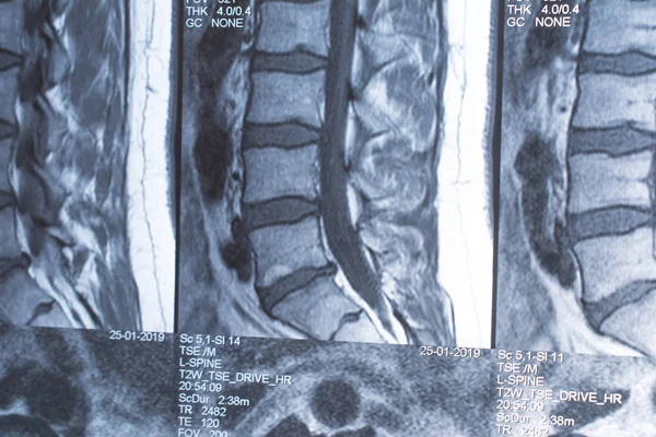
MRI Of Lumbar Spine Showing Cervical Spondylosis With Mild To Moderate Spinal Cord Compression At C4-5 And C5-6 With Myelopathy
Image, 5.74MB, 6000 × 4000 jpg

Doctor Analyzes The Results Of Magnetic Resonance Imaging Of A Patient Spine With Chronic Back Pain. The MRI Shows Degenerative Changes Of Spines, Lumbar Discs Herniation And Nerve Roots Compression.
Image, 2.17MB, 3840 × 2160 jpg


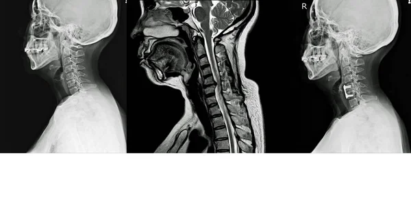

A Neck Xray Image Of A Patient Suffering From Car Accident Showing A Bursted Fracture Of C 5 Cervical Spine.
Image, 8.7MB, 6240 × 3902 jpg
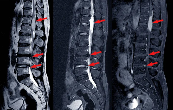
MRI Scan Of Lumbar Spines Of A Patient Finding Spinal Mass At Lt.side T12-L1 Level Severe Bulging Disc L3-4 Causing Bilateral L4 Nerve Root Compression And Spinal Stenosis On Arrow Point.
Image, 6.3MB, 6181 × 3928 jpg

Doctor Analyzes The Results Of Magnetic Resonance Imaging Of A Patient Spine With Chronic Back Pain. The MRI Shows Degenerative Changes Of Spines, Lumbar Discs Herniation And Nerve Roots Compression.
Image, 1.08MB, 3770 × 2121 jpg

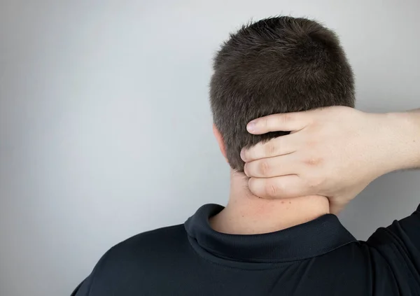
A Man Suffers From Pain In The Neck, Massages The Cervical Spine With His Hand. Osteochondrosis, Hernia, Or Nerve Injury Due To Sedentary Work Or Physical Stress
Image, 6.87MB, 5276 × 3725 jpg


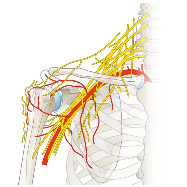
The Shoulder Region Houses A Complex Network Of Nerves And Vessels, Including The Brachial Plexus, Arteries, And Veins, Essential For Limb Innervation And Blood Supply, Facilitating Movement And Function.
Image, 4.77MB, 5000 × 5000 jpg
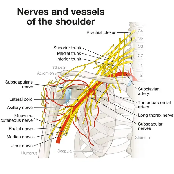
The Shoulder Region Houses A Complex Network Of Nerves And Vessels, Including The Brachial Plexus, Arteries, And Veins, Essential For Limb Innervation And Blood Supply, Facilitating Movement And Function.
Image, 4.85MB, 5000 × 5000 jpg
Previous << Page 2 >> Next