photo free
Home Images Vectors Videos

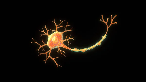
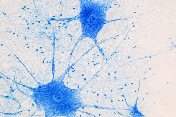
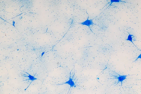
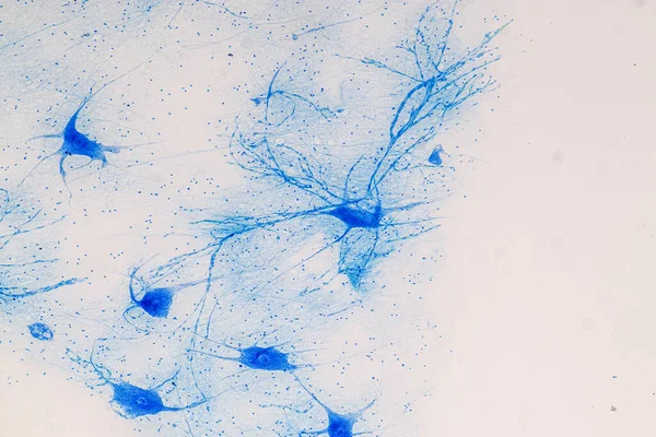
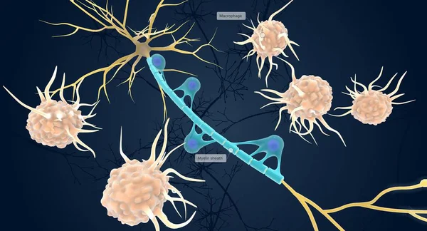
Stock image Nervous Tissue

stock image selective focus of upset woman looking at picture frame, crying and wiping tears at home

Selective Focus Of Upset Woman Looking At Picture Frame, Crying And Wiping Tears At Home

Image, 15.03MB, 7360 × 4912 jpg
stock image Neurons cells from the brain under the microscope view for education.

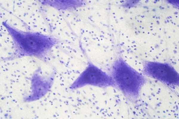
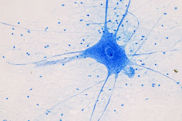
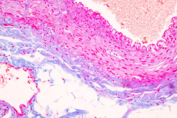
Neurons Cells From The Brain Under The Microscope View For Education.

Image, 10.06MB, 5812 × 3875 jpg
stock image woman lying in bed with tissue box, crying and wiping tears

Woman Lying In Bed With Tissue Box, Crying And Wiping Tears

Image, 6.02MB, 4655 × 6975 jpg
stock image A single astrocyte supports a great number of neurons. Protoplasmic astrocytes play an active role in neuronal communication through synapses and regulation of neural circuit function.

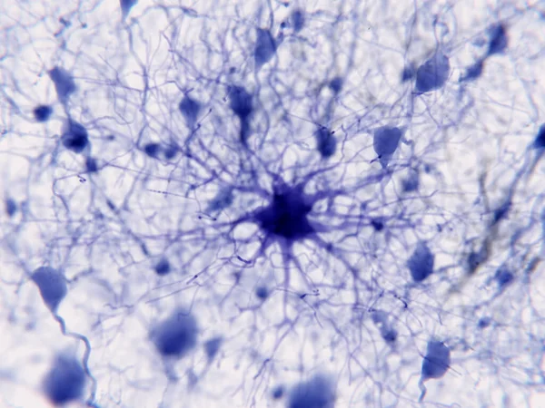
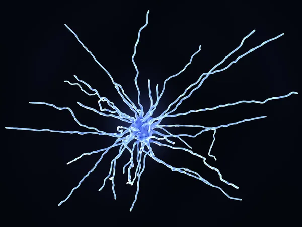
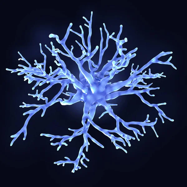
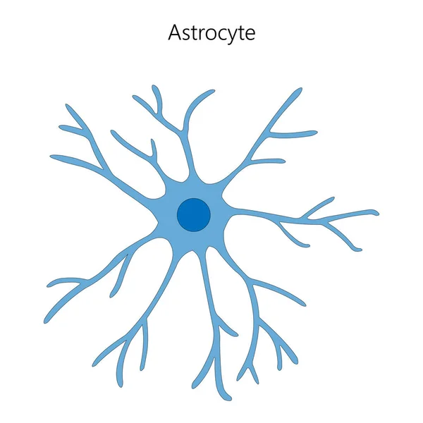
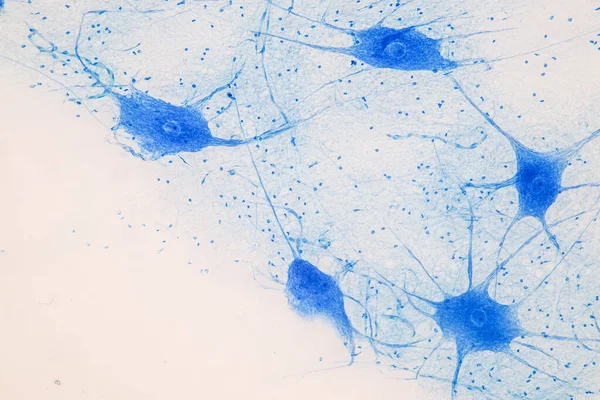
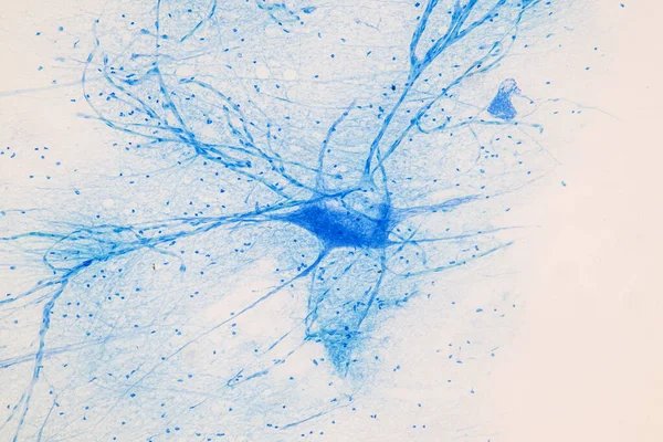
A Single Astrocyte Supports A Great Number Of Neurons. Protoplasmic Astrocytes Play An Active Role In Neuronal Communication Through Synapses And Regulation Of Neural Circuit Function.

Image, 5.6MB, 8000 × 6000 jpg
stock image selective focus of upset woman holding picture frame and crying at home

Selective Focus Of Upset Woman Holding Picture Frame And Crying At Home

Image, 17.66MB, 7360 × 4912 jpg
stock image cells of the human body under the microscope. Microbiology and m

Cells Of The Human Body Under The Microscope. Microbiology And M

Image, 6.9MB, 4184 × 2789 jpg
stock image upset woman hugging knees, looking at camera and sitting on bed at home

Upset Woman Hugging Knees, Looking At Camera And Sitting On Bed At Home

Image, 8.96MB, 7360 × 4912 jpg
stock vector Neurons and neuroglial cells. Glial cells are non-neuronal cells in brain. There are different types of glial cells: oligodendrocyte, microglia, astrocytes and Schwann cells. Vector diagram for educational, medical, biological and science use

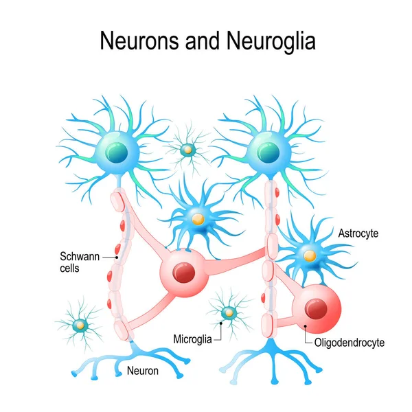
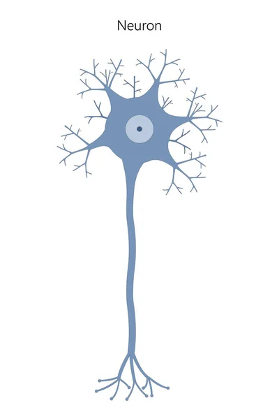
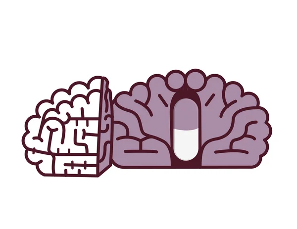
Neurons And Neuroglial Cells. Glial Cells Are Non-neuronal Cells In Brain. There Are Different Types Of Glial Cells: Oligodendrocyte, Microglia, Astrocytes And Schwann Cells. Vector Diagram For Educational, Medical, Biological And Science Use

Vector, 5.27MB, 4808 × 4808 eps
stock image upset woman with tissue box lying in bed and holding wedding rings

Upset Woman With Tissue Box Lying In Bed And Holding Wedding Rings

Image, 6.5MB, 6727 × 4489 jpg
stock image Nerve cell under the microscope - Abstract blue dots on white ba

Nerve Cell Under The Microscope - Abstract Blue Dots On White Ba

Image, 12.84MB, 4658 × 3105 jpg
stock image partial view of woman lying in bed and holding wedding rings while reaching for tissue

Partial View Of Woman Lying In Bed And Holding Wedding Rings While Reaching For Tissue

Image, 6.16MB, 7360 × 4912 jpg
stock image Neurons in the brain

Neurons In The Brain

Image, 3.48MB, 3500 × 2625 jpg
stock image cropped view of upset woman holding picture frame and crying at home

Cropped View Of Upset Woman Holding Picture Frame And Crying At Home

Image, 12.54MB, 7360 × 4912 jpg
stock image Fibrous astrocyte

Fibrous Astrocyte

Image, 4.44MB, 8000 × 6000 jpg
stock image Neuron anatomy isolated in black background.3d illustration

Neuron Anatomy Isolated In Black Background.3d Illustration

Image, 1.56MB, 3840 × 2160 jpg
stock image Protoplasmic astrocytes are found in the gray matter and the fibrous in the white matter of the brain. They support neurons in a metabolic and a structural way and regulate the ion concentration in the extracellular space.

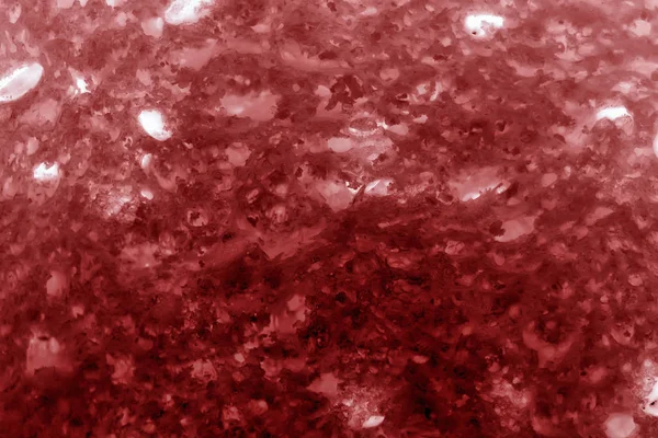
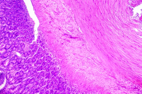
Protoplasmic Astrocytes Are Found In The Gray Matter And The Fibrous In The White Matter Of The Brain. They Support Neurons In A Metabolic And A Structural Way And Regulate The Ion Concentration In The Extracellular Space.

Image, 1.71MB, 8000 × 6000 jpg
stock image Spinal Stenosis Concept

Spinal Stenosis Concept

Image, 3.31MB, 3927 × 3927 jpg
stock image Protoplasmic astrocyte

Protoplasmic Astrocyte

Image, 7.42MB, 8000 × 8000 jpg
stock image Education Spinal cord, Nerve, Cerebellum, Cortex and Motor Neuron Human under the microscope in Lab.

Education Spinal Cord, Nerve, Cerebellum, Cortex And Motor Neuron Human Under The Microscope In Lab.

Image, 19.22MB, 6720 × 4480 jpg
stock image Protoplasmic astrocytes (violet) play an active role in neuronal communication through synapses and regulation of neural circuit function.

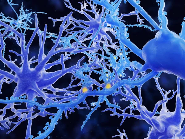
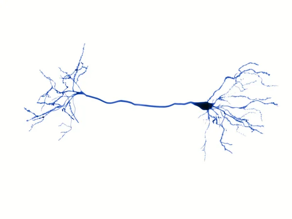
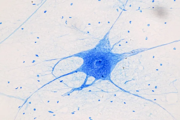
Protoplasmic Astrocytes (violet) Play An Active Role In Neuronal Communication Through Synapses And Regulation Of Neural Circuit Function.

Image, 6.27MB, 8000 × 6000 jpg
stock vector  Neuron. Neuronal structure. Vector illustration

Neuron. Neuronal Structure. Vector Illustration

Vector, 5.27MB, 8333 × 12500 eps
stock vector Neuron. Neuronal structure. Vector illustration

Neuron. Neuronal Structure. Vector Illustration

Vector, 5.27MB, 8333 × 12500 eps
stock image Spinal cord

Spinal Cord

Image, 1.28MB, 6480 × 4096 jpg
stock vector Astrocyte. Glial cell. Vector illustration.

Astrocyte. Glial Cell. Vector Illustration.

Vector, 5.37MB, 8333 × 8333 eps
stock vector Doctor found pill in brain. Medical research concept.

Doctor Found Pill In Brain. Medical Research Concept.

Vector, 1.2MB, 6046 × 4000 eps
stock image Amazing vascular plant fine roots looking like a neural network

Amazing Vascular Plant Fine Roots Looking Like A Neural Network

Image, 5.91MB, 2816 × 2112 jpg
stock image Education Spinal cord  and Motor Neuron under the microscope in Lab.

Education Spinal Cord And Motor Neuron Under The Microscope In Lab.

Image, 12.06MB, 6720 × 4480 jpg
stock image Education Spinal cord  and Motor Neuron under the microscope in Lab.

Education Spinal Cord And Motor Neuron Under The Microscope In Lab.

Image, 9.7MB, 6720 × 4480 jpg
stock image Spinal Stenosis medical concept as a degenerative illness in the human vertebrae causing compressed spine nerves human body disease as a 3D illustration.

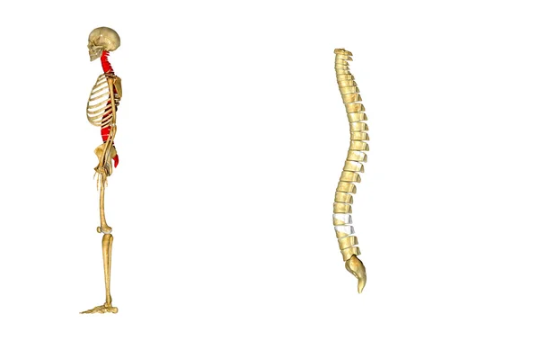
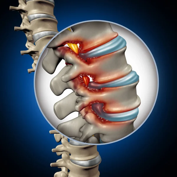
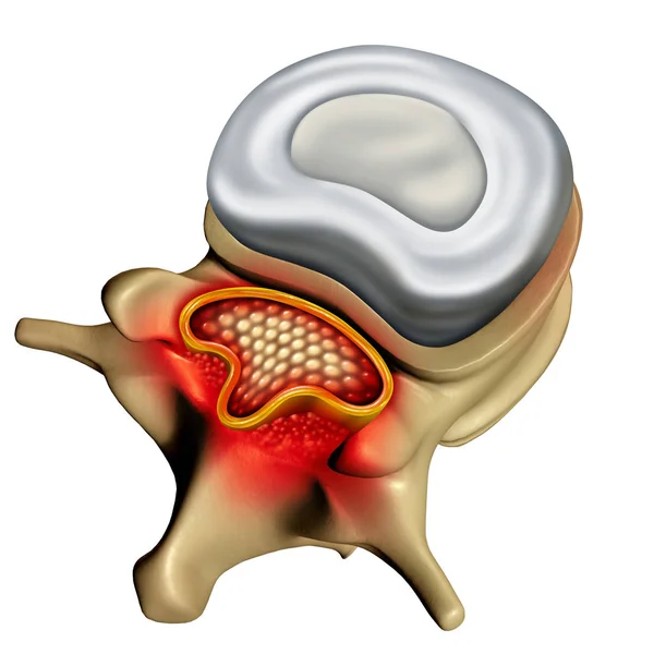
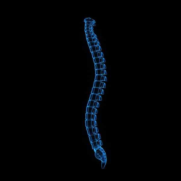
Spinal Stenosis Medical Concept As A Degenerative Illness In The Human Vertebrae Causing Compressed Spine Nerves Human Body Disease As A 3D Illustration.

Image, 5.57MB, 4224 × 4224 jpg
stock image Education Spinal cord  and Motor Neuron under the microscope in Lab.

Education Spinal Cord And Motor Neuron Under The Microscope In Lab.

Image, 12.9MB, 6720 × 4480 jpg
stock image Spinal Cord

Spinal Cord

Image, 3MB, 3618 × 3475 jpg
stock image Spinal Stenosis

Spinal Stenosis

Image, 2.96MB, 3226 × 3226 jpg
stock image Education Spinal cord  and Motor Neuron under the microscope in Lab.

Education Spinal Cord And Motor Neuron Under The Microscope In Lab.

Image, 11.03MB, 6720 × 4480 jpg
stock image Education Spinal cord  and Motor Neuron under the microscope in Lab.

Education Spinal Cord And Motor Neuron Under The Microscope In Lab.

Image, 11.77MB, 6720 × 4480 jpg
stock image Education Spinal cord  and Motor Neuron under the microscope in Lab.

Education Spinal Cord And Motor Neuron Under The Microscope In Lab.

Image, 9.43MB, 6720 × 4480 jpg
stock image Education Spinal cord  and Motor Neuron under the microscope in Lab.

Education Spinal Cord And Motor Neuron Under The Microscope In Lab.

Image, 12.65MB, 6720 × 4480 jpg
stock image Education Spinal cord  and Motor Neuron under the microscope in Lab.

Education Spinal Cord And Motor Neuron Under The Microscope In Lab.

Image, 11.83MB, 6720 × 4480 jpg
stock image Spine therapy and spinal stenosis medical surgery concept as a degenerative illness surgery in the human vertebrae as a doctor treating and diagnosing the anatomy with 3D illustration elements.

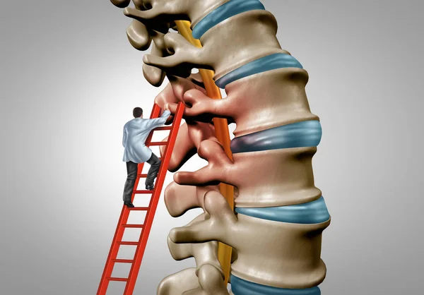
Spine Therapy And Spinal Stenosis Medical Surgery Concept As A Degenerative Illness Surgery In The Human Vertebrae As A Doctor Treating And Diagnosing The Anatomy With 3D Illustration Elements.

Image, 5.8MB, 5151 × 3590 jpg
stock vector Pill in brain illustration.Placebo concept.

Pill In Brain Illustration.Placebo Concept.

Vector, 0.66MB, 5176 × 4000 eps
stock image Education Spinal cord  and Motor Neuron under the microscope in Lab.

Education Spinal Cord And Motor Neuron Under The Microscope In Lab.

Image, 20.37MB, 6720 × 4480 jpg
stock image Education Spinal cord  and Motor Neuron under the microscope in Lab.

Education Spinal Cord And Motor Neuron Under The Microscope In Lab.

Image, 10.13MB, 6720 × 4480 jpg
stock image Spinal Cord

Spinal Cord

Image, 2.41MB, 4096 × 4096 jpg
stock image spinal cord cross section

Spinal Cord Cross Section

Image, 11.48MB, 3825 × 2696 jpg
stock image Spinal cord

Spinal Cord

Image, 2.2MB, 4096 × 4096 jpg
stock image Education Spinal cord  and Motor Neuron under the microscope in Lab.

Education Spinal Cord And Motor Neuron Under The Microscope In Lab.

Image, 10.95MB, 6720 × 4480 jpg
stock image Education anatomy and Histological sample of Human under the microscope.

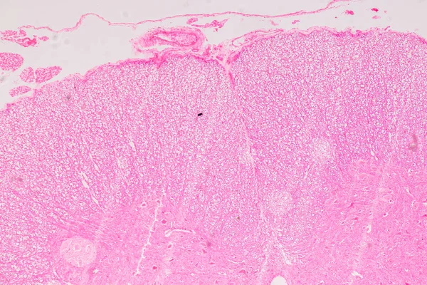
Education Anatomy And Histological Sample Of Human Under The Microscope.

Image, 21.93MB, 6720 × 4480 jpg
stock image Autoimmune Attack on Myelin Sheath 3D rendering

Autoimmune Attack On Myelin Sheath 3D Rendering

Image, 10.21MB, 7258 × 3920 jpg
stock image Human nervous system under the microscope in Lab.

Human Nervous System Under The Microscope In Lab.

Image, 13.19MB, 6000 × 4000 jpg
stock image Motor Neuron, Spinal cord, Nerve fibres and nerve cells under the microscope in Lab.

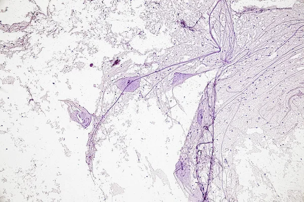
Motor Neuron, Spinal Cord, Nerve Fibres And Nerve Cells Under The Microscope In Lab.

Image, 13.05MB, 6000 × 4000 jpg
stock image Light microscope micrograph showing neurons in a dorsal root ganglion stained with cresyl violet. They are pseudounipolar neurons of rounded soma showing small, thin Nissl bodies characteristic of sensitive neurons and a large nucleus with a prominen

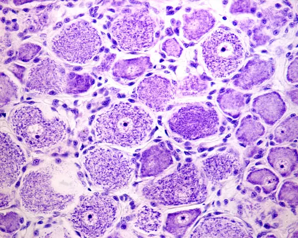
Light Microscope Micrograph Showing Neurons In A Dorsal Root Ganglion Stained With Cresyl Violet. They Are Pseudounipolar Neurons Of Rounded Soma Showing Small, Thin Nissl Bodies Characteristic Of Sensitive Neurons And A Large Nucleus With A Prominen

Image, 12.15MB, 3840 × 3072 jpg
Page 1 >> Next