
Stock image Neuroanatomy

Pituitary Gland Or Neurohypophysis X-ray View 3D Rendering Illustration. Human Brain, Nervous And Endocrine System Anatomy, Medical, Healthcare, Science, Neuroscience, Neurology Concepts.
Image, 1.98MB, 3300 × 2200 jpg

Nucleus Accumbens Lateral X-ray View 3D Rendering Illustration. Human Brain And Basal Ganglia Anatomy, Medical, Healthcare, Biology, Science, Neuroscience, Neurology Concepts.
Image, 2.34MB, 3300 × 2200 jpg
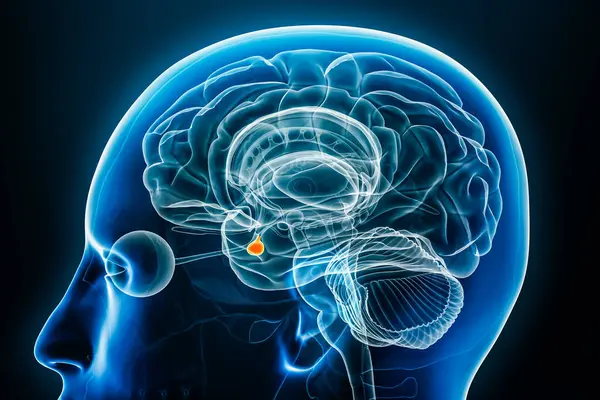
Xray Lateral Or Profile View Of The Amygdala 3D Rendering Illustration With Male Body Contours. Human Brain Anatomy, Medical, Biology, Neuroscience, Neurology Concepts.
Image, 2.28MB, 3300 × 2200 jpg

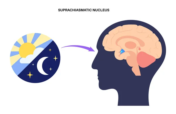
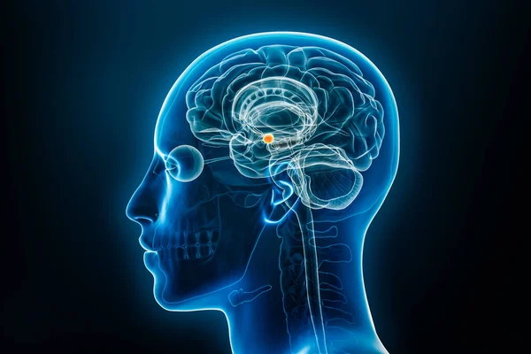
Xray Profile View Of The Hypothalamus 3D Rendering Illustration. Human Brain And Body Anatomy, Medical, Biology, Science, Neuroscience, Neurology Concepts.
Image, 2.28MB, 3300 × 2200 jpg

Xray Profile View Of The Brain Stem Or Brainstem With Medulla, Pons And Midbrain 3D Rendering Illustration. Human Body Anatomy, Medical, Biology, Science, Neuroscience, Neurology Concepts.
Image, 2.26MB, 3300 × 2200 jpg

Pathway Of Vagus Nerve Through Human Body, Including Its Connection To The Brain, Heart, And Lungs Structure Diagram Hand Drawn Schematic Raster Illustration. Medical Science Educational Illustration
Image, 5.08MB, 6000 × 6000 jpg

Ventricles And Cerebral Aqueduct Lateral X-ray View 3D Rendering Illustration. Human Brain And Ventricular System Anatomy, Medical, Healthcare, Science, Neuroscience, Neurology, Biology Concepts.
Image, 2.08MB, 3300 × 2200 jpg
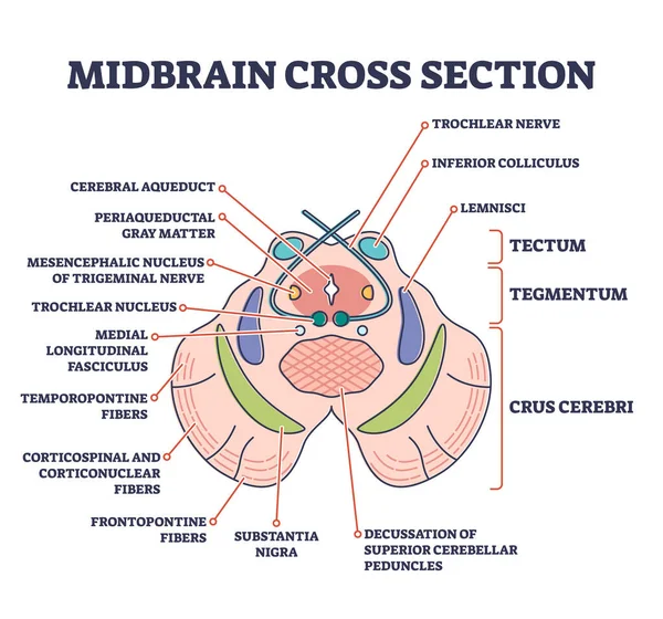
Midbrain Cross Section With Labeled Brain Structure Parts Outline Diagram
Vector, 5.89MB, 4500 × 4200 eps


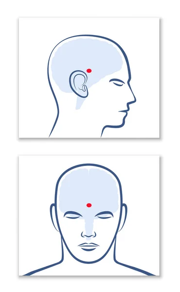
PINEAL GLAND Or THIRD EYE. Lateral And Frontal View With Position In The Human Brain. Isolated Vector Graphic Illustration On White Background.
Vector, 5.37MB, 5551 × 9000 eps

The Human Brain. Side View. Human Anatomy . Medical 3d Vector Illustration Isolated On White Background.
Vector, 6.88MB, 8334 × 8334 eps
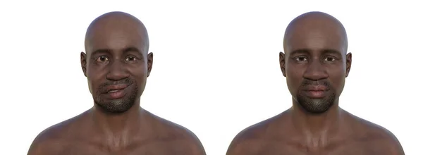
Facial Palsy In An African Man And The Same Healthy Man, Photorealistic 3D Illustration Highlighting The Asymmetry And Drooping Of The Facial Muscles On One Side Of The Face
Image, 26.79MB, 13789 × 5067 jpg
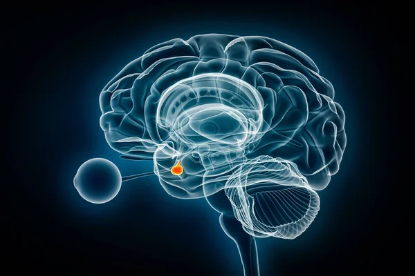
Pituitary Gland X-ray Profile Close-up View 3D Rendering Illustration With Body Contours. Human Brain, Nervous And Endocrine System Anatomy, Medical, Science, Neuroscience, Neurology Concepts.
Image, 2.76MB, 3276 × 2184 jpg

Nucleus Accumbens X-ray Profile Close-up View 3D Rendering Illustration With Body Contours. Human Brain And Basal Ganglia Anatomy, Medical, Biology, Science, Neuroscience, Neurology Concepts.
Image, 2.94MB, 3260 × 2173 jpg


Xray Lateral Or Profile View Of The Cingulate Gyrus Or Cortex 3D Rendering Illustration With Body Contours. Human Brain And Limbic System Anatomy, Medical, Biology, Neuroscience, Neurology Concepts.
Image, 2.24MB, 3300 × 2200 jpg

Xray Profile View Of The Cerebral Cortex Or Cerebrum 3D Rendering Illustration With Body Contours. Human Brain Anatomy, Medical, Biology, Science, Neuroscience, Neurology Concepts.
Image, 2.09MB, 3300 × 2200 jpg
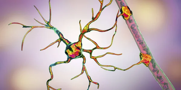
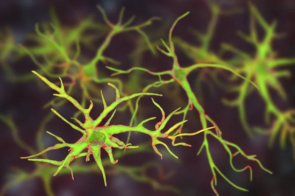
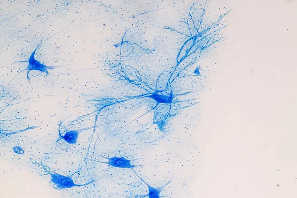
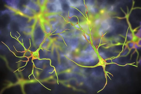
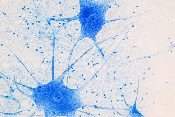
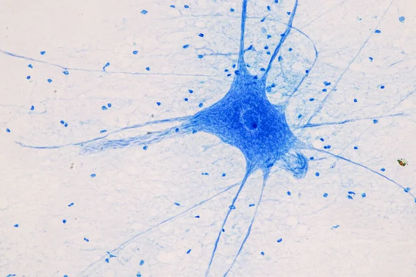
Protoplasmic Astrocytes Are Found In The Gray Matter And The Fibrous In The White Matter Of The Brain. They Support Neurons In A Metabolic And A Structural Way And Regulate The Ion Concentration In The Extracellular Space.
Image, 1.71MB, 8000 × 6000 jpg
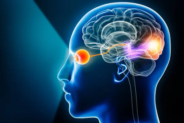
Visual Pathway With Eye, Optic Nerve And Visual Cortex 3D Rendering Illustration With Copy Space. Human Brain And Sensory System Anatomy, Medical, Neuroscience, Neurology, Ophthalmology Concepts.
Image, 2.59MB, 3300 × 2200 jpg

Upper Limbs Showing The Cutaneous Nerve Innervation, With Labels For Specific Nerves And Regions Diagram Hand Drawn Schematic Raster Illustration. Medical Science Educational Illustration
Image, 3.41MB, 6000 × 4500 jpg

A Healthcare Professional Examines Multiple MRI Brain Scans Displayed On A Screen, Focusing On Neurological Analysis And Diagnosis.
Image, 2.22MB, 6016 × 4016 jpg

Facial Palsy In A Man, Photorealistic 3D Illustration Highlighting The Asymmetry And Drooping Of The Facial Muscles On One Side Of The Face.
Image, 13.55MB, 5000 × 5000 jpg

Fornix Profile X-ray View 3D Rendering Illustration. Human Brain And Limbic System Anatomy, Medical, Healthcare, Biology, Science, Neuroscience, Neurology Concepts.
Image, 1.96MB, 3300 × 2200 jpg
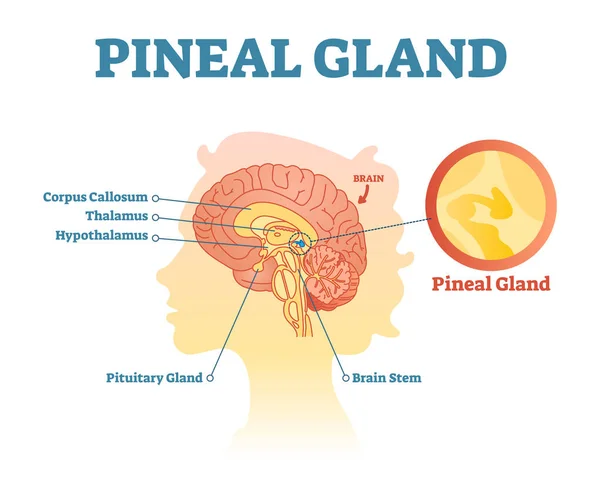
Pineal Gland Anatomical Cross Section Vector Illustration Diagram With Human Brains.
Vector, 2.1MB, 4605 × 3678 eps

Detailed Human Brain Vector Illustration, Neuroscience Brain Diagram Vector.
Vector, 5.24MB, 8334 × 8334 eps
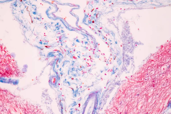
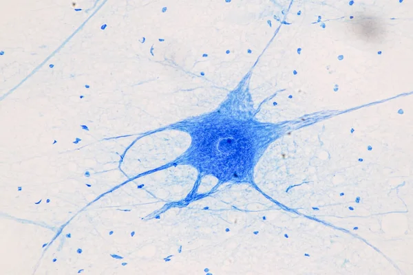
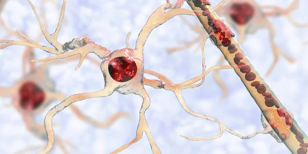
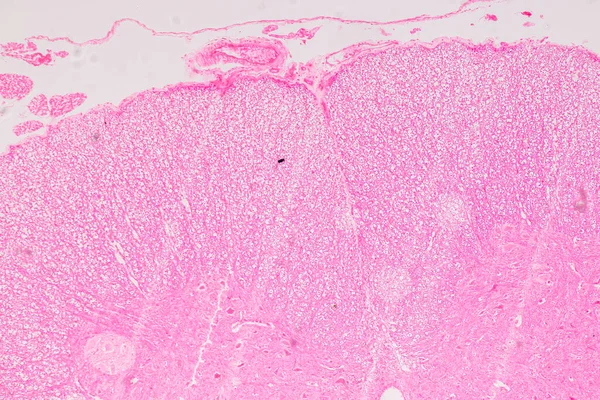
Cerebellum, Thalamus, Medulla Oblongata, Spinal Cord And Motor Neuron Human Under The Microscope In Lab.
Image, 12.22MB, 6000 × 4000 jpg
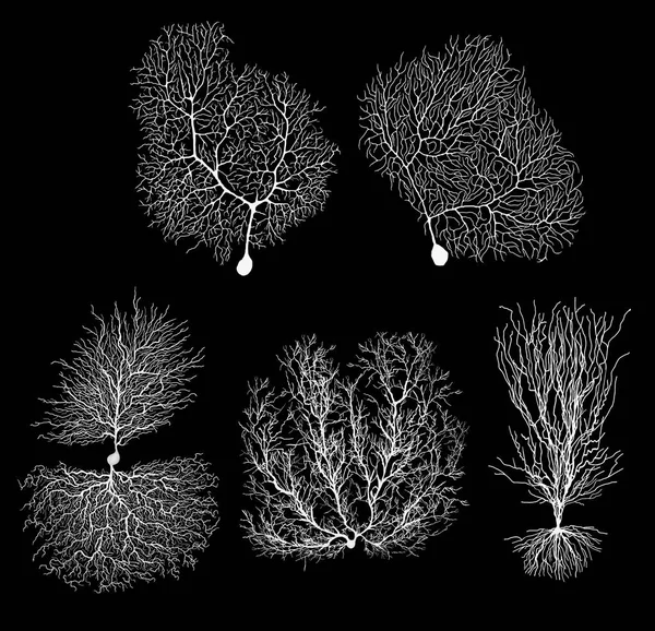
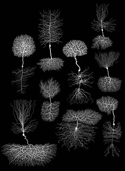

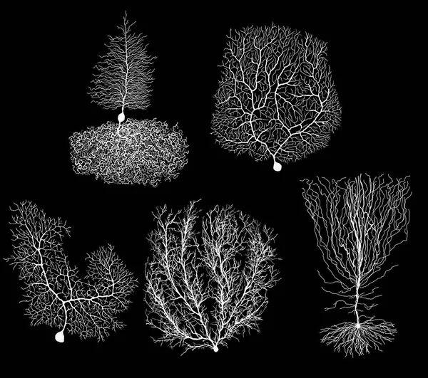
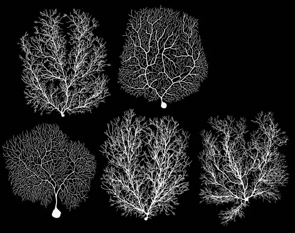
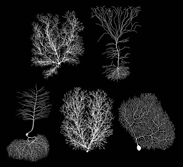
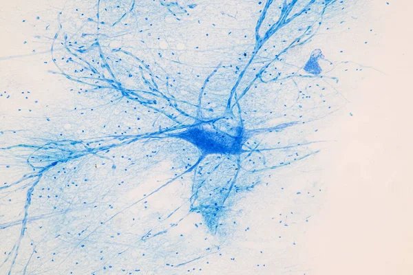
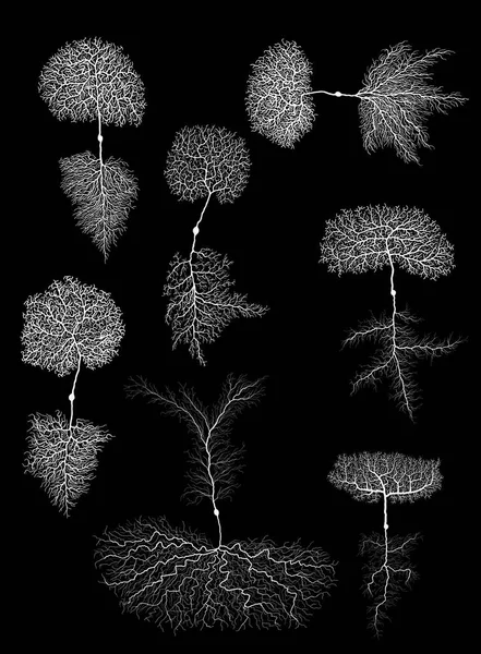
Purkinje Neuron, GABAergic Neuron Located In The Cerebellum, Isolated On Black Background, 3D Illustration
Image, 6.1MB, 6000 × 4000 jpg

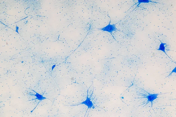
Cerebellum, Thalamus, Medulla Oblongata, Spinal Cord And Motor Neuron Human Under The Microscope In Lab.
Image, 13.45MB, 6000 × 4000 jpg

Xray Lateral Or Profile View Of The Thalamus 3D Rendering Illustration With Male Body Contours. Human Brain And Limbi System Anatomy, Medical, Biology, Neuroscience, Neurology Concepts.
Image, 2.26MB, 3300 × 2200 jpg
Page 1 >> Next