
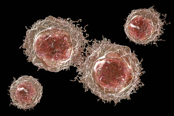
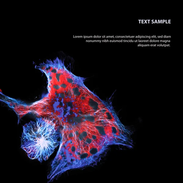
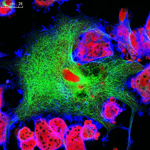
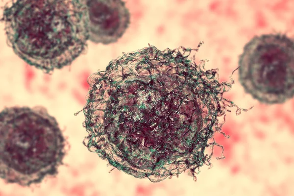
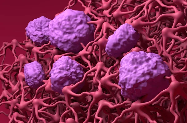
Stock image Neuroblastoma
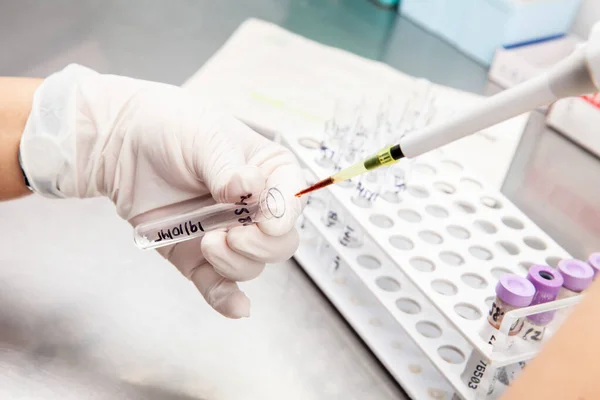
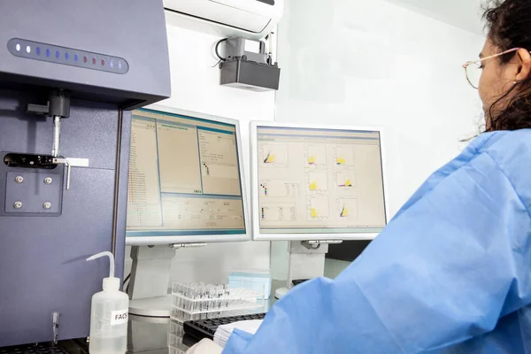
Scientist Preparing Samples For Flow Cytometric Analysis In The Laboratory
Image, 4.53MB, 5049 × 3366 jpg


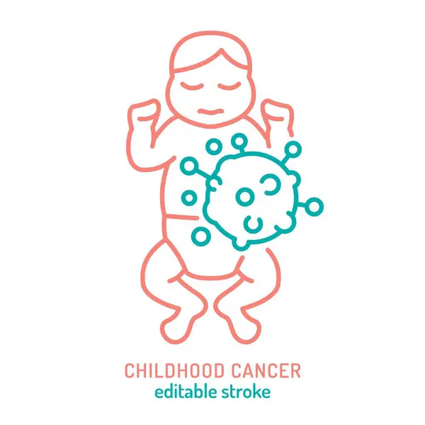
Childhood Cancer, Tumor In Kids International Month. Landscape Poster, Banner With Oncological Sign. Medical Line Concept. Pediatric Oncology. Editable Vector Illustration Isolated On White Background
Vector, 6.46MB, 6811 × 3671 eps

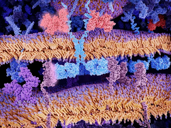
Engineered Receptors (light Blue) On The Surface Of A T-lymphocyte Bind Specifically To CD19-antigen Molecules (magenta) On A Leukemia Cell. This Activates A Signal Cascade In The T-cell Leading To The Apoptosis Of The Cancer Cell.
Image, 11.66MB, 8000 × 6000 jpg

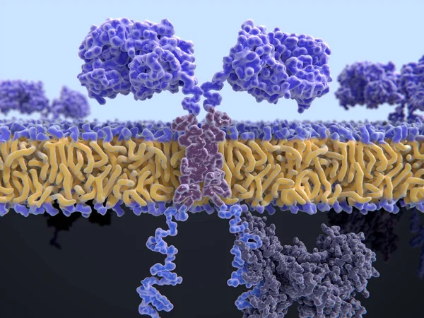
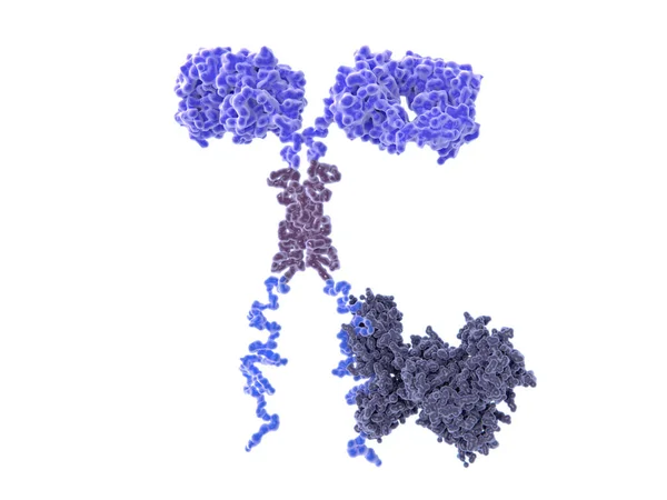
T-cell Receptors Are Similar To One Arm Of An Antibody. Like Antibodies, They Are Composed Of Two Chains. The Binding Site Is At The Tip Of The Molecule,
Image, 2.5MB, 8000 × 6000 jpg


Test Tube Containing A Patient Sample Loaded On The Flow Cytometer Ready For Analysis. Flow Cytometer
Image, 8.63MB, 5365 × 3577 jpg

Neuroblastoma Can Separate A Person From The World And Lock In An Isolation That Limits - Pictured As A Human Figure Locked Inside A Glass With A Phrase Neuroblastoma, 3d Illustration
Image, 3.83MB, 6800 × 4320 jpg

Childhood Cancer Awareness Month In September. Gold Color Ribbon Cancer Awareness Products. Vector Illustration.
Vector, 6.92MB, 9355 × 6615 eps
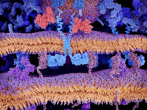
Engineered Receptors (light Blue) On The Surface Of A T-lymphocyte Bind Specifically To CD19-antigen Molecules (magenta) On A Leukemia Cell. This Activates A Signal Cascade In The T-cell Leading To The Segregation Of Vesicles That Contain Perforin An
Image, 11.44MB, 8000 × 6000 jpg

Childhood Cancer, Malignant Melanoma In Kids Outline Icon. Oncological Sign. Medical Linear Pictogram. Pediatric Oncology. Brain Tumor. Editable Vector Illustration Isolated On White Background
Vector, 5.71MB, 5000 × 5000 eps

3d Computer Illustration Of A Chimeric Antigen Receptor. CARs Are Engineered Cell Receptors That Allow T Cells To Recognize And Attack Cancer Cells In A Specific Way. They Are Built By Connecting Several Functional Parts From Different Proteins.
Image, 8.45MB, 8000 × 6000 jpg
Childhood Cancer, Malignant Melanoma In Kids Outline Icon. Oncological Sign. Medical Linear Pictogram. Pediatric Oncology. Brain Tumor. Editable Vector Illustration Isolated On White Background
Vector, 5.72MB, 5000 × 5000 eps

Childhood Cancer Awareness With Gold Ribbon Symbolic Color (isolated With Clipping Path)
Image, 3.18MB, 6000 × 4000 jpg
Scientist Performing A Flow Cytometric Analysis In The Laboratory. Flow Cytometer.
Image, 7.51MB, 5616 × 3744 jpg

Childhood Cancer, Tumor In Kids International Month. Landscape Poster, Banner With Oncological Sign. Medical Line Concept. Pediatric Oncology. Editable Vector Illustration Isolated On White Background
Vector, 6.34MB, 5930 × 4216 eps

Childhood Cancer, Tumor In Kids International Month. Vertical Poster, Banner With Oncological Sign. Medical Line Concept. Pediatric Oncology. Editable Vector Illustration Isolated On White Background
Vector, 6.53MB, 4341 × 5760 eps

Childhood Cancer Awareness With Gold Ribbon Symbolic Color (isolated With Clipping Path)
Image, 3.59MB, 6000 × 4000 jpg


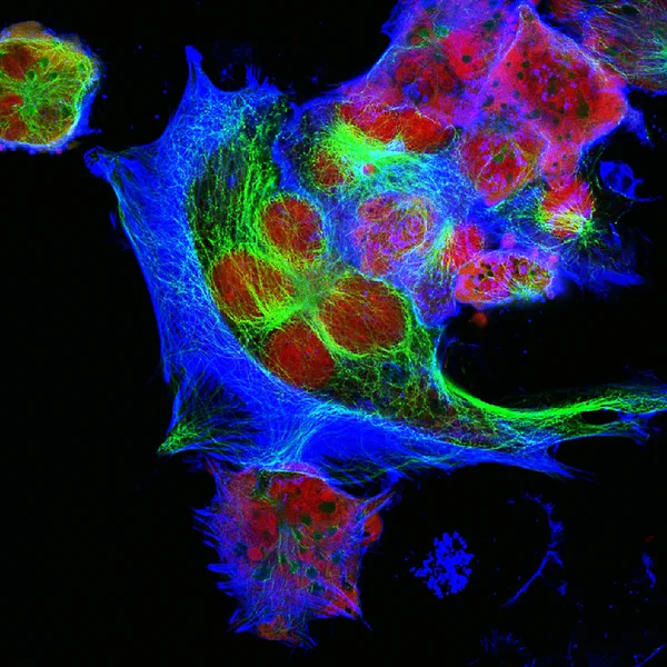
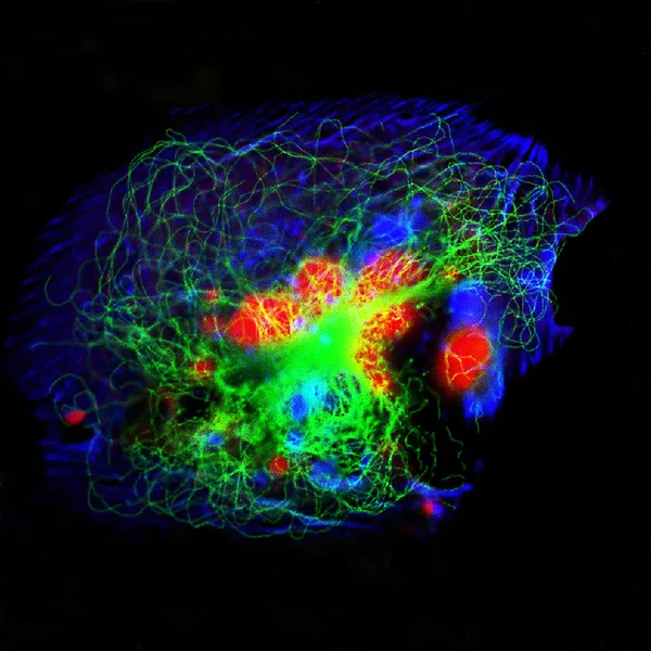
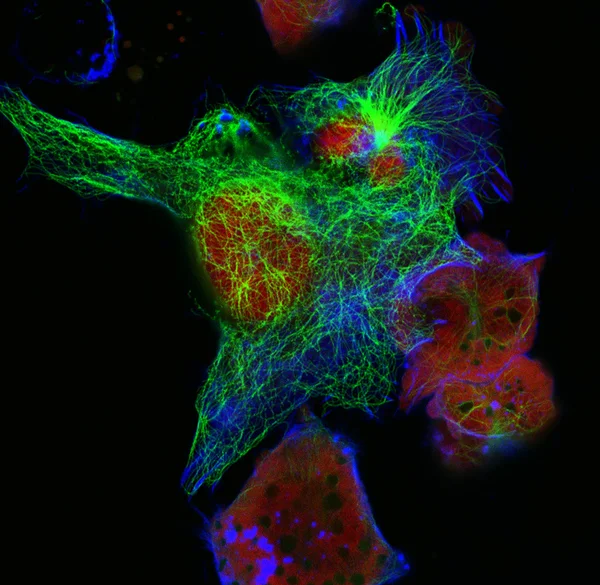
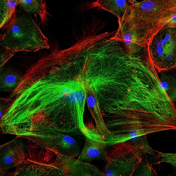
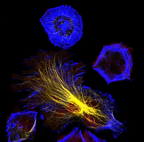
3d Rendering Of Astrocytes With Blood Vessel, Found Abundantly Throughout The Brain And Spinal Cord.
Image, 0.35MB, 3840 × 2160 jpg


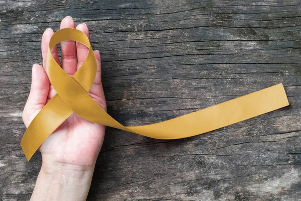
Childhood Cancer Awareness With Gold Ribbon Symbolic Color On Helping Hand On Old Aged Wood
Image, 14.3MB, 6000 × 4000 jpg


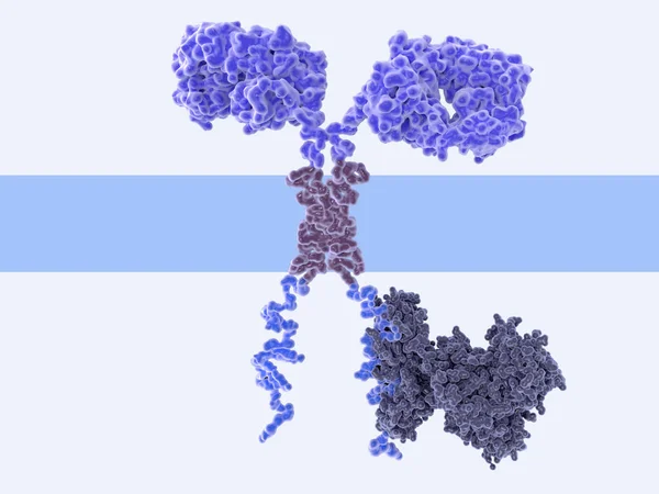
3d Computer Illustration Of A Chimeric Antigen Receptor. CARs Are Engineered Cell Receptors That Allow T Cells To Recognize/attack Specifically Cancer Cells. A Signal Protein Is Attached To The Intracellular Domain.
Image, 2.19MB, 8000 × 6000 jpg
3d Computer Illustration Of A Chimeric Antigen Receptor. CARs Are Engineered Cell Receptors That Allow T Cells To Recognize/attack Specifically Cancer Cells. A Signal Protein Is Attached To The Intracellular Domain.
Image, 3.45MB, 8000 × 6000 jpg

Health Care Concept Meaning Neuroblastoma With Inscription On The Piece Of Paper
Image, 8.74MB, 5000 × 3750 jpg


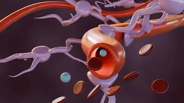
Blood-brain Barrier (BBB) In The Human Brain - Closeup View 3d Illustration
Image, 7.46MB, 10000 × 6600 jpg
Page 1 >> Next