
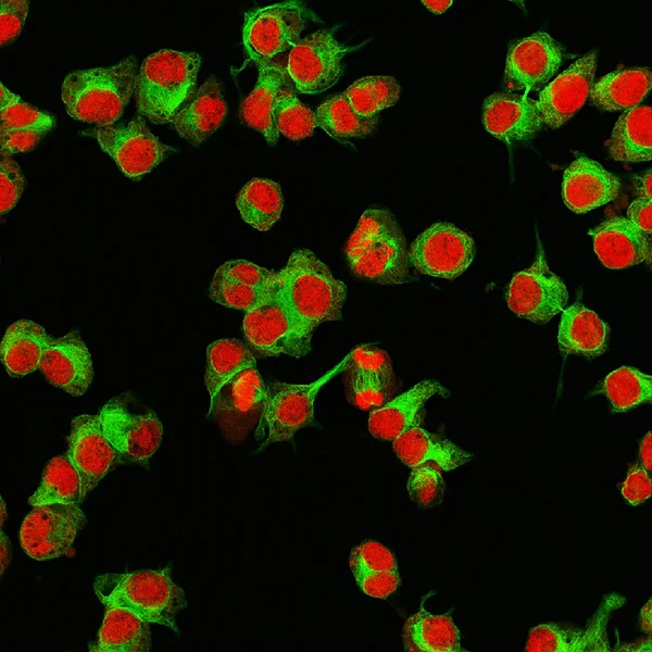
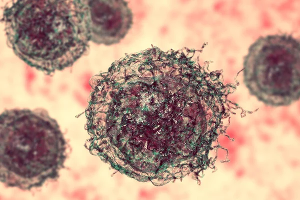
Stock image Neuroblastoma Cell
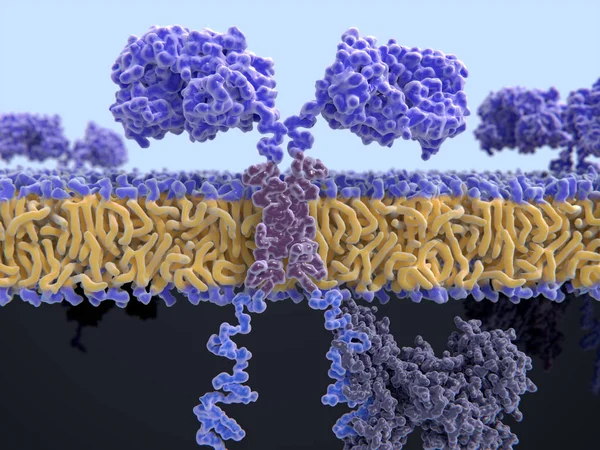
3d Computer Illustration Of A Chimeric Antigen Receptor. CARs Are Engineered Cell Receptors That Allow T Cells To Recognize And Attack Cancer Cells In A Specific Way. They Are Built By Connecting Several Functional Parts From Different Proteins.
Image, 8.45MB, 8000 × 6000 jpg
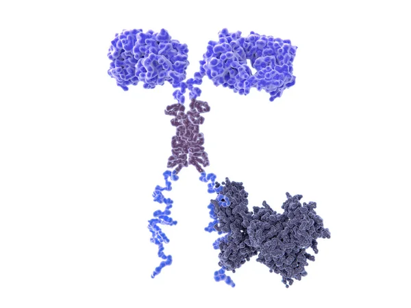
3d Computer Illustration Of A Chimeric Antigen Receptor. CARs Are Engineered Cell Receptors That Allow T Cells To Recognize/attack Specifically Cancer Cells. A Signal Protein Is Attached To The Intracellular Domain.
Image, 3.45MB, 8000 × 6000 jpg

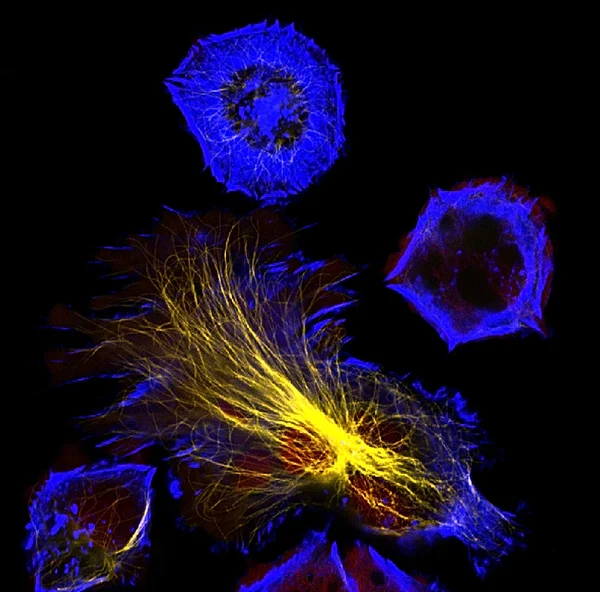
T-cell Receptors Are Similar To One Arm Of An Antibody. Like Antibodies, They Are Composed Of Two Chains. The Binding Site Is At The Tip Of The Molecule,
Image, 2.5MB, 8000 × 6000 jpg
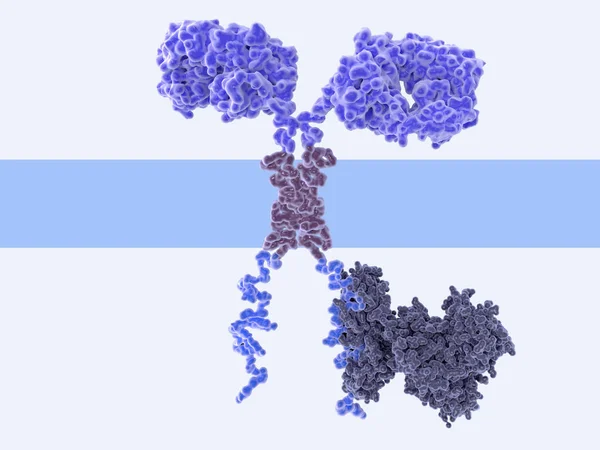
3d Computer Illustration Of A Chimeric Antigen Receptor. CARs Are Engineered Cell Receptors That Allow T Cells To Recognize/attack Specifically Cancer Cells. A Signal Protein Is Attached To The Intracellular Domain.
Image, 2.19MB, 8000 × 6000 jpg
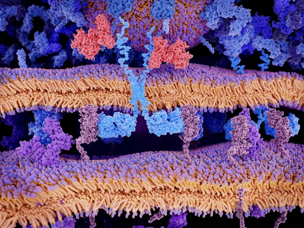
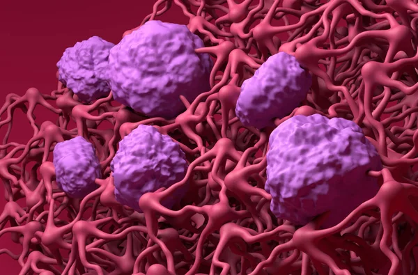
Engineered Receptors (light Blue) On The Surface Of A T-lymphocyte Bind Specifically To CD19-antigen Molecules (magenta) On A Leukemia Cell. This Activates A Signal Cascade In The T-cell Leading To The Segregation Of Vesicles That Contain Perforin An
Image, 11.44MB, 8000 × 6000 jpg
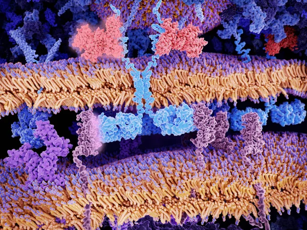
Engineered Receptors (light Blue) On The Surface Of A T-lymphocyte Bind Specifically To CD19-antigen Molecules (magenta) On A Leukemia Cell. This Activates A Signal Cascade In The T-cell Leading To The Apoptosis Of The Cancer Cell.
Image, 11.66MB, 8000 × 6000 jpg
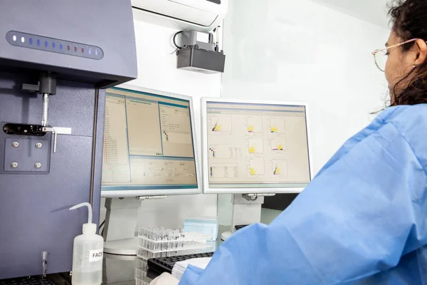
Scientist Preparing Samples For Flow Cytometric Analysis In The Laboratory
Image, 4.53MB, 5049 × 3366 jpg


Blood-brain Barrier (BBB) In The Human Brain - Closeup View 3d Illustration
Image, 7.46MB, 10000 × 6600 jpg


Scientist Preparing Samples For Flow Cytometric Analysis In The Laboratory. Cancer Diagnosis
Image, 5.03MB, 5049 × 3366 jpg
Scientist Performing A Flow Cytometric Analysis In The Laboratory. Flow Cytometer.
Image, 7.51MB, 5616 × 3744 jpg

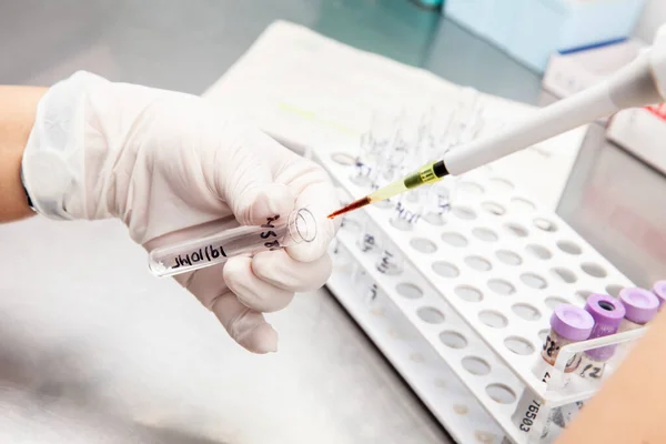
Test Tube Containing A Patient Sample Loaded On The Flow Cytometer Ready For Analysis. Flow Cytometer
Image, 8.63MB, 5365 × 3577 jpg
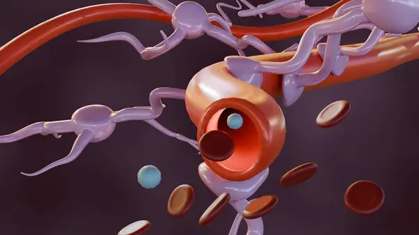
3d Rendering Of Astrocytes With Blood Vessel, Found Abundantly Throughout The Brain And Spinal Cord.
Image, 0.35MB, 3840 × 2160 jpg
Page 1 >> Next