
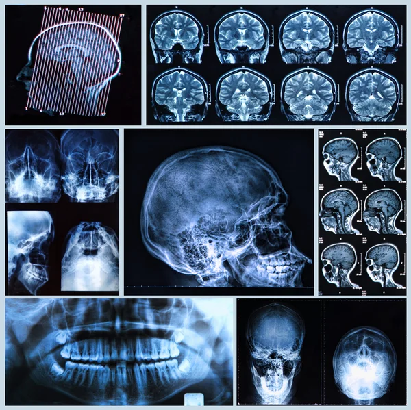
Stock image Neurodegeneration
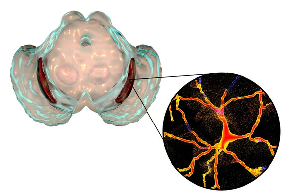
Black Substance, Basal Banglia Of The Midbrain, In Parkinson's Disease, 3D Illustration Showing Decrease Of Its Volume And Degeneration Of Dopaminergic Neurons In The Pars Compacta Of The Black Substance
Image, 3.06MB, 6000 × 4000 jpg


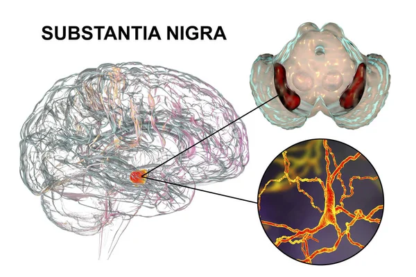
Black Substance Of The Midbrain And Its Dopaminergic Neurons, 3D Illustration. Black Substance Regulates Movement And Reward, Its Degeneration Is A Key Step In Development Of Parkinson's Disease
Image, 11.59MB, 7926 × 5284 jpg
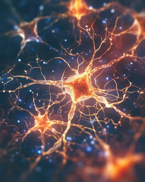
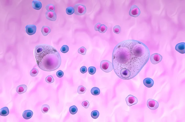
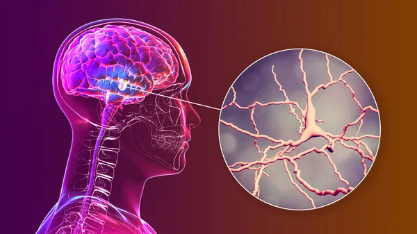
Neuron Conceptual Image Of Human Nervous System. 3D Illustration Of Neurons With Vivid Colors.
Image, 16.7MB, 3880 × 4850 jpg

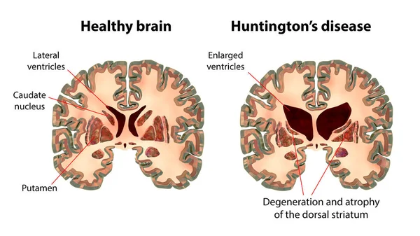
Coronal Sections Of A Healthy Brain And A Brain In Huntington's Disease Showing Enlarged Anterior Horns Of The Lateral Ventricles, Degeneration And Atrophy Of The Dorsal Striatum, 3D Illustration
Image, 10.57MB, 11104 × 6246 jpg
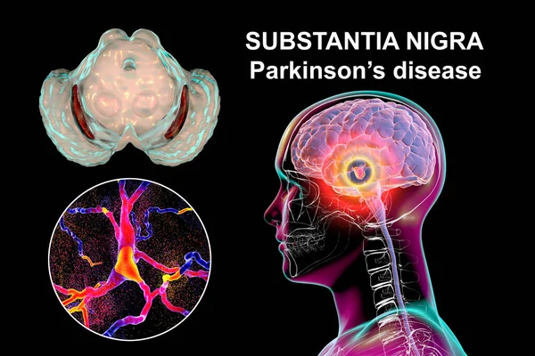
Black Substance Of The Midbrain In Parkinson's Disease, 3D Illustration Showing Decrease Of Its Volume And Accumulation Of Lewy Bodies In Dopaminergic Neurons Of The Black Substance
Image, 8.37MB, 7711 × 5140 jpg
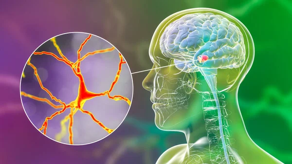
Black Substance Of The Midbrain And Its Dopaminergic Neurons, 3D Illustration. Black Substance Regulates Movement And Reward, Its Degeneration Is A Key Step In Development Of Parkinson's Disease
Image, 7.38MB, 6456 × 3632 jpg

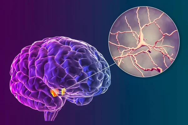
Dorsal Striatum, Caudate Nucleus And Putamen, Highlighted In The Brain Of A Person With Huntington's Disease And Close-up View Of Neuronal Degradation, Conceptual 3D Illustration
Image, 12.32MB, 7814 × 5210 jpg

Nicotinamide Mononucleotide Molecule. Skeletal Formula. Precursor Of NAD. Vector Illustration
Vector, 13.13MB, 4722 × 4722 eps


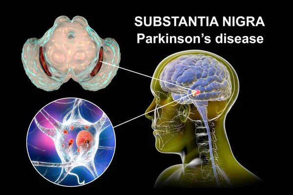
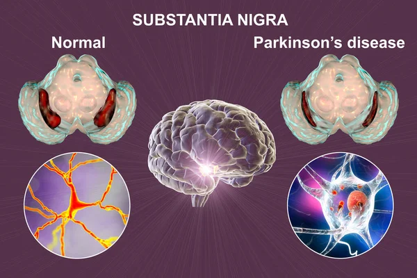
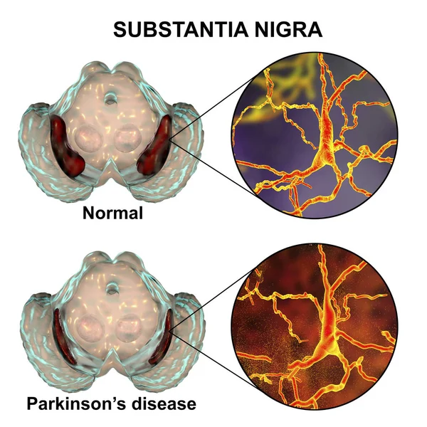
Substantia Nigra In Norm And In Parkinson's Disease, 3D Illustration Showing Decrease Of Its Volume. There Is Degeneration Of Dopaminergic Neurons In The Pars Compacta Of The Substantia Nigra
Image, 1.94MB, 6000 × 4000 jpg
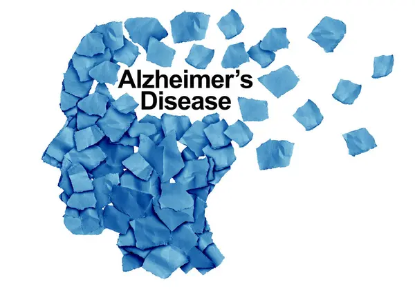
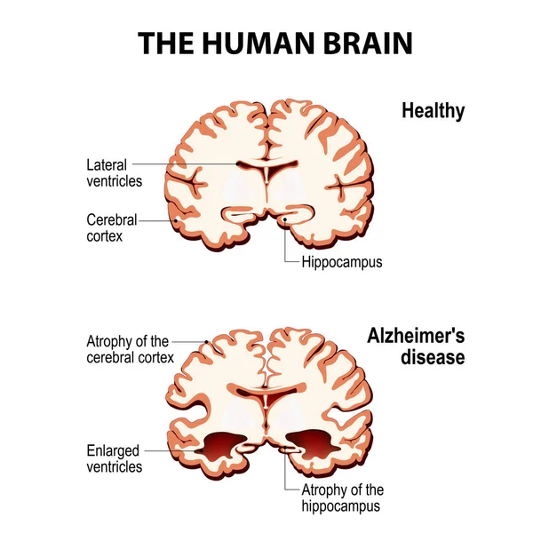
Alzheimer Disease As A Cognitive Decline As A Degenerative Dementia Brain Illness Resulting In Memory Loss As A Neurology Symbol For Aging Of The Mind.
Image, 9.61MB, 6277 × 4421 jpg


Black Substance Of The Midbrain And Its Dopaminergic Neurons, 3D Illustration. Black Substance Regulates Movement And Reward, Its Degeneration Is A Key Step In Development Of Parkinson's Disease
Image, 5.22MB, 6456 × 3632 jpg

Alzheimer's Disease. Comparison Of Neurons In A Healthy Brain And Nerve Cells In Neurodegenerative Disease. Dementia. Close-up Of Neurons With Neurofibrillary Tangles And Amyloid Plaques. Vector Illustration
Vector, 11.61MB, 5000 × 3472 eps

Dementia And Alzheimer Disease In Elderly Man Experiencing Brain Confusion And Pain In Head. Old Grandfather Needs Doctor Help Due To Symptoms Of Dementia And Sensitivity To Weather.
Vector, 5.21MB, 7500 × 5014 eps
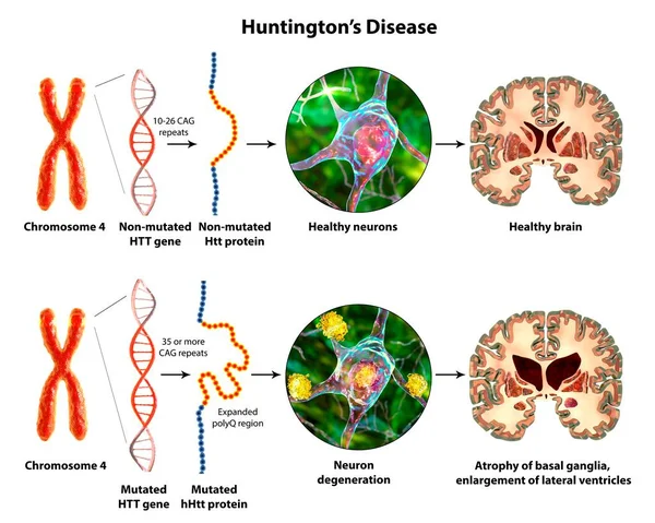
Molecular Genesis Of Huntington's Disease, 3D Illustration. Expansion Of The CAG Trinucleotide Sequence In The Htt Gene Causes Production Of Mutated Huntingtin Protein Leading To Neurodegeneration
Image, 17.01MB, 12941 × 10352 jpg

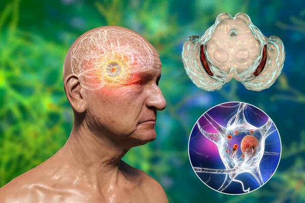
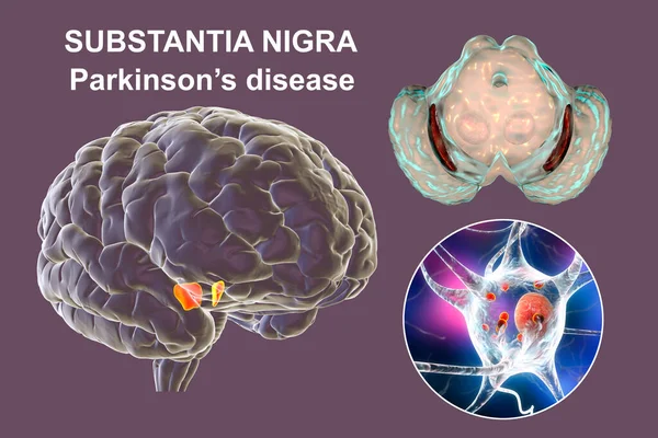
An Old Man With Parkinson's Disease And Highlighted Black Substance Of The Midbrain. 3D Illustration Shows Decrease Of Substantia Volume And Accumulation Of Lewy Bodies In Its Dopaminergic Neurons
Image, 11.03MB, 6306 × 4203 jpg

Kainic Acid Or Kainate Molecule. It Is Neuroexcitatory Amino Acid. Structural Chemical Formula, Molecule Model. Vector Illustration
Vector, 0.35MB, 5735 × 4359 eps


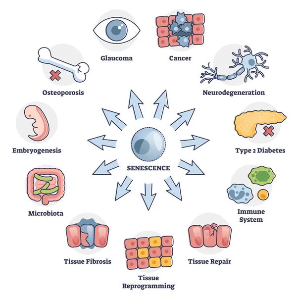
Senescence Cell Aging Problem With Human Health Risks In Outline Diagram
Vector, 6.67MB, 4000 × 4000 eps

Kainic Acid Or Kainate Molecule. It Is Neuroexcitatory Amino Acid. Molecular Model. 3D Rendering. Illustration
Image, 1.96MB, 4120 × 3370 jpg

Elderly Woman With Severe Dementia Sitting In A Chair, Having A Conversation With Herself, Illustrating The Impact Of Alzheimer's On Communication And Cognitive Function.
Image, 4.96MB, 3541 × 2833 jpg
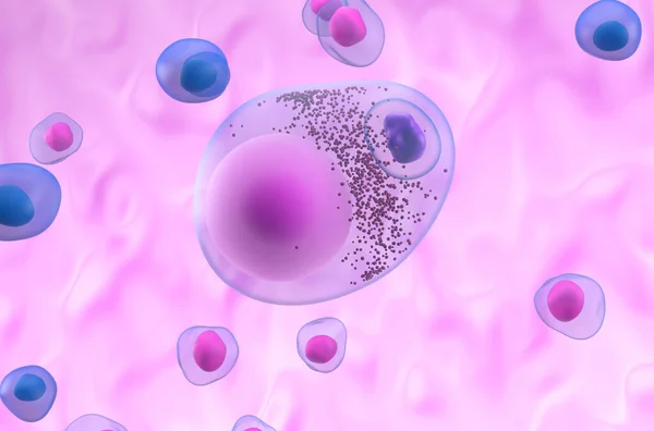
Lewy Bodies In Parkinson's Disease (PD) Or Dementia (LBD) - Isometric 3d Illustration
Image, 5.18MB, 10000 × 6600 jpg
Black Substance Of The Midbrain And Its Dopaminergic Neurons In Normal State And In Parkinson's Disease. 3D Illustration Showing Volume Decrease And Accumulation Of Lewy Bodies In Neurons
Image, 20.15MB, 9200 × 6133 jpg


Dorsal Striatum, Caudate Nucleus And Putamen, Highlighted In The Brain Of A Person With Huntington's Disease And Close-up View Of Neuronal Inclusions, Conceptual 3D Illustration
Image, 7.98MB, 6000 × 6000 jpg
Black Substance Of The Midbrain In Parkinson's Disease, 3D Illustration Showing Decrease Of Its Volume And Degeneration Of Dopaminergic Neurons In The Pars Compacta Of The Black Substance
Image, 12.3MB, 7711 × 5140 jpg

Kainic Acid Or Kainate Molecule. It Is Neuroexcitatory Amino Acid. Structural Chemical Formula, Dark Blue Background. Vector Illustration
Vector, 0.3MB, 5000 × 5000 eps

Kainic Acid Or Kainate Molecule. It Is Neuroexcitatory Amino Acid. Skeletal Chemical Formula. Vector Illustration
Vector, 0.28MB, 5000 × 5000 eps
Black Substance Of The Midbrain And Its Dopaminergic Neurons, 3D Illustration. Black Substance Regulates Movement And Reward, Its Degeneration Is A Key Step In Development Of Parkinson's Disease
Image, 11.07MB, 7395 × 4160 jpg
Lewy Body In Parkinson's Disease (PD) Or Dementia (LBD) - Closeup 3d Illustration
Image, 4.62MB, 10000 × 6600 jpg
Substantia Nigra In Norm And In Parkinson's Disease, 3D Illustration Showing Decrease Of Its Volume. There Is Degeneration Of Dopaminergic Neurons In The Pars Compacta Of The Substantia Nigra
Image, 4.57MB, 6000 × 6000 jpg
Black Substance Of The Midbrain In Parkinson's Disease, 3D Illustration Showing Decrease Of Its Volume And Accumulation Of Lewy Bodies In Dopaminergic Neurons Of The Black Substance
Image, 8.32MB, 7711 × 5140 jpg

Black Substance Of The Midbrain And Its Dopaminergic Neurons, 3D Illustration. Black Substance Regulates Movement And Reward, Its Degeneration Is A Key Step In Development Of Parkinson's Disease
Image, 10.21MB, 7395 × 4160 jpg

Black Substance Of The Midbrain And Its Dopaminergic Neurons, 3D Illustration. Black Substance Regulates Movement And Reward, Its Degeneration Is A Key Step In Development Of Parkinson's Disease
Image, 7.91MB, 7711 × 5140 jpg
Black Substance Of The Midbrain And Its Dopaminergic Neurons, 3D Illustration. Black Substance Regulates Movement And Reward, Its Degeneration Is A Key Step In Development Of Parkinson's Disease
Image, 11.11MB, 7711 × 5140 jpg

Laboratory Black Mouse Is Navigating In A Plastic 3D Printed Labyrinth In Lab Experiments To Study Spatial Learning, Memory And Brain Navigation Skills, Details, Closeup
Image, 2.61MB, 1668 × 2500 jpg


Nicotinamide Mononucleotide Molecule. Skeletal Formula. Precursor Of NAD. Vector Stock Illustration.
Vector, 4.87MB, 7020 × 4723 eps
Page 1 >> Next