
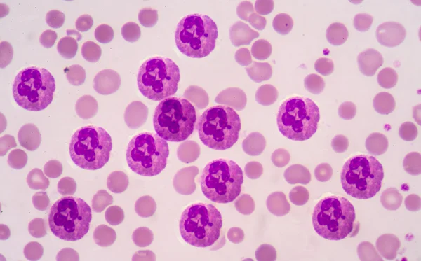
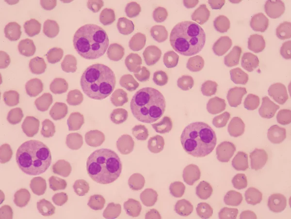
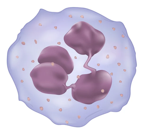
Stock image Neutrophil


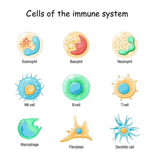
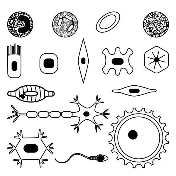
Cells Of The Immune System. White Blood Cells Or Leukocytes: Eosinophil, Neutrophil, Basophil, Macrophage, Fibroblast, And Dendritic Cell. Vector Diagram
Vector, 1.83MB, 4444 × 4444 eps
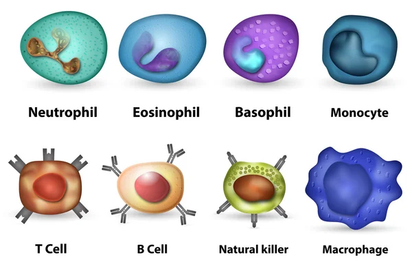

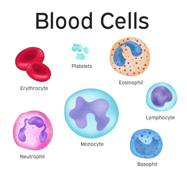
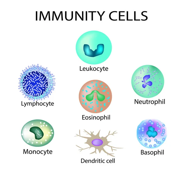
Cells Of Immunity. Set. Leukocyte, Lymphocyte, Eosinophil, Neutrophil, Monocyte, Basophil, Dendritic Cell. Vector Illustration On Isolated Background.
Vector, 5.85MB, 5000 × 4649 eps
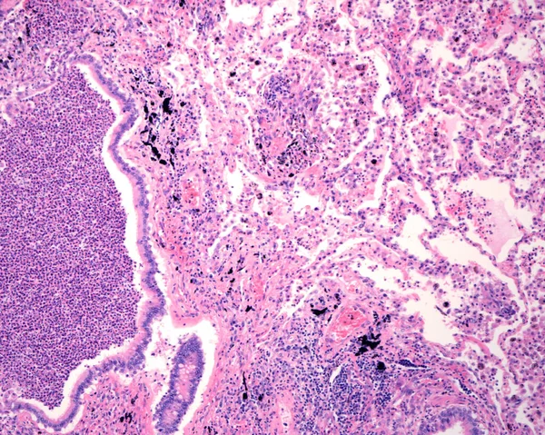
Human Lung Affected By An Acute Bronchopneumonia, Commonly A Hospital-acquired Bacterial Pneumonia. The Lumen Of Alveoli Is Occupied By Liquid Of Oedema Which Contains Acute Inflammatory Infiltrates (with Predominance Of Neutrophil Granulocytes). On
Image, 11.33MB, 3840 × 3072 jpg

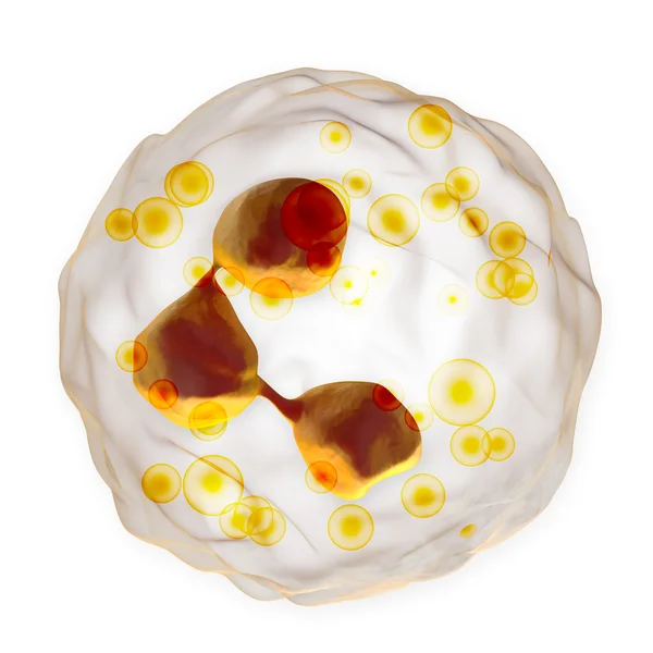
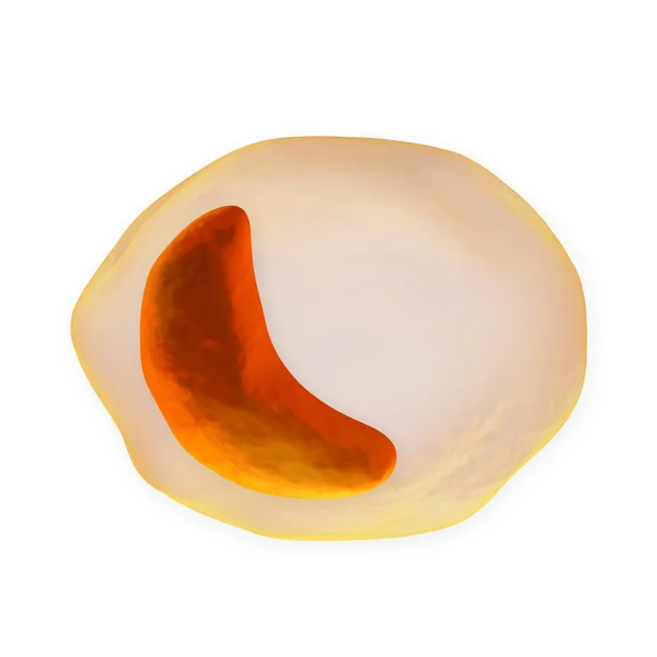
Eosinophilic Cationic Protein Is Incorporated Into The Membranes Of Helminth Cells And Disrupts Their Integrity. Eosinophil Is A Blood Cell. Vector Illustration On Isolated Background
Vector, 16.6MB, 5000 × 5000 eps

Diagnosis Of Lymphocytopenia. Decreased Lymphocytes In The Blood. Infographics. Vector Illustration On Isolated Background.
Vector, 33.73MB, 5000 × 4868 eps


Symptoms Of Lymphocytopenia. Decreased Lymphocytes In The Blood. Infographics. Vector Illustration On Isolated Background.
Vector, 33.17MB, 5000 × 4868 eps


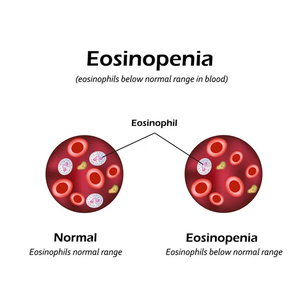
Eosinophils Below Normal Range In Blood. Eosinopenia. Infographics. Vector Illustration
Vector, 6.45MB, 5000 × 5000 eps

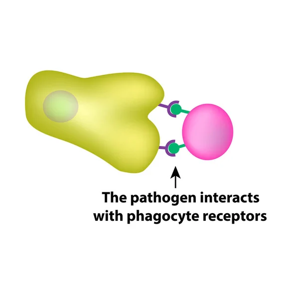
Innate Immunity. Adaptive Specific . Phagocytosis. Infographics. Vector Illustration
Vector, 1.45MB, 5000 × 4734 eps
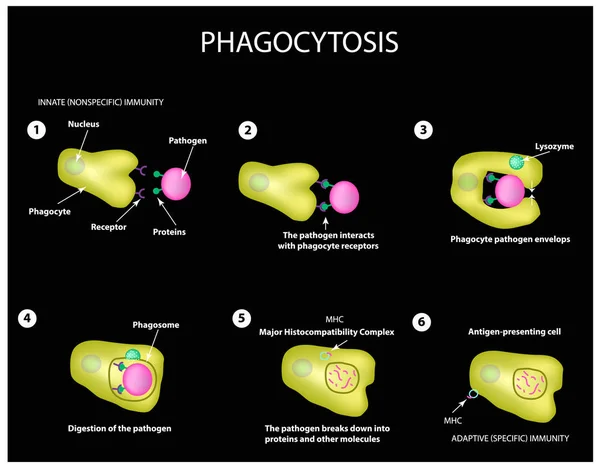
Innate Immunity. Adaptive Specific . Phagocytosis. Infographics. Vector
Vector, 3.99MB, 5000 × 3911 eps
Primary Myelofibrosis (PMF) Cells In Blood Flow - Closeup View 3d Illustration
Image, 6.32MB, 10000 × 6600 jpg
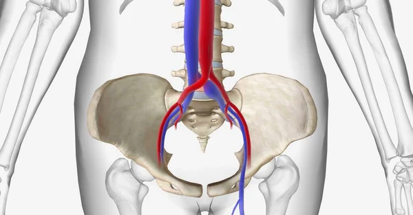
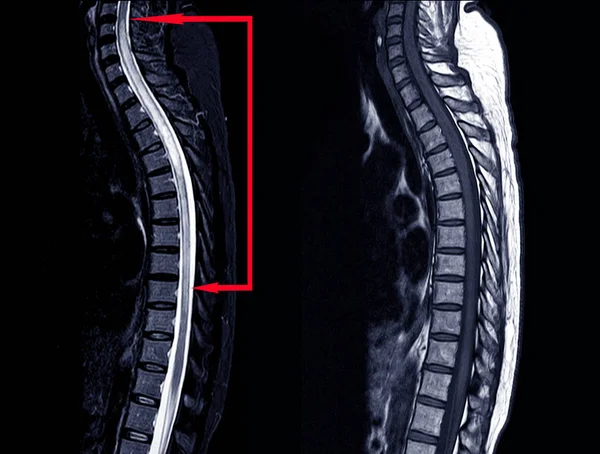
May Thurner Syndrome Is Compression Of The Left Common Iliac Vein Between The Right Common Iliac Artery And The 5th Lumbar Vertebra Of The Spine. 3D Rendering
Image, 16.81MB, 11115 × 5802 jpg
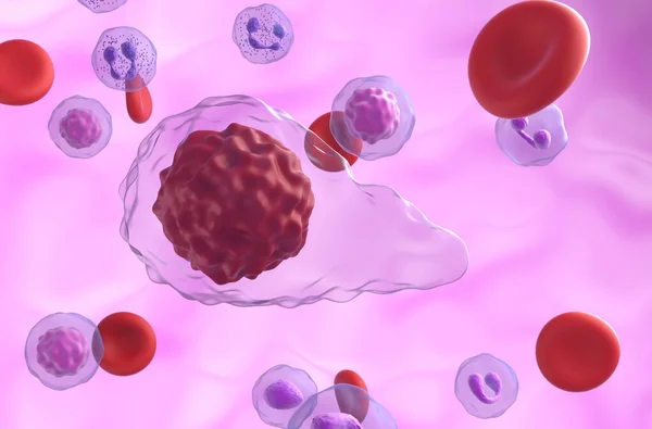
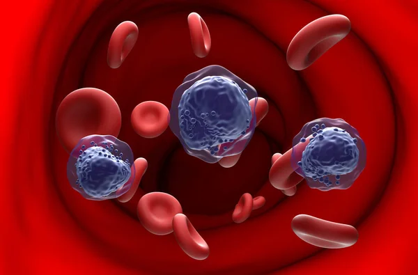
Lymphocytosis, Leukocytosis, 3D Illustration Showing Abundant White Blood Cells Inside Blood Vessel
Image, 9.19MB, 7200 × 4050 jpg


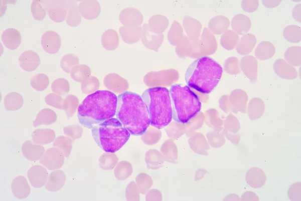
Auer Rods (or Auer Bodies) In Acute Hypergranular Promyelocytic Leukemia (APL) - 3d Illustration Closeup View
Image, 6.32MB, 10000 × 6600 jpg
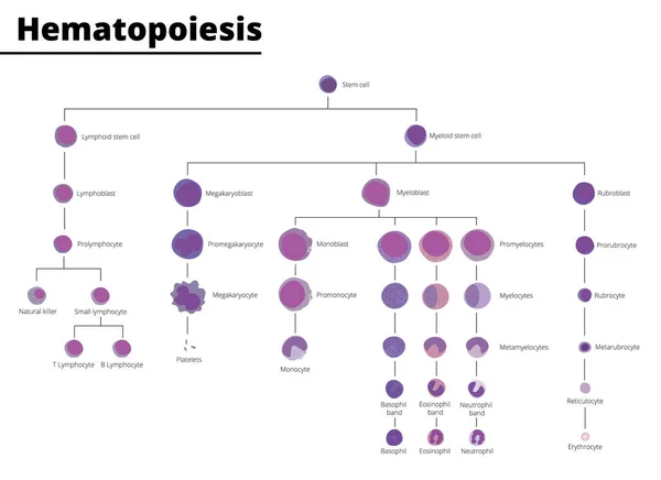
Hematopoiesis Differentiation Of Blood Cell Types Infographic Stem Cell Derived Blood Cells And Immune Cells. Vector Illustration. Didatic Illustration.
Vector, 1.2MB, 5500 × 4000 ai
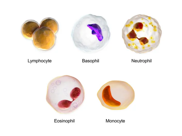
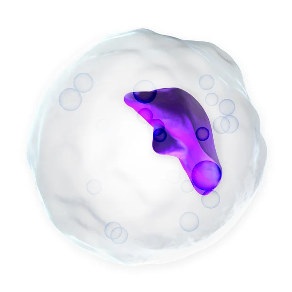
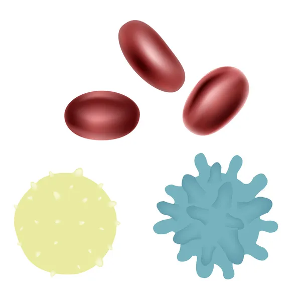
White Blood Cells - Lymphocyte, Basophil, Neutrophil, Eosinophil, Monocyte
Image, 6.39MB, 8000 × 6000 jpg
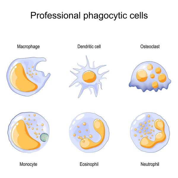
Phagocytosis. Professional Phagocytic Cells. Neutrophils, Macrophages, Monocytes, Dendritic Cells, Osteoclasts And Eosinophils Are Immune Response To Most Infections. Vector Illustration. Medical Poster.
Vector, 7.09MB, 4444 × 4444 eps
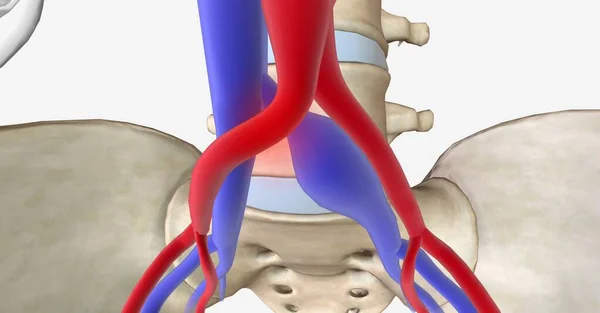
May Thurner Syndrome Is Compression Of The Left Common Iliac Vein Between The Right Common Iliac Artery And The 5th Lumbar Vertebra Of The Spine. 3D Rendering
Image, 19.34MB, 11115 × 5802 jpg
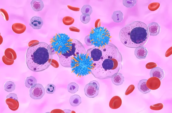
CAR T Cell Therapy In Multiple Myeloma (MM) - Isometric View 3d Illustration
Image, 7.7MB, 10000 × 6600 jpg
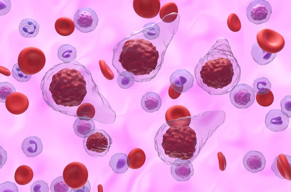
Primary Myelofibrosis (PMF) Cells In Blood Flow - Isometric View 3d Illustration
Image, 7.02MB, 10000 × 6600 jpg

Acute Myeloid Leukemia (AML) Cells In Blood Flow - Section View 3d Illustration
Image, 7.29MB, 10000 × 6600 jpg

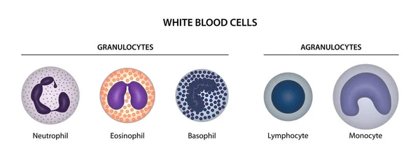
White Blood Cells (WBCs) Or Leukocytes: Granulocytes (neutrophil, Eosinophil, Basophil) And Agranulocytes (lymphocyte, Monocyte).
Image, 9.6MB, 16667 × 6250 jpg

CAR T Cell Therapy In Hodgkin Lymphoma (HL) - Section View 3d Illustration
Image, 5.46MB, 10000 × 6600 jpg

Page 1 >> Next