
Stock image Ophthalmic Disease page 2
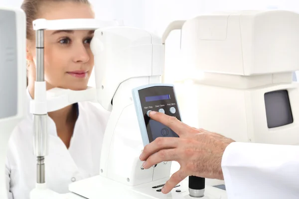


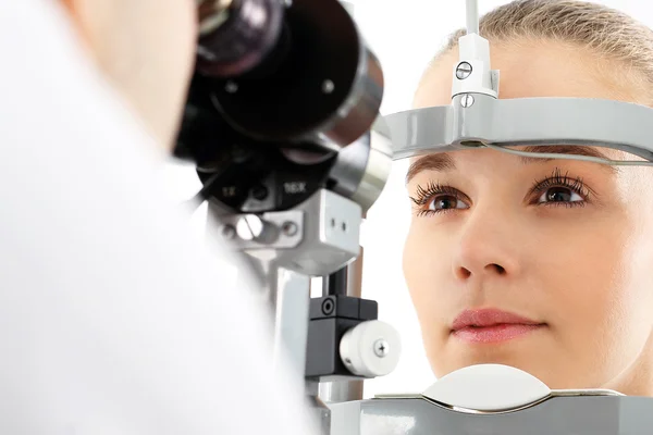
Childs Optometry - Little Girl Hecks Eyesight In Eye Ophthalmological Clinic
Image, 2.7MB, 3840 × 2160 jpg


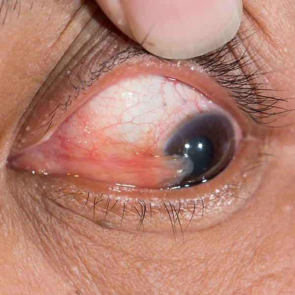


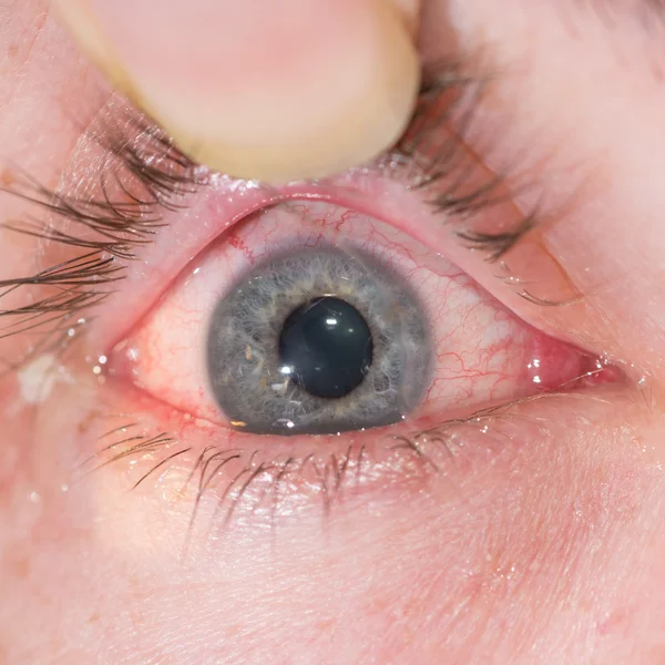


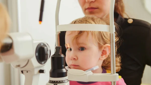
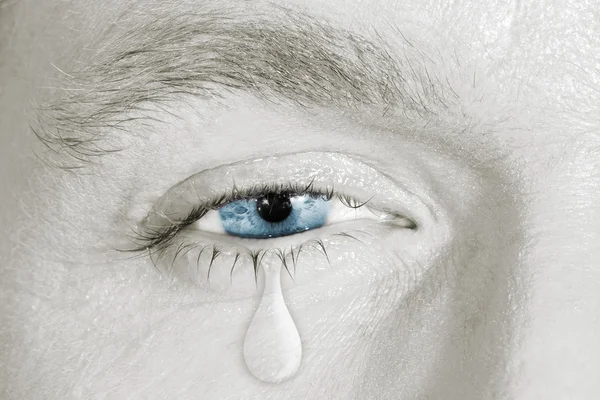
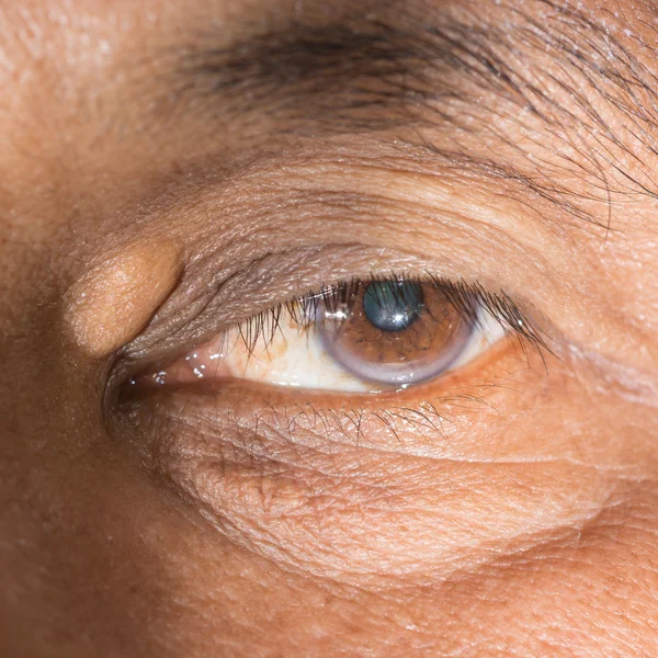
A Man Suffers From Pain In The Eye. Patient With Ophthalmic Disease, Uveitis, Optic Neuritis, Conjunctivitis, Or Eye Injury
Image, 6.26MB, 5282 × 3521 jpg


Proliferative Diabetic Retinopathy, Illustration Showing Neovascularization In The Disk And Other Sites, Macula Edema And Hard Exudates. Fundoscopic Examination Of The Eye Retina In Diabetes Mellitus
Image, 3.05MB, 5000 × 5000 jpg
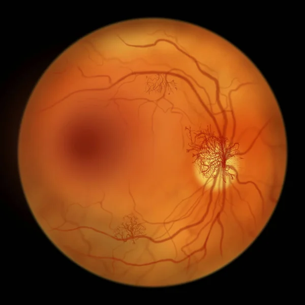
Proliferative Diabetic Retinopathy, Illustration Showing Neovascularization In The Disk And Other Sites, And Macula Edema. Fundoscopic Examination Of The Eye Retina In Diabetes Mellitus
Image, 2.9MB, 5000 × 5000 jpg

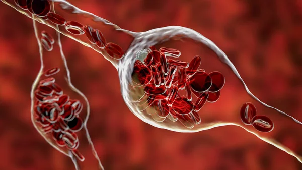
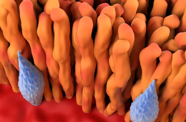
Microaneurysms, Microscopic Buldges In The Artery Walls Filled With Blood, 3D Illustration. Found In The Eye Retina In Diabetic Retinopathy, And Also In Brain (Charcot-Bouchard Aneurysms)
Image, 6.95MB, 7200 × 4050 jpg


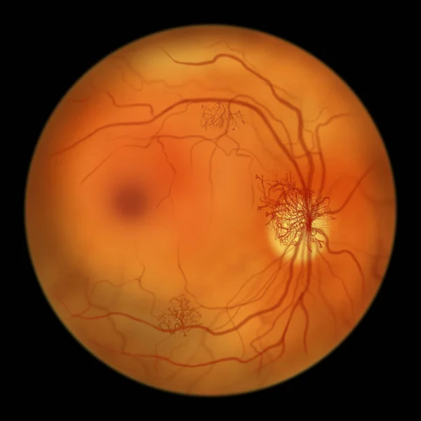
Proliferative Diabetic Retinopathy, Illustration Showing Neovascularization (formation Of New Vessels) In The Optic Disk And Other Sites. Fundoscopic Examination Of The Eye Retina In Diabetes Mellitus
Image, 2.86MB, 5000 × 5000 jpg

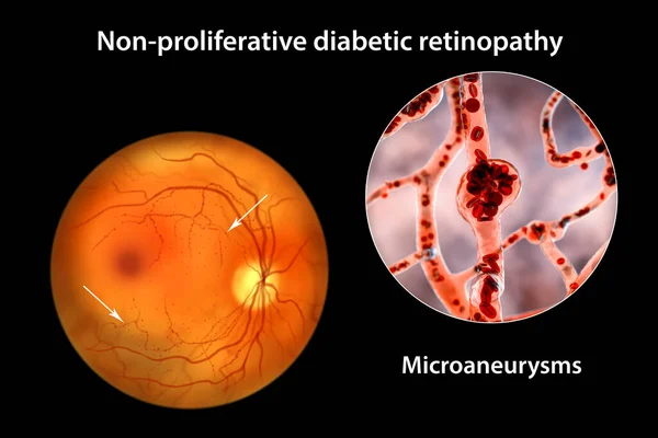
Non-proliferative Diabetic Retinopathy, 3D Illustration Showing Multiple Microaneurysms On The Eye Retina And Closeup View Of Microaneurysms, Microscopic Buldges In The Artery Walls Filled With Blood
Image, 10.46MB, 10431 × 6954 jpg
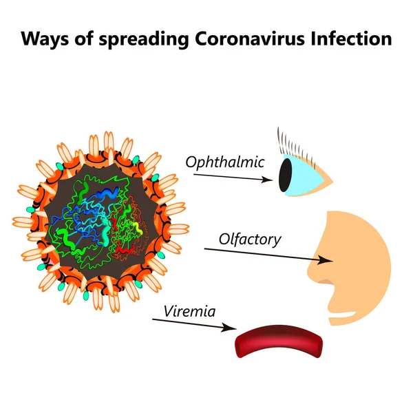
Ways Of Spreading Coronavirus Infection. Olfactory Transmission Of Covid 19. Ophthalmic, Viremia Coronavirus. Vector Illustration On Isolated Background
Vector, 2.17MB, 5000 × 5000 eps
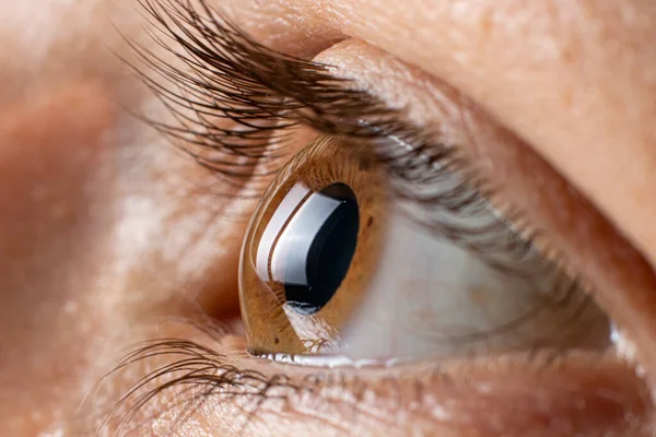
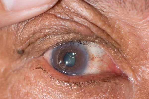
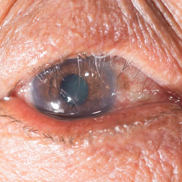
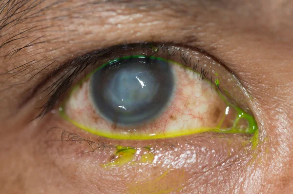
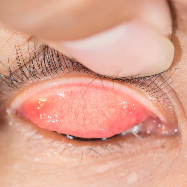
Keratoconus Of Eye, 3th Degree. Contortion Of The Cornea In The Form Of A Cone, Deterioration Of Vision, Astigmatism. Macro Close Up
Image, 16.59MB, 6000 × 4000 jpg
Previous << Page 2 >> Next