
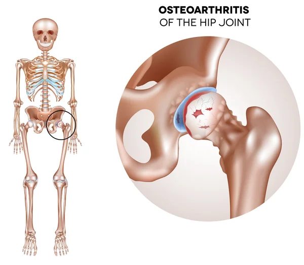
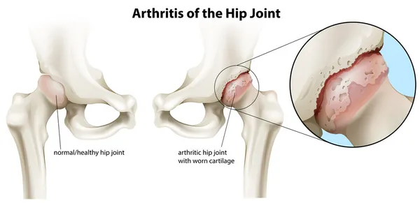
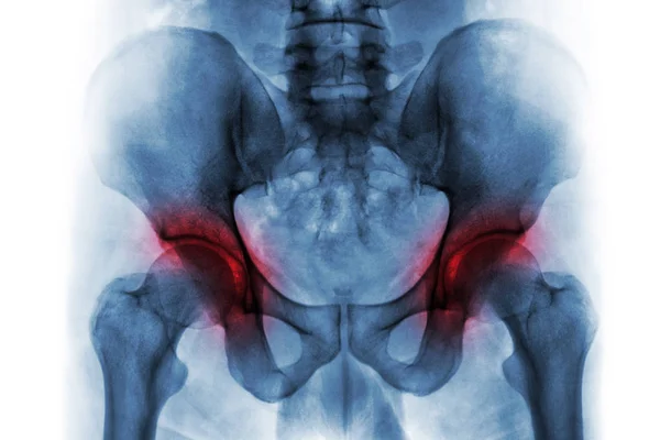
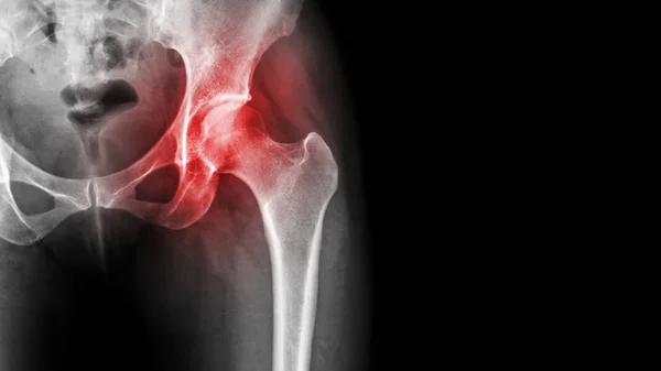
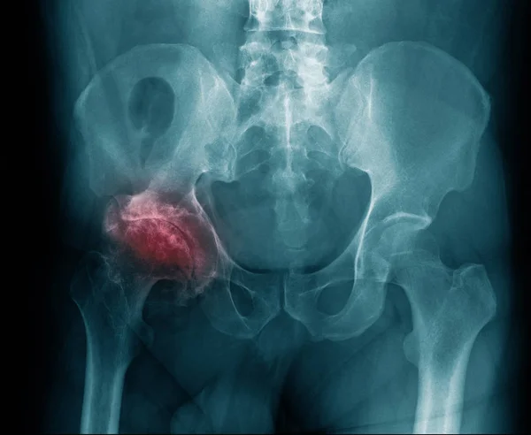
Stock image Osteoarthritis Of Hip
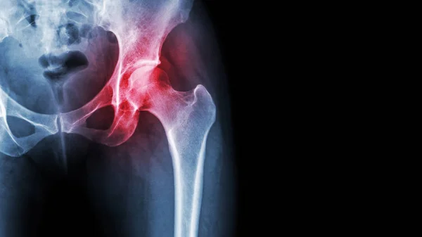
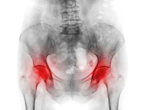
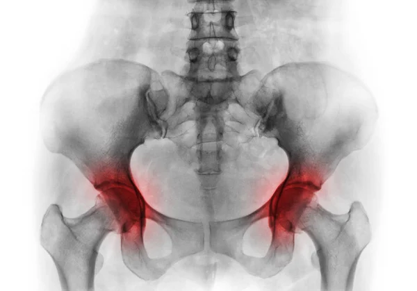
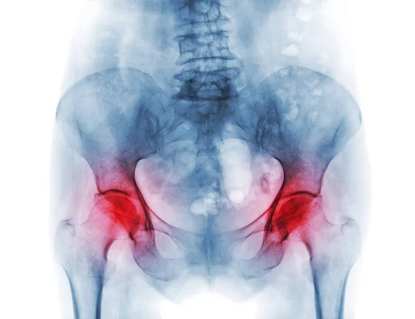
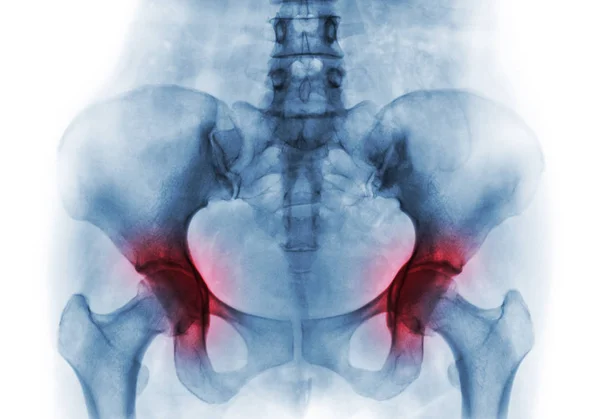
Arthritis At Hip Joint . Film X-ray Show Inflamed Of Hip Joint And Blank Area At Right Side . Avascular Necrosis Concept
Image, 5.61MB, 5815 × 3271 jpg
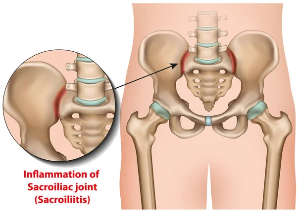
Sacroiliac Joint Inflammation 3d Medical Vector Illustration Sacroiliitis
Vector, 6.7MB, 7000 × 5000 eps
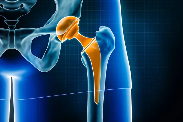
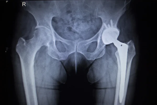
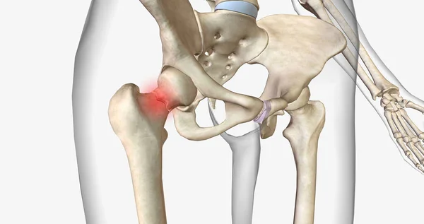
Hip Prosthesis X-ray 3D Rendering Illustration. Total Hip Joint Replacement Surgery Or Arthroplasty, Medical And Healthcare, Arthritis, Pathology, Science, Osteology, Orthopedics Concepts.
Image, 3.21MB, 3000 × 2000 jpg


Hip Osteoarthritis Bone Disease With Painful Skeletal Spurs Outline Diagram. Labeled Educational Scheme With Osteophytes Damaged Pelvis And Femur Anatomy Vector Illustration. Inflammation Illness.
Vector, 9.43MB, 5200 × 3328 eps
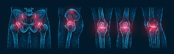
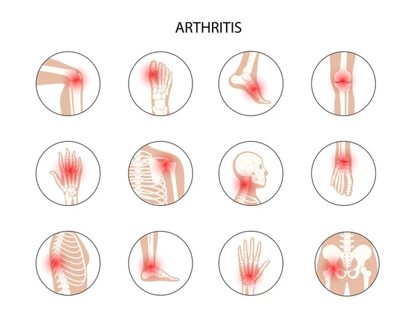
Polygonal Vector Illustration Of Pain Or Inflammation Of The Bones In The Pelvis, Hip Joint, And Knee Joints Isolated On A Dark Blue Background. Orthopedic Diseases Medical Template.
Vector, 5.56MB, 8000 × 2500 eps
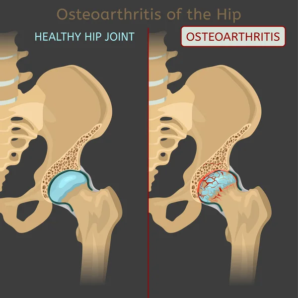
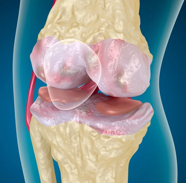
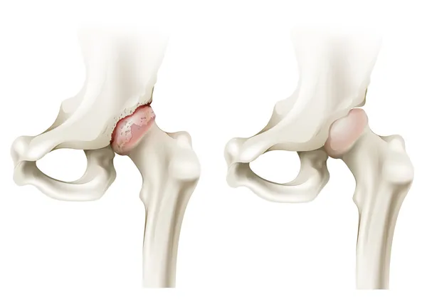
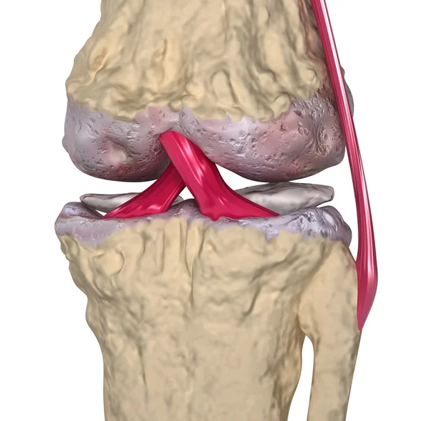
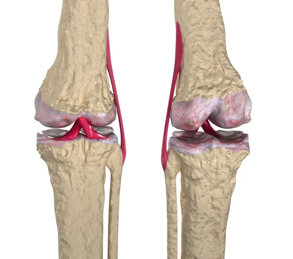
Hip Osteoarthritis. Synovial Joints Degenerative Disease. Editable Vector Illustration In Realistic Style Isolated On A Grey Background. Medical, Healthcare, Physiology Concept. Scientific Infographic
Vector, 9.1MB, 4725 × 4724 eps


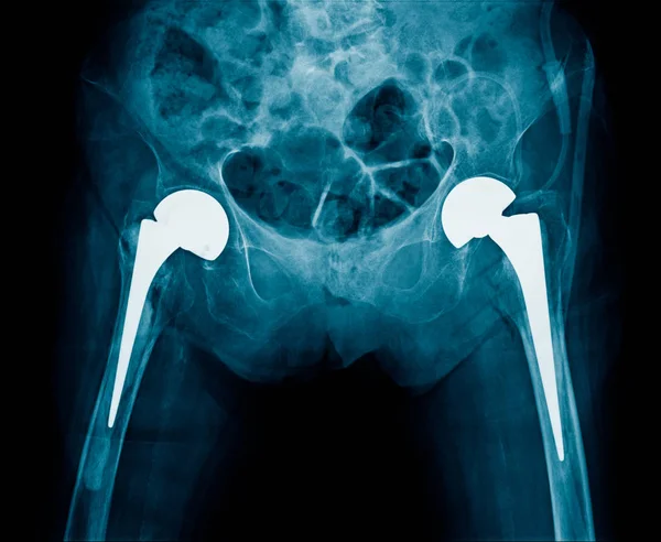
Bilateral Hip Replacement Of Patient, Hight Qulity X-ray Image Of Hip Joint Replacement Both Side In Blue Tone With Black Background
Image, 2.91MB, 2428 × 1994 jpg

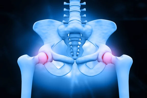
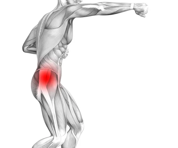
Conceptual Hip Human Anatomy With Red Hot Spot Inflammation Articular Joint Pain For Leg Health Care Therapy Or Sport Muscle Concepts. 3D Illustration Man Arthritis Or Bone Sore Osteoporosis Disease
Image, 1.45MB, 4432 × 2645 jpg

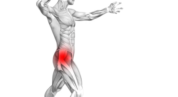
Conceptual Hip Human Anatomy With Red Hot Spot Inflammation Articular Joint Pain For Leg Health Care Therapy Or Sport Muscle Concepts. 3D Illustration Man Arthritis Or Bone Sore Osteoporosis Disease
Image, 1.31MB, 4456 × 2549 jpg

A Femoral Neck Fracture Is A Type Of Hip Fracture That Occurs In The Section Of The Femur Closest To The Pelvis.3D Rendering
Image, 2.83MB, 7340 × 3884 jpg

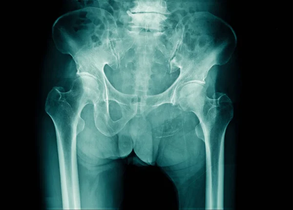
High Quality X-ray Hip Replacement In Old Man, Degenerative Change Of Hip And Post Operation Femur Head Fixation
Image, 1.54MB, 2428 × 1784 jpg
X-ray Image Pelvic Bone And Part Of L-spine With Compression Of Spine Or Degenertive Change
Image, 2.48MB, 2792 × 2010 jpg
Conceptual Hip Human Anatomy With Red Hot Spot Inflammation Articular Joint Pain For Leg Health Care Therapy Or Sport Muscle Concepts. 3D Illustration Man Arthritis Or Bone Sore Osteoporosis Disease
Image, 1.61MB, 3596 × 2985 jpg
Arthritis At Hip Joint . Film X-ray Show Inflamed Of Hip Joint And Blank Area At Right Side . Avascular Necrosis Concept
Image, 4.8MB, 5815 × 3271 jpg
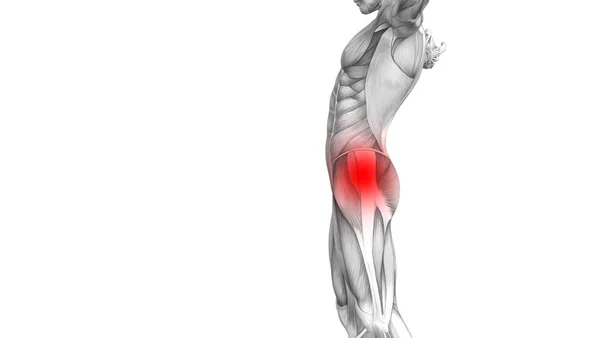
Conceptual Hip Human Anatomy With Red Hot Spot Inflammation Articular Joint Pain For Leg Health Care Therapy Or Sport Muscle Concepts. 3D Illustration Man Arthritis Or Bone Sore Osteoporosis Disease
Image, 1.3MB, 4270 × 2339 jpg
Conceptual Hip Human Anatomy With Red Hot Spot Inflammation Articular Joint Pain For Leg, 3D Illustration Man Arthritis Or Bone Sore Osteoporosis Disease
Image, 1.02MB, 4043 × 2279 jpg
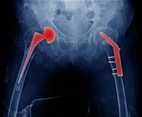
High Quality X-ray Hip Replacement In Old Man, Degenerative Change Of Hip And Post Operation Femur Head Fixation, Bilateral Hip Replacement And Fixation Of Head Of Femur
Image, 2.24MB, 2448 × 2010 jpg

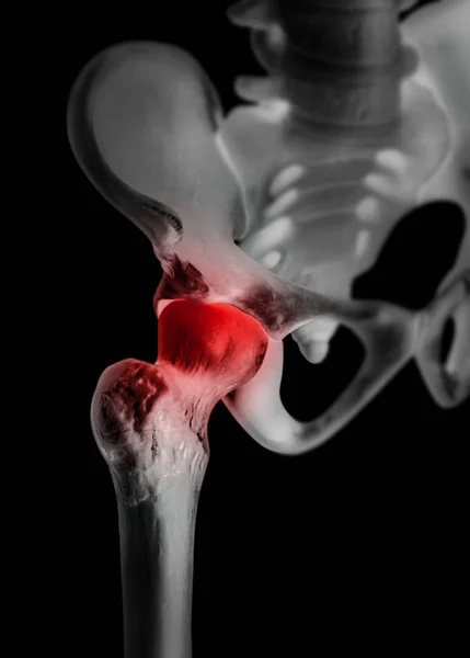
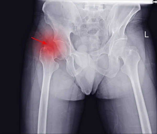
Human Hip Joint With Red Highlight On Pain Area - X Ray Film-Healthcare-Human Anatomy And Medical Concept-Isolated On Black Background.
Image, 7.01MB, 4856 × 6789 jpg
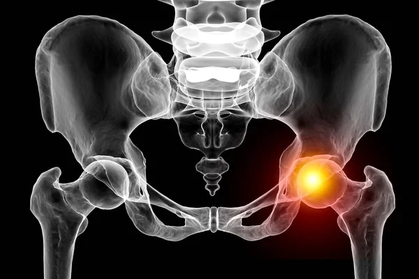
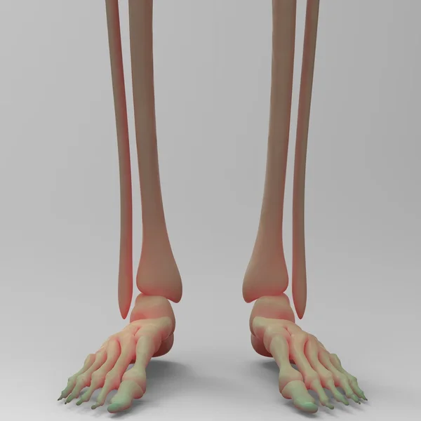
Hip Joint Pain. A 3D Medical Illustration Depicting The Pelvis And Femur Bones With The Femur Head Highlighted In Red, Illustrating Femur Joint Pain Concept.
Image, 4.88MB, 7000 × 4667 jpg
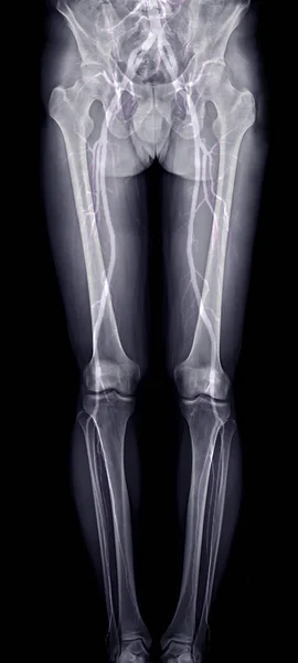
Scanogram Of Lower Limb Or X-ray Image With Merge CTA Femoral Run Off Showing Bone And Vessel Of Lower Limb.
Image, 2.19MB, 1936 × 4303 jpg

X-ray Image Lumbar Spine And Degenerative Change Of Spine, L-spondylosis X-ray Image In Blue Tone
Image, 2.03MB, 1996 × 2428 jpg

X-ray Image Of Spine Show Degeneration Of Lumbar Spine With Red High Light, Spur Or Calcification At Body Of Lumbar Spine
Image, 1.81MB, 1996 × 2428 jpg

Conceptual Hip Human Anatomy With Red Hot Spot Inflammation Articular Joint Pain For Leg Health Care Therapy Or Sport Muscle Concepts. 3D Illustration Man Arthritis Or Bone Sore Osteoporosis Disease
Image, 1.67MB, 4720 × 2700 jpg

Conceptual Hip Human Anatomy With Red Hot Spot Inflammation Articular Joint Pain For Leg Health Care Therapy Or Sport Muscle Concepts. 3D Illustration Man Arthritis Or Bone Sore Osteoporosis Disease
Image, 1.64MB, 4014 × 2968 jpg
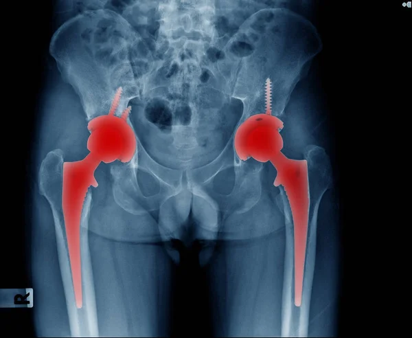
X-ray Bilateral Hip Replacement, Post Operation Total Hip Arthroplasty Both Side Of Old Man
Image, 2.8MB, 2448 × 2010 jpg

Page 1 >> Next