
Stock image Ovarian Tumor


Doctor Surgeon Holds A Surgical Scalpel Against The Background Of The Layout Of The Female Reproductive System. Concept Of Vaginal Plastics, Abortion, Surgical Operations To Remove Polyps And Cysts In Women, Hysteroscopy
Image, 1.66MB, 5304 × 3450 jpg


Doctor Holding A Test Blood Sample Tube With Inhibin A Test On The Background Of Medical Test Tubes With Analyzes.
Image, 8.65MB, 5789 × 3859 jpg

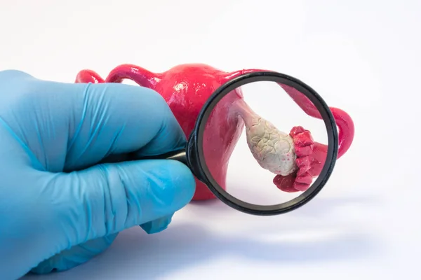
Search Disease, Abnormalities Or Pathology Of Ovary Concept Photo. Doctor Holding Magnifying Glass And Examines Model Of Ovaries, Conducting Diagnostics For Disease Like Cancer, Apoplexy, Cyst, POS
Image, 5.94MB, 6000 × 4000 jpg


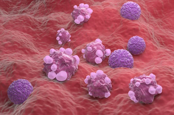
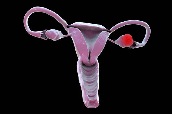
Ovarian Cancer, 3D Illustration Showing Malignant Tumor In The Left Ovary
Image, 10.01MB, 6000 × 4000 jpg

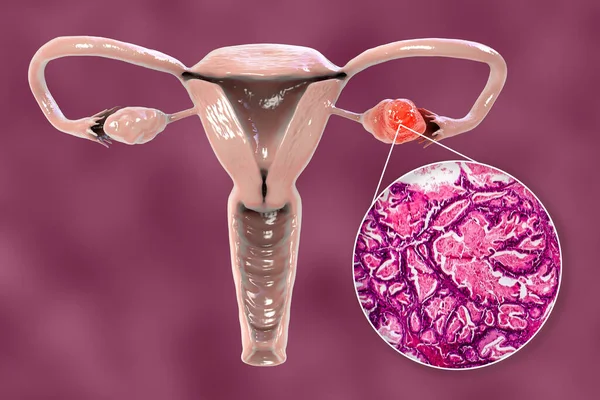
Ovarian Cancer, 3D Illustration Showing Malignant Tumor In The Left Ovary And Light Photomicrograph Showing Histopathology Of Ovarian Cancer
Image, 9.11MB, 6146 × 4097 jpg

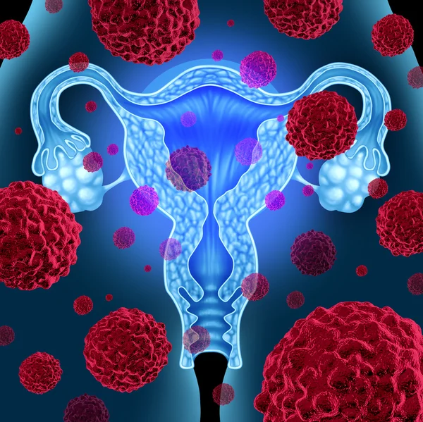
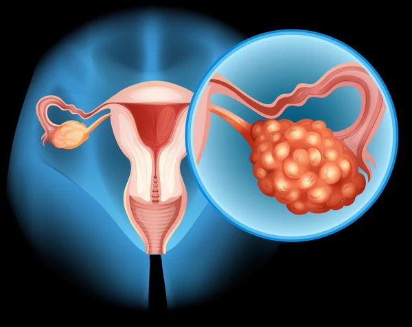
Stage II Ovarian Tumor Has Spread To Nearby Organs, Such As The Uterus And The Body Of The Fallopian Tubes. 3D Rendering
Image, 1.88MB, 7340 × 3884 jpg
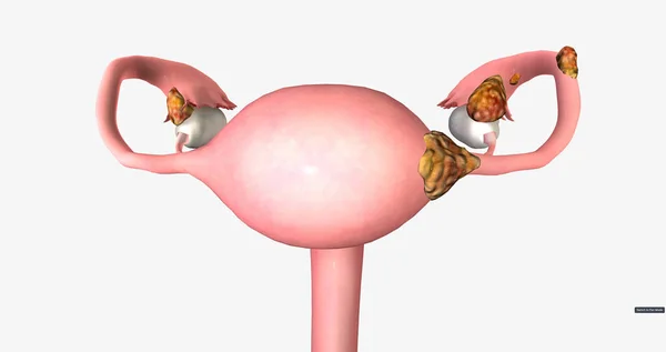
Stage II Ovarian Tumor Has Spread To Nearby Organs, Such As The Uterus And The Body Of The Fallopian Tubes. 3D Rendering
Image, 1.86MB, 7340 × 3884 jpg
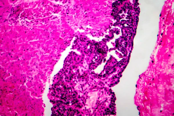
Papillary Serous Ovarian Adenocarcinoma, Cancer Of Ovary, Light Micrograph, Photo Under Microscope
Image, 13.36MB, 4084 × 2722 jpg
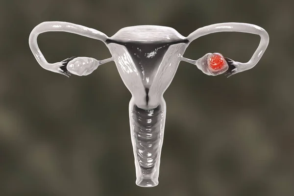
Ovarian Cancer, 3D Illustration Showing Malignant Tumor In The Left Ovary
Image, 2.44MB, 6000 × 4000 jpg

Ovarian Cancer, 3D Illustration Showing Malignant Tumor In The Left Ovary
Image, 6.51MB, 6000 × 4000 jpg
Ultrasound Upper Abdomen Or Ultrasound Kidney Showing Renal Function.
Image, 2.35MB, 4464 × 2608 jpg
Ultrasound Upper Abdomen Showing Anatomical Of Hepatobiliary System And Liver And Gall Bladder For Screening Hepatic Cell Carcinoma Of Hcc. Clipping Path
Image, 3.55MB, 6000 × 4000 jpg
Ovarian Cancer, 3D Illustration Showing Malignant Tumor In The Left Ovary
Image, 4.45MB, 6000 × 4000 jpg
Papillary Serous Ovarian Adenocarcinoma, Cancer Of Ovary, Light Micrograph, Photo Under Microscope
Image, 14.06MB, 4152 × 2768 jpg
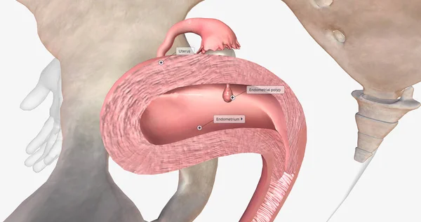
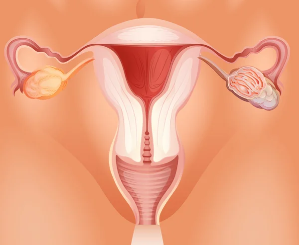
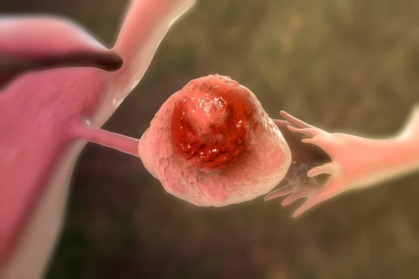
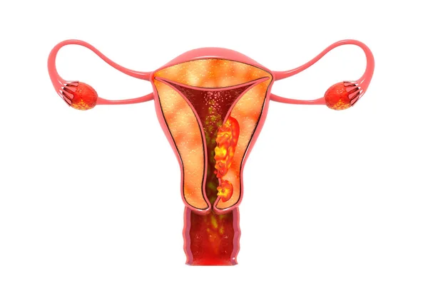
Endometrial Polyps Are Abnormal Growths Of The Inner Lining Of The Uterus, Known As The Endometrium. 3D Rendering
Image, 3.45MB, 7340 × 3884 jpg
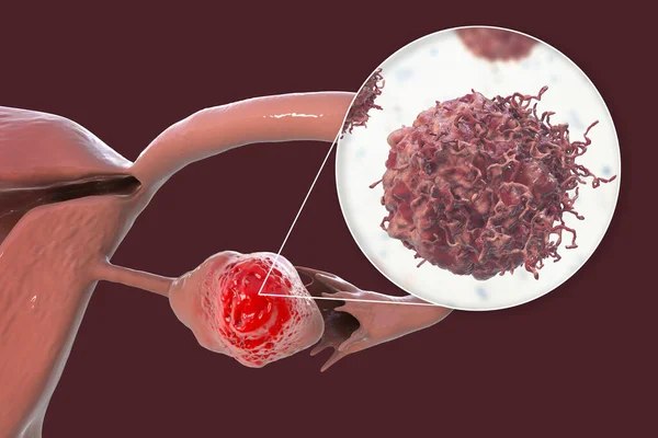
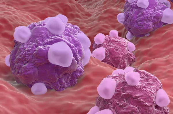
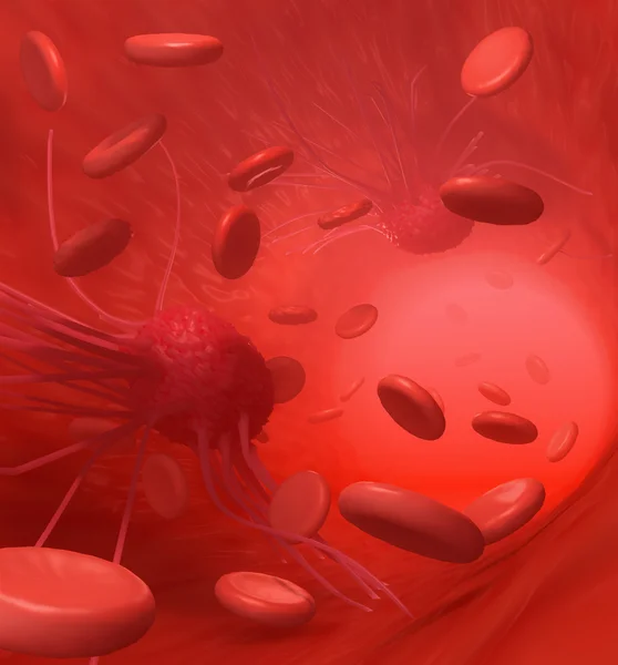
Ovarian Cancer, 3D Illustration Showing Malignant Tumor In The Left Ovary And Close-up View Of Cancer Cells
Image, 8.04MB, 7039 × 4693 jpg

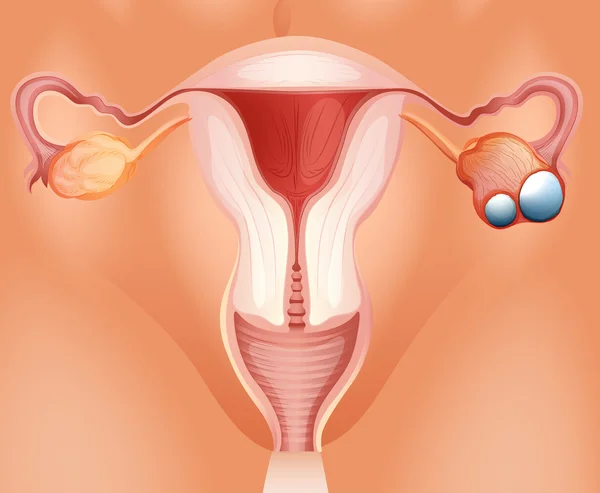
Symptoms Of Ovarian Cyst. Ovaries Structure. Infographics. Vector Illustration On Isolated Background
Vector, 0.94MB, 5000 × 5000 eps

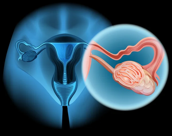
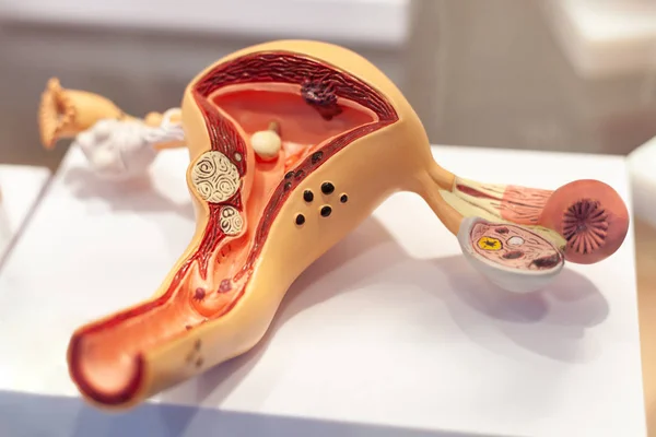
Natural Anatomical 3D Uterus With Ovaries Model With Placard Inscripted SOS Referring To Patient Or Doctor For Help. Conceived For All Symptoms, Syndromes, Diseases And Pathologies Of Female Organs
Image, 4.49MB, 6016 × 4000 jpg
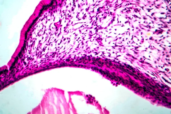
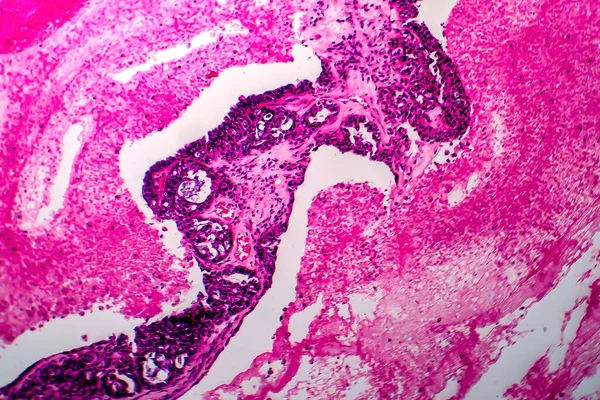
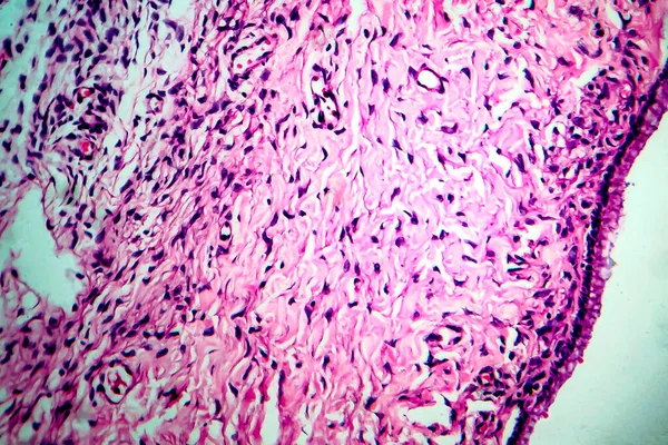
Ovarian Mucinous Cystadenoma, A Benign Tumor Of Ovary, Light Micrograph, Photo Under Microscope
Image, 9.7MB, 4568 × 3045 jpg
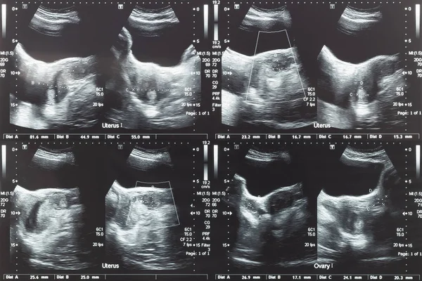
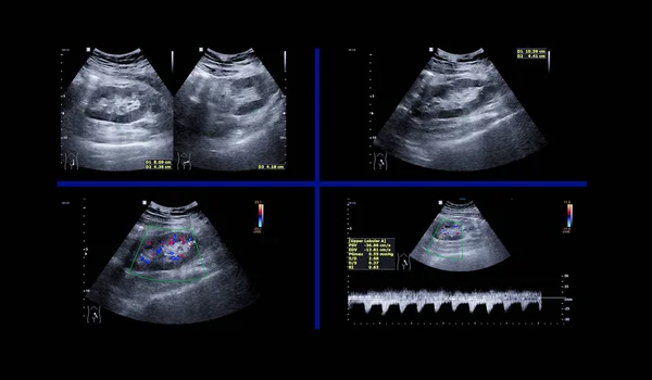
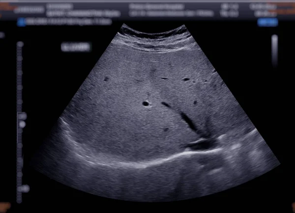
Ultrasound Image Of Lower Abdomen, Ovary And Uterus With Tumor Or Uterine Fibroid, Leiomyoma Of Female Woman Patient For Gynecological Medical Exam, Analysis And Test
Image, 3.91MB, 3246 × 2164 jpg
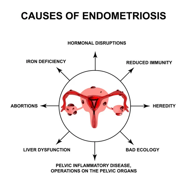
Causes Of Endometriosis. Adenomyosis. The Structure Of The Pelvic Organs With Endometriosis. Infographics. Vector Illustration On Isolated Background.
Vector, 1.9MB, 5034 × 5000 eps


Ovarian Endometriomas Chocolate Cysts Female Reproductive System Uterus Problem Diagram With Inscriptions. Human Medical Anatomy Internal Organs Location Scheme Flat Icon Vector Illustration Isolated
Vector, 0.57MB, 10682 × 5001 eps

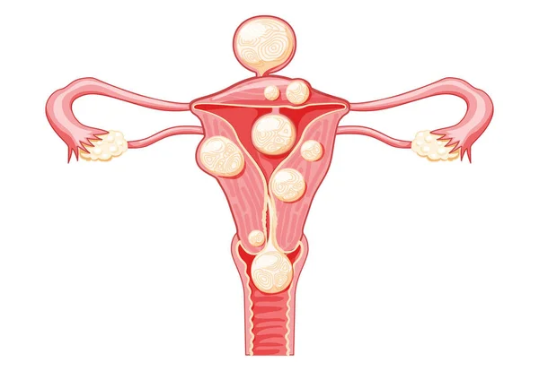
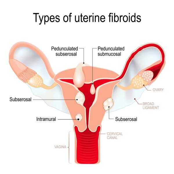
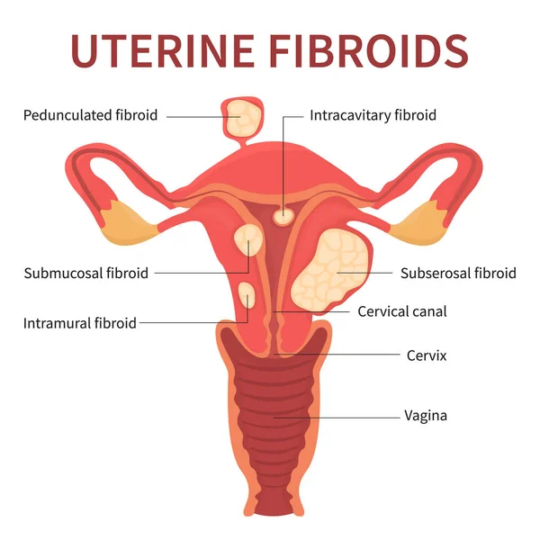
Uterine Fibroids Female Leiomyomas Reproductive System Uterus. Front View. Human Anatomy Medical Illustration Isolated Internal Organs Location Scheme, Cervix, Ovary, Fallopian Tube Flat Style Icon
Vector, 1.04MB, 7239 × 5001 eps

Menstrual Cycle. Changes In The Endometrium During The Menstrual Cycle. Uterus Lining From Menstruation, Proliferative Phase To Ovulation And Secretory Phase. Luteal And Follicular Phase. Vector Poster
Vector, 4.4MB, 5403 × 3000 eps
Ovarian Mucinous Cystadenoma, A Benign Tumor Of Ovary, Light Micrograph, Photo Under Microscope
Image, 11.27MB, 4436 × 2957 jpg

Uterine Polyps Removal. Endometrial Disease. Overgrowth Of Cells In The Uterus And Endometrium. Woman Health Concept. Cause Of Irregular Menstrual Bleeding And Infertility Flat Vector Illustration.
Vector, 1.52MB, 4584 × 3790 eps
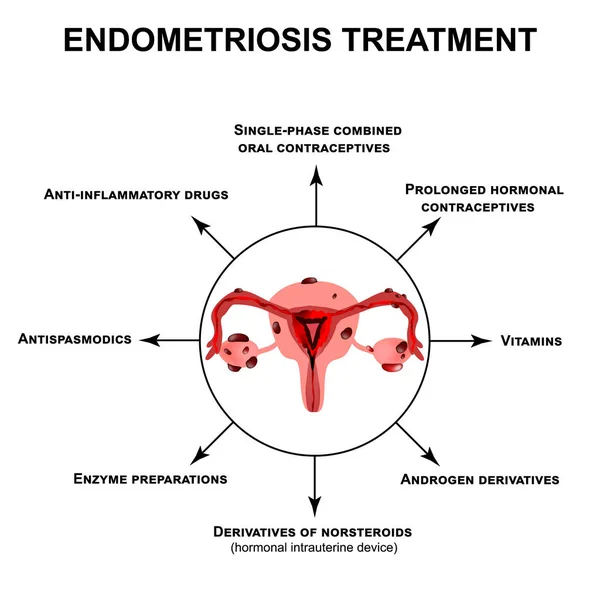
Treatment Of Endometriosis. Adenomyosis. The Structure Of The Pelvic Organs With Endometriosis. Infographics. Vector Illustration On Isolated Background.
Vector, 2.02MB, 5000 × 4967 eps
Page 1 >> Next