photo free
Home Images Vectors Videos

Stock image Patella Tendon page 2

stock vector Knee anatomy. Structure of leg joint. Major parts. Vector poster with text label for medical education

Knee Anatomy. Structure Of Leg Joint. Major Parts. Vector Poster With Text Label For Medical Education

Vector, 8.29MB, 4444 × 4444 eps
stock vector bone edema of knee medical poster

Bone Edema Of Knee Medical Poster

Vector, 2.73MB, 6378 × 4252 eps
stock vector Anatomy of the knee joint front view, template for training a medical surgical poster, traumatology page. Vector illustration.

Anatomy Of The Knee Joint Front View, Template For Training A Medical Surgical Poster, Traumatology Page. Vector Illustration.

Vector, 5MB, 5000 × 5000 eps
stock vector Medial knee injuries. joint anatomy. Vector illustration for biological, medical, science and educational use

Medial Knee Injuries. Joint Anatomy. Vector Illustration For Biological, Medical, Science And Educational Use

Vector, 5.78MB, 5811 × 5811 eps
stock vector Fibular collateral ligament injury. joint anatomy. Vector illustration for biological, medical, science and educational use

Fibular Collateral Ligament Injury. Joint Anatomy. Vector Illustration For Biological, Medical, Science And Educational Use

Vector, 8.67MB, 5470 × 5470 eps
stock vector Ligaments of the knee. Anterior and Posterior cruciate ligaments, Patellar and Quadriceps,  tendons, Medial and Lateral collateral ligaments. joint anatomy. Vector illustration for biological, medical, science and educational use

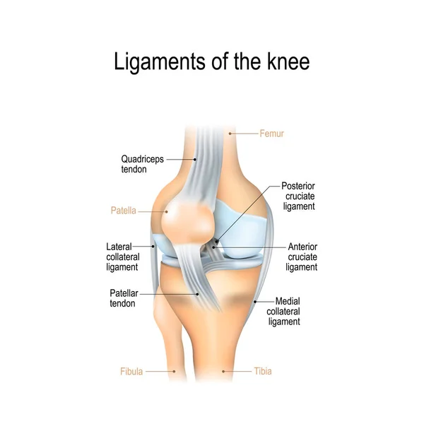
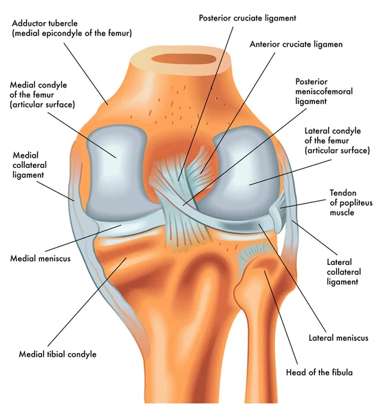
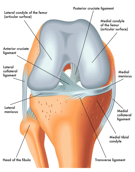
Ligaments Of The Knee. Anterior And Posterior Cruciate Ligaments, Patellar And Quadriceps, Tendons, Medial And Lateral Collateral Ligaments. Joint Anatomy. Vector Illustration For Biological, Medical, Science And Educational Use

Vector, 7.27MB, 4062 × 4062 eps
stock image Woman holding her knee with red pain

Woman Holding Her Knee With Red Pain

Image, 11.47MB, 6000 × 4004 jpg
stock vector Knee Dislocations - medical vector illustration diagrams. Anatomical knee injury types scheme.

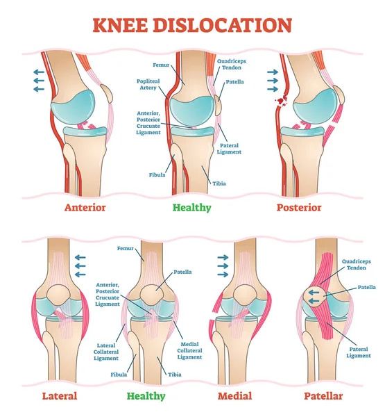
Knee Dislocations - Medical Vector Illustration Diagrams. Anatomical Knee Injury Types Scheme.

Vector, 7.05MB, 4528 × 4891 eps
stock vector Medial knee ligament sprain medical vector illustration isolated on white background infographic

Medial Knee Ligament Sprain Medical Vector Illustration Isolated On White Background Infographic

Vector, 7.79MB, 6000 × 5000 eps
stock vector bursitis. Prepatellar bursitis. inflammation of sacs with synovial fluid. Front and side view of human knee joint. Lateral and anterior section of the knee. vector illustration.

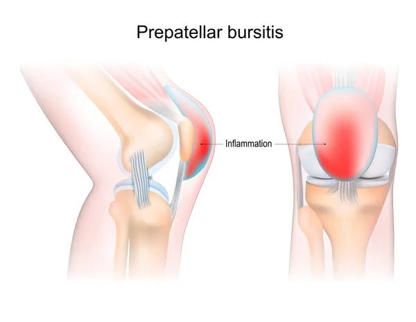
Bursitis. Prepatellar Bursitis. Inflammation Of Sacs With Synovial Fluid. Front And Side View Of Human Knee Joint. Lateral And Anterior Section Of The Knee. Vector Illustration.

Vector, 3.98MB, 5162 × 4000 eps
stock vector Anatomy. Structure knee joint vector

Anatomy. Structure Knee Joint Vector

Vector, 0.74MB, 5333 × 4000 eps
stock vector Normal position of kneecap and patella displaced

Normal Position Of Kneecap And Patella Displaced

Vector, 0.75MB, 4380 × 4381 eps
stock vector knee anatomy. side and front view. Cross section of the joint showing the main parts: femur, fibula, articular capsule, menisci, muscles and ligaments. vector illustration

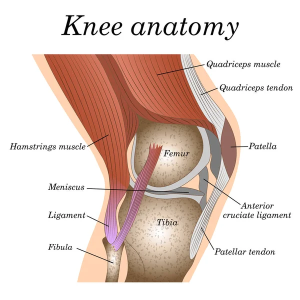
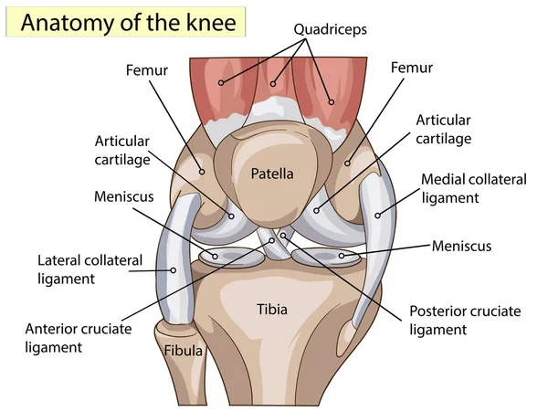
Knee Anatomy. Side And Front View. Cross Section Of The Joint Showing The Main Parts: Femur, Fibula, Articular Capsule, Menisci, Muscles And Ligaments. Vector Illustration

Vector, 3.91MB, 4444 × 3973 eps
stock image Injury from workout concept : Asian man use hands hold on his an

Injury From Workout Concept : Asian Man Use Hands Hold On His An

Image, 8.28MB, 5472 × 3648 jpg
stock vector Anterior cruciate ligament injury. joint anatomy. Vector illustration for biological, medical, science and educational use

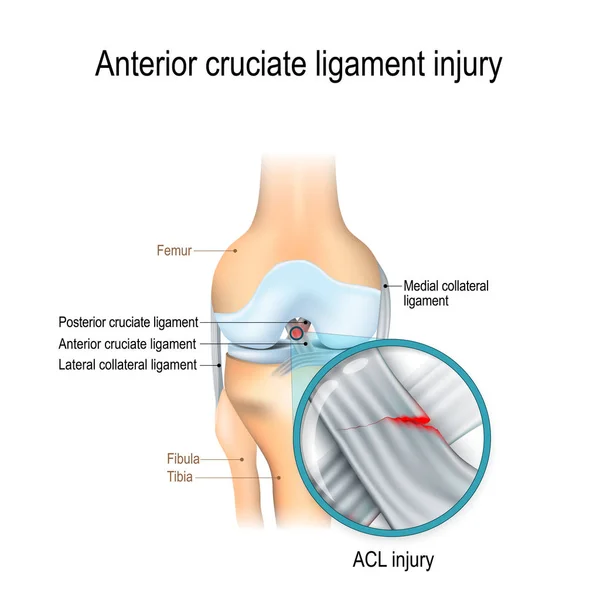
Anterior Cruciate Ligament Injury. Joint Anatomy. Vector Illustration For Biological, Medical, Science And Educational Use

Vector, 10.53MB, 5073 × 5073 eps
stock vector Posterior view of the right knee in extension

Posterior View Of The Right Knee In Extension

Vector, 1.63MB, 4618 × 5000 eps
stock vector Knee anatomy. Human joint structure. Lateral and Front aspects of right knee. Vector poster

Knee Anatomy. Human Joint Structure. Lateral And Front Aspects Of Right Knee. Vector Poster

Vector, 3.95MB, 4000 × 4888 eps
stock vector Dislocated knee, medial collateral ligament tear medical vector illustration isolated on white background

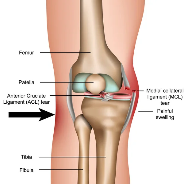
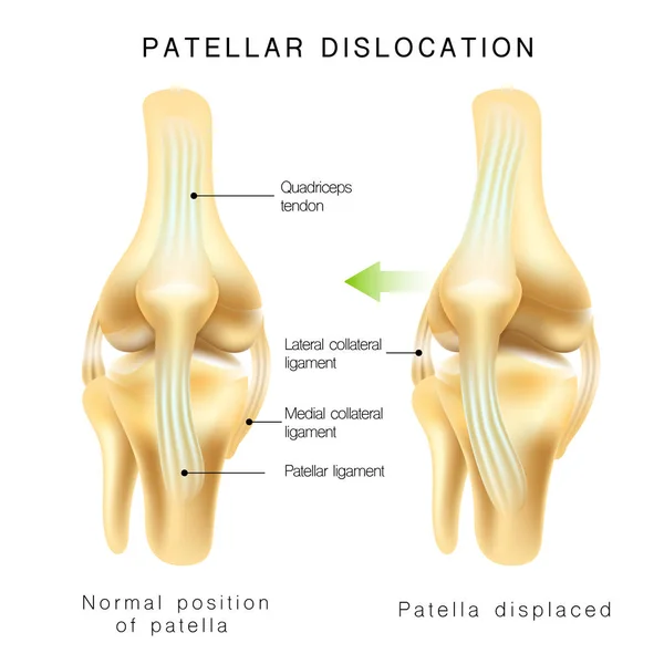
Dislocated Knee, Medial Collateral Ligament Tear Medical Vector Illustration Isolated On White Background

Vector, 5.34MB, 5000 × 5000 eps
stock vector Anatomy of the knee joint side view, template for training a medical surgical poster, traumatology page. Vector illustration.

Anatomy Of The Knee Joint Side View, Template For Training A Medical Surgical Poster, Traumatology Page. Vector Illustration.

Vector, 2.75MB, 5000 × 5000 eps
stock vector Knee and meniscus anatomy medical vector illustration isolated on white background eps 10

Knee And Meniscus Anatomy Medical Vector Illustration Isolated On White Background Eps 10

Vector, 26.81MB, 5000 × 5000 eps
stock image  Young woman suffering from an ankle injury while exercising and running

Young Woman Suffering From An Ankle Injury While Exercising And Running

Image, 9.85MB, 6016 × 4000 jpg
stock vector synovitis of a Knee. Close-up of joint with inflammation of the synovial membrane. Signs and symptoms of the disease. Synovial joint anatomy. Frontal and side view of human knee joint. Vector illustration

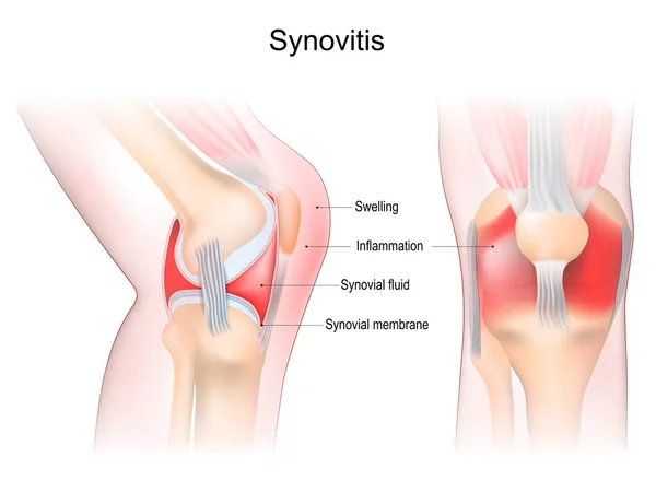
Synovitis Of A Knee. Close-up Of Joint With Inflammation Of The Synovial Membrane. Signs And Symptoms Of The Disease. Synovial Joint Anatomy. Frontal And Side View Of Human Knee Joint. Vector Illustration

Vector, 8.37MB, 5241 × 3930 eps
stock vector Osgood-schlatter disease (knee joint disease) illustration

Osgood-schlatter Disease (knee Joint Disease) Illustration

Vector, 1.85MB, 6583 × 4558 eps
stock vector Medical illustration of a knee with an inflamed prepatellar bursa (BURSITIS) viewed frontally and laterally, with annotations.

Medical Illustration Of A Knee With An Inflamed Prepatellar Bursa (BURSITIS) Viewed Frontally And Laterally, With Annotations.

Vector, 6.57MB, 7000 × 4118 eps
stock vector ACL injury or trauma as tear or sprain of anterior cruciate outline concept

ACL Injury Or Trauma As Tear Or Sprain Of Anterior Cruciate Outline Concept

Vector, 5.83MB, 4200 × 3818 eps
stock vector Chondromalacia patella knee breakdown compared with healthy outline diagram. Labeled educational kneecap tissue damage with cartilage problem and anatomical leg joint structure vector illustration.

Chondromalacia Patella Knee Breakdown Compared With Healthy Outline Diagram. Labeled Educational Kneecap Tissue Damage With Cartilage Problem And Anatomical Leg Joint Structure Vector Illustration.

Vector, 7.11MB, 4200 × 3990 eps
stock vector Osgood-schlatter disease (knee joint disease) illustration (Japanese)

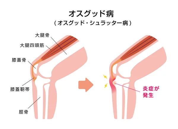
Osgood-schlatter Disease (knee Joint Disease) Illustration (Japanese)

Vector, 1.47MB, 6250 × 4529 eps
stock image Injury from workout concept : Asian man use hands hold on his kn

Injury From Workout Concept : Asian Man Use Hands Hold On His Kn

Image, 8.38MB, 5472 × 3648 jpg
stock image Injury from workout concept : Asian man use hands hold on his an

Injury From Workout Concept : Asian Man Use Hands Hold On His An

Image, 7.5MB, 5472 × 3648 jpg
stock image Red AIDS awareness ribbon holding by man on grey background. World Aids Day, Healthcare and medical concept.

Red AIDS Awareness Ribbon Holding By Man On Grey Background. World Aids Day, Healthcare And Medical Concept.

Image, 11.42MB, 6000 × 4000 jpg
stock vector Front view of the right knee in flexion

Front View Of The Right Knee In Flexion

Vector, 3.66MB, 3923 × 5000 eps
stock image Injury from workout concept : Asian man use hands hold on his an

Injury From Workout Concept : Asian Man Use Hands Hold On His An

Image, 7.38MB, 5472 × 3648 jpg
stock image Injury from workout concept : Asian man use hands hold on his an

Injury From Workout Concept : Asian Man Use Hands Hold On His An

Image, 8.31MB, 5158 × 3439 jpg
stock image Injury from workout concept : Asian man use hands hold on his an

Injury From Workout Concept : Asian Man Use Hands Hold On His An

Image, 8.48MB, 5472 × 3648 jpg
stock image Man with blue runing shorts use hands hold on his knee after running on road in morning time with copy space for text or design

Man With Blue Runing Shorts Use Hands Hold On His Knee After Running On Road In Morning Time With Copy Space For Text Or Design

Image, 12.33MB, 6240 × 4160 jpg
stock vector Osgood-Schlatter disease. Healthy Joint and Apophysitis of the tibial tubercle. Vector illustration. Poster for medical use

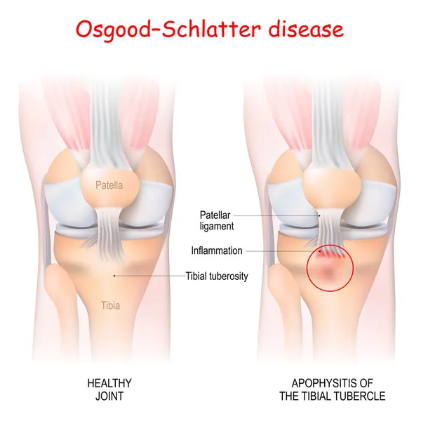
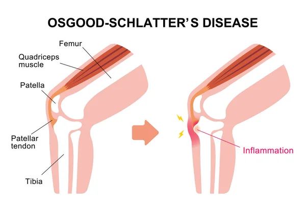
Osgood-Schlatter Disease. Healthy Joint And Apophysitis Of The Tibial Tubercle. Vector Illustration. Poster For Medical Use

Vector, 3.08MB, 4444 × 4445 eps
stock vector Knee joint section illustration

Knee Joint Section Illustration

Vector, 1.44MB, 4817 × 4558 eps
stock image Injury from workout concept : Asian man use hands hold on his kn

Injury From Workout Concept : Asian Man Use Hands Hold On His Kn

Image, 9.62MB, 5472 × 3648 jpg
stock image Injury from workout concept : Asian man use hands hold on his an

Injury From Workout Concept : Asian Man Use Hands Hold On His An

Image, 8.3MB, 5175 × 3450 jpg
stock image Wrapping knee injury

Wrapping Knee Injury

Image, 6.86MB, 4288 × 2848 jpg
stock vector Knee joint section illustration

Knee Joint Section Illustration

Vector, 1.06MB, 7225 × 6838 eps
stock image Knee joint

Knee Joint

Image, 3.58MB, 5400 × 4096 jpg
stock vector Structure of the human knee joint with the name and description of all sites. Lateral view. Medical science anatomy poster. Vector illustration isolated on white background.

Structure Of The Human Knee Joint With The Name And Description Of All Sites. Lateral View. Medical Science Anatomy Poster. Vector Illustration Isolated On White Background.

Vector, 6.98MB, 8334 × 8334 eps
stock image Knee joint anatomy

Knee Joint Anatomy

Image, 4.1MB, 5400 × 4096 jpg
stock image Knee joint scheme

Knee Joint Scheme

Image, 4.1MB, 5400 × 4096 jpg
stock image Sportsman suffering for ankle pain during training session

Sportsman Suffering For Ankle Pain During Training Session

Image, 14.56MB, 6000 × 4000 jpg
stock vector anatomy of the front of the right knee in flexion

Anatomy Of The Front Of The Right Knee In Flexion

Vector, 0MB, 4304 × 4616 zip
stock image Orthopedic diagnosing painful knee

Orthopedic Diagnosing Painful Knee

Image, 8.41MB, 5760 × 3840 jpg
stock vector Rectus femoris muscle as one of quadriceps muscular group outline diagram

Rectus Femoris Muscle As One Of Quadriceps Muscular Group Outline Diagram

Vector, 10.13MB, 4000 × 4571 eps
stock vector Anatomy. Structure knee joint vector

Anatomy. Structure Knee Joint Vector

Vector, 0.75MB, 5333 × 4000 eps
Previous << Page 2 >> Next