
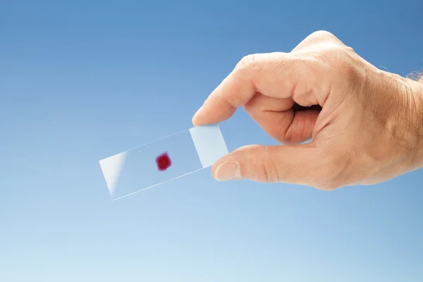
Stock image Pathology Slides

Photomicrograph Of Basal Cell Carcinoma, Displaying Malignant Basal Cells Typical Of The Most Common Skin Cancer.
Image, 21.12MB, 6345 × 4231 jpg

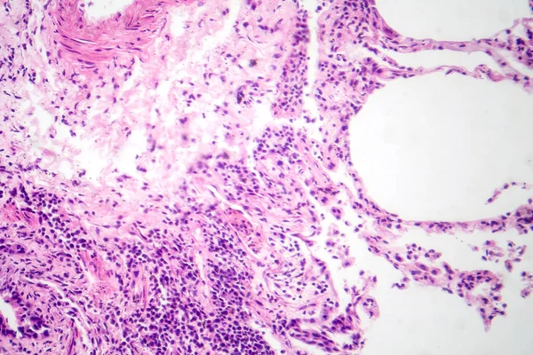
Photomicrograph Of Interstitial Pneumonia, Showing Inflammation And Fibrosis In The Lung's Interstitial Tissue.
Image, 26.23MB, 7124 × 4749 jpg
Photomicrograph Of Interstitial Pneumonia, Showing Inflammation And Fibrosis In The Lung's Interstitial Tissue.
Image, 20.38MB, 7124 × 4749 jpg
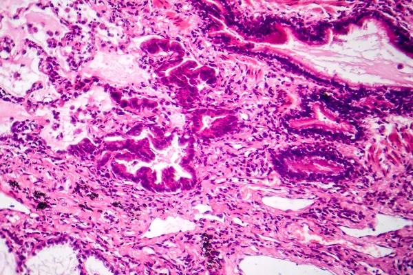
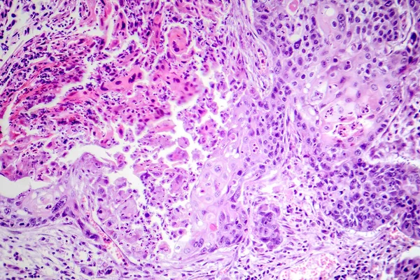
Photomicrograph Of Lung Adenocarcinoma, Displaying Malignant Glandular Cells Indicative Of The Most Common Type Of Lung Cancer.
Image, 26.24MB, 7161 × 4775 jpg
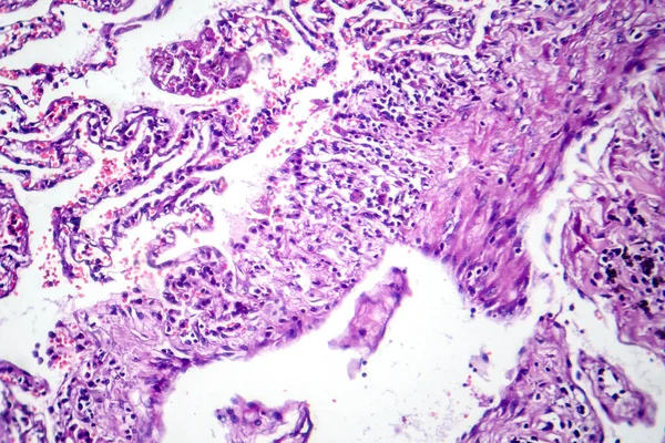
Photomicrograph Of Smoker's Lung, Revealing Characteristic Changes And Damage Associated With Long-term Smoking Habits.
Image, 20.02MB, 7173 × 4782 jpg


Laboratory Assistant Works On A Rotary Microtome Section And Making Microscope Slides
Image, 9.76MB, 6046 × 3969 jpg

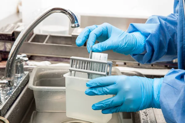
Young Female Scientist Preparing Slides With Paraffin-embedded Sections For Pathological Analysis .
Image, 8.88MB, 5105 × 3403 jpg
Scientist Preparing Slides With Paraffin Embedded Tissue Samples For Immunohistochemistry Assay In The Laboratory.
Image, 7.39MB, 5616 × 3744 jpg

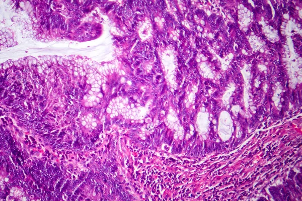
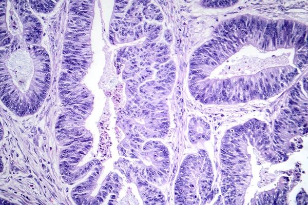
Photomicrograph Of Colon Adenocarcinoma, Illustrating Malignant Glandular Cells Characteristic Of Colon Cancer.
Image, 27.78MB, 7124 × 4749 jpg

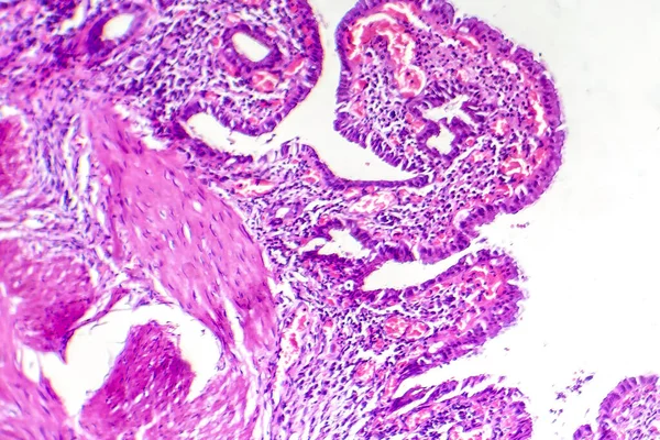
Photomicrograph Of Nasal Polyps, Displaying Abnormal Tissue Growth In The Nasal Passages Often Causing Congestion And Discomfort.
Image, 16.6MB, 6150 × 4100 jpg


Photomicrograph Of Lung Cancer Tissue, Revealing Malignant Cells And The Abnormal Growth Characteristic Of Lung Malignancy.
Image, 22.68MB, 7008 × 4673 jpg
Photomicrograph Of Squamous Cell Carcinoma Of The Lung, Showing Malignant Squamous Cells In Lung Tissue.
Image, 29.5MB, 7208 × 4805 jpg
Photomicrograph Of Esophageal Squamous Cell Carcinoma, Showing Malignant Squamous Cells Characteristic Of Esophageal Cancer.
Image, 28.7MB, 7134 × 4757 jpg

Photomicrograph Of Nasal Polyps, Displaying Abnormal Tissue Growth In The Nasal Passages Often Causing Congestion And Discomfort.
Image, 18.16MB, 6363 × 4242 jpg

Photomicrograph Of Chronic Cholecystitis, Showcasing Inflammation And Structural Changes In The Gallbladder Wall.
Image, 9.89MB, 4444 × 2963 jpg
Page 1 >> Next