
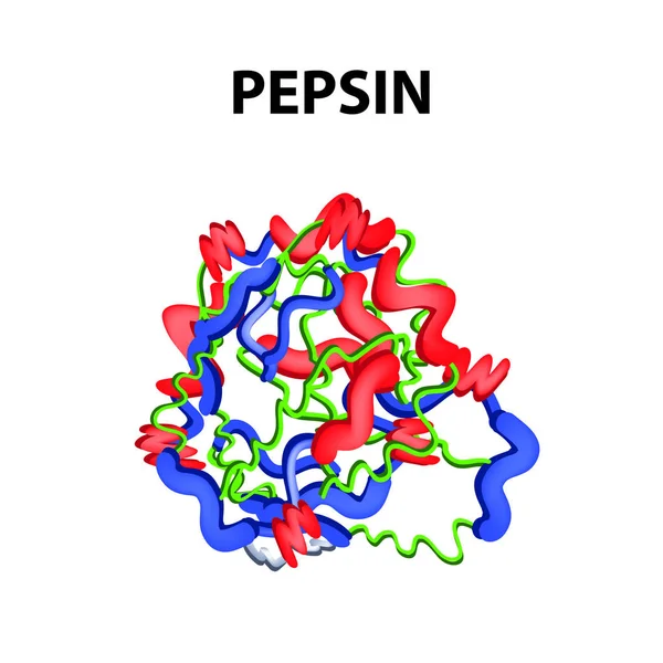
Stock image Pepsin
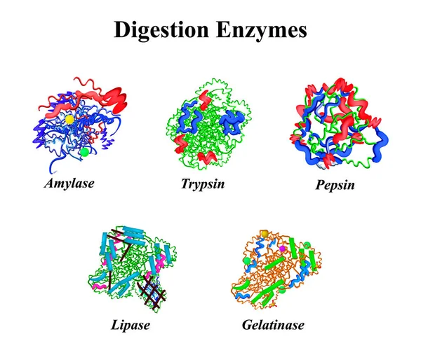
Digestion Enzymes Set. Chemical Molecular Formula. Amylase, Trypsin, Gelatinase, Pepsin, Lipase. Infographics. Vector Illustration On Isolated Background.
Vector, 7.37MB, 5000 × 4231 eps

Protein Digestion. Protein Metabolism. Digestion In The Gastrointestinal Tract. Infographics. Vector Illustration On Isolated Background.
Vector, 4.19MB, 5000 × 5000 eps

Several Vials With Soluble Protease Proteins For Activation Of The Coronavirus Severe Acute Respiratory Syndrome (SARS) Trypsin-like Protein In Human Respiratory Tract In A Hospital, Spain
Image, 7.23MB, 5184 × 3600 jpg
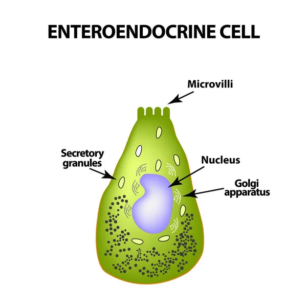
Enteroendocrine Cell. Cell Of The Intestines. Vector Illustration On Isolated Background
Vector, 0.97MB, 5000 × 5000 eps

Monoclonal Antibody Therapy In Helicobacter Pylori - Closeup View 3d Illustration
Image, 8.24MB, 10000 × 6600 jpg

CAR T Cell Therapy In Stomach Cancer (gastric Cancer) - Isometric View 3d Illustration
Image, 11.53MB, 10000 × 6600 jpg

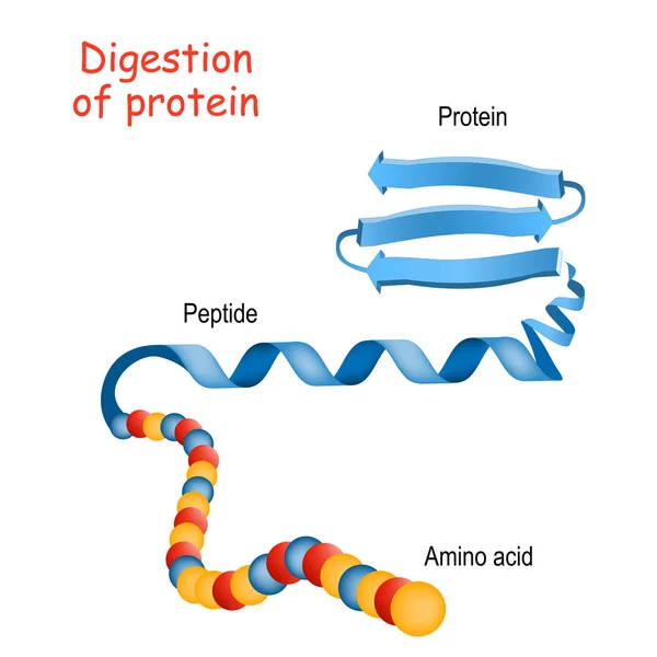
Digestion Of Protein. Breaking The Complex Molecule First Into Peptides Then Into Individual Amino Acids. The Pepsins Are Enzymes Secreted By The Stomach That Breaks Down Proteins. Vector Illustration
Vector, 5.26MB, 6000 × 3321 eps
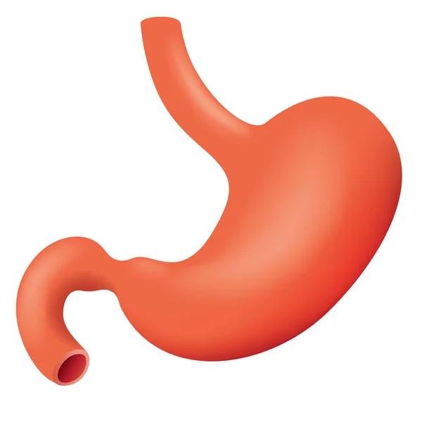
Illustration Representing Human Stomach Stomach Of The Digestive System, Anatomy. Ideal For Medical And Educational Materials
Vector, 3.22MB, 5000 × 5000 eps
Early Stage Gastric Cancer Tumor On The Stomach Wall Isometric View 3d Illustration
Image, 5.85MB, 10000 × 6600 jpg

Protein Digestion. Enzymes Proteases Are Digestion Breaks The Protein Into Single Amino Acids, Which Are Absorbed Into The Blood. Vector Illustration
Vector, 1.47MB, 4444 × 4444 eps

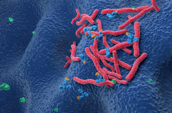
Stomach Ulcer And Closeup View Of Bacteria Helicobacter Pylori, Associated With Ulcer Formation, 3D Illustration
Image, 10.85MB, 7200 × 4050 jpg
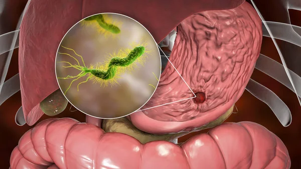
Stomach Ulcer And Closeup View Of Bacteria Helicobacter Pylori, Associated With Ulcer Formation, 3D Illustration
Image, 13.09MB, 7200 × 4050 jpg
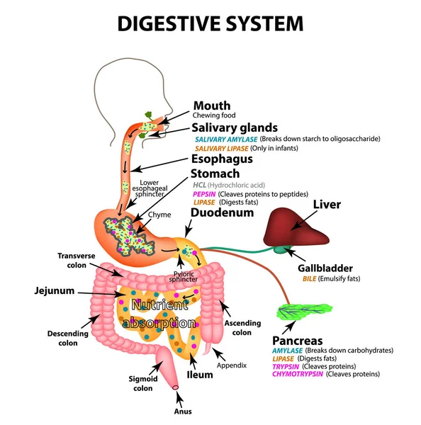
The Human Digestive System. Anatomical Structure. Digestion Of Carbohydrates, Fats And Proteins. Enzymes Of The Gastrointestinal Tract, Pancreas, Liver, Gallbladder. Metabolism. Infographics. Vector.
Vector, 3.01MB, 5000 × 5050 eps


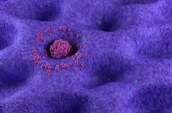
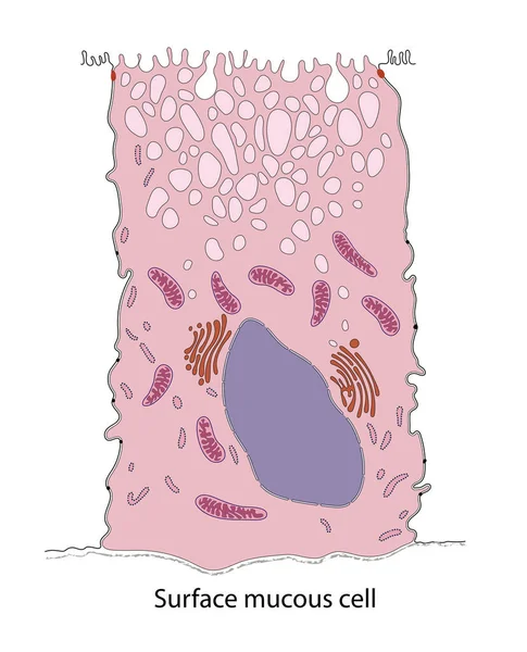
Gastric Glands And Cell Types. Sectional View Of Stomach Mucosa. Stomach Anatomy
Image, 10.89MB, 8858 × 7806 jpg

Protein Digestion. Protein Metabolism. Digestion In The Gastrointestinal Tract. Infographics. Vector Illustration On Isolated Background.
Vector, 4.18MB, 5000 × 5000 eps
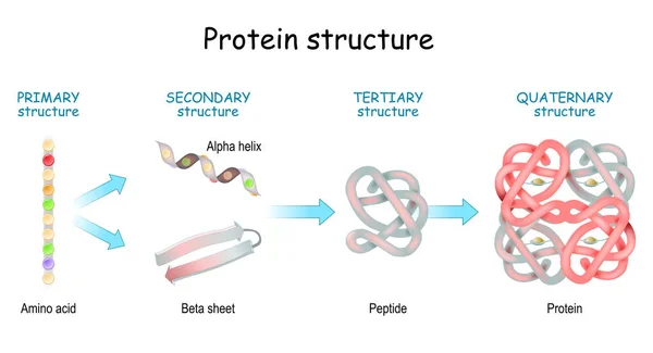
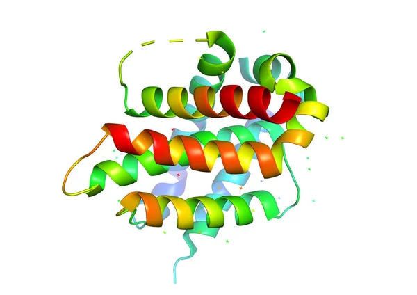
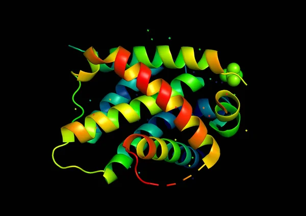
Protein Structure Levels: Primary, Secondary, Tertiary, And Quaternary. From Amino Acid To Alpha Helix, Beta Sheet, Peptide, And Protein Molecule. Concept. Vector Illustration.
Vector, 13.21MB, 5555 × 3018 eps
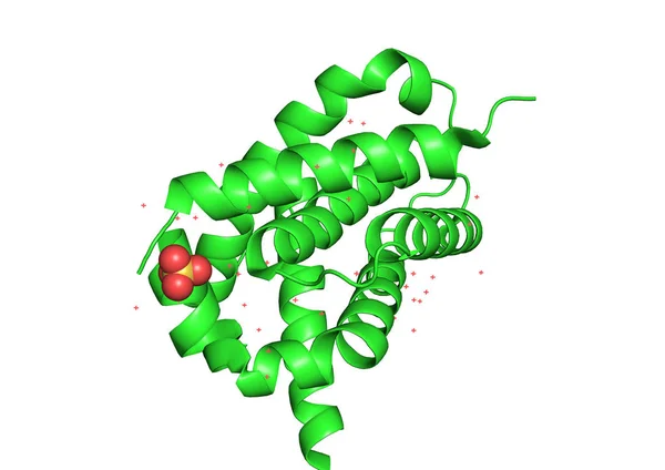

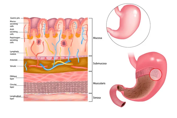
Diagram Of The Alkaline Mucous Layer In The Stomach. Diagram Of The Histological Cross-section Of The Stomach Layers Of The Stomach.
Vector, 5.66MB, 5001 × 3334 eps

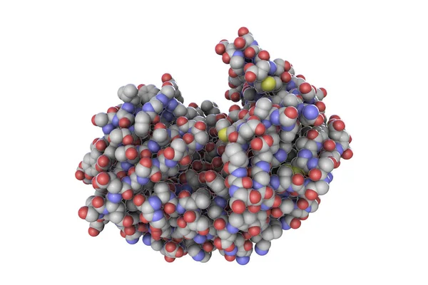
Biochemical Structure Of Amino Acids, Peptides And Proteins Molecular Model, 3d Rendering
Image, 2.89MB, 6000 × 4000 jpg
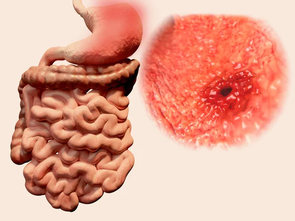
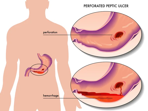
Occurrence Of Gastric Ulcera In The Gastrointestinal Tract. Illustration
Image, 1.5MB, 4000 × 3000 jpg

Stomach Ulcer And Closeup View Of Bacteria Helicobacter Pylori, Associated With Ulcer Formation, 3D Illustration
Image, 6.55MB, 7200 × 4050 jpg
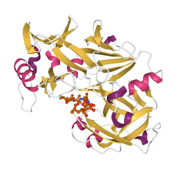
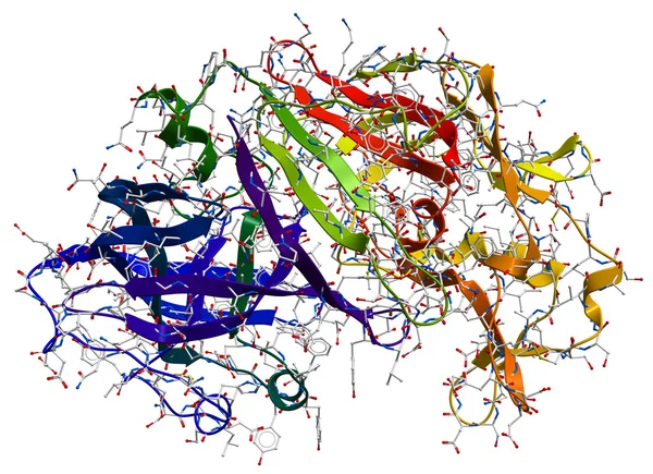
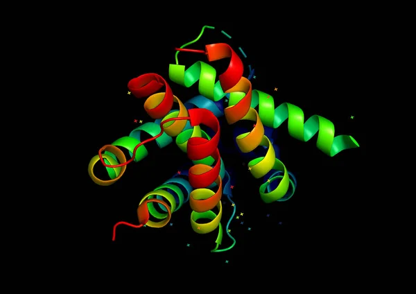
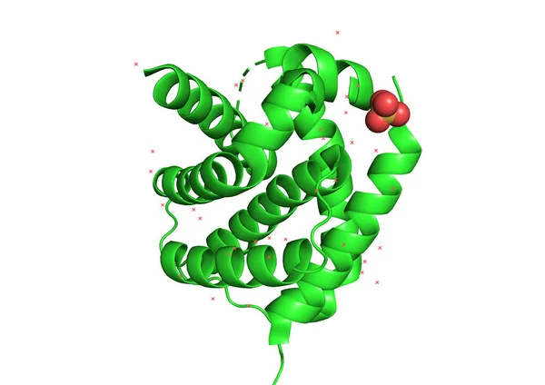
Structure Of Human Pepsin Complexed With Inhibitor Pepstatin, 3D Cartoon Model Isolated, White Background
Image, 2.07MB, 4096 × 4096 jpg
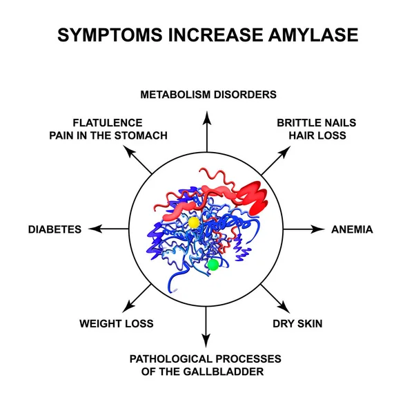
Symptoms Of Increased Amylase. The Enzyme Amylase. Infographics. Vector Illustration On Isolated Background.
Vector, 2.14MB, 5000 × 4830 eps
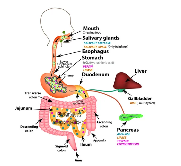
The Human Digestive System. Anatomical Structure. Digestion Of Carbohydrates, Fats And Proteins. Enzymes Of The Gastrointestinal Tract, Pancreas, Liver, Gallbladder. Metabolism. Infographics. Vector.
Vector, 2.89MB, 5000 × 4572 eps
Molecules, Bold Line Icons. The Illustrations Are A Vector, Editable Stroke, 48x48 Pixel Perfect Files. Crafted With Precision And Eye For Quality.
Vector, 0.48MB, 5613 × 5000 eps

Monoclonal Antibody Treatment In Stomach Cancer (gastric) - Closeup View 3d Illustration
Image, 6.14MB, 10000 × 6600 jpg


Protein Structure. Amino Acids, Alpha Helix, Polypeptide Chains, And Complex Of Protein Molecule. Protein Is A Polymer (polypeptide) That Formed From Sequences Of Amino Acids. Isometric Flat Vector Illustration.
Vector, 6.23MB, 5000 × 3500 eps

Stomach Ulcer And Closeup View Of Bacteria Helicobacter Pylori, Associated With Ulcer Formation, 3D Illustration
Image, 13.96MB, 7200 × 4050 jpg


Stomach Ulcer And Closeup View Of Bacteria Helicobacter Pylori, Associated With Ulcer Formation, 3D Illustration
Image, 14.74MB, 7200 × 4050 jpg
Monoclonal Antibody Therapy In Helicobacter Pylori - Isometric View 3d Illustration
Image, 10.55MB, 10000 × 6600 jpg

CAR T Cell Therapy In Stomach Cancer (gastric Cancer) - Closeup View 3d Illustration
Image, 9.31MB, 10000 × 6600 jpg

Close-up Of Betaine HCl+ Tablets In The Jar. Dietary Concept. Dietary Supplement
Image, 15.8MB, 5536 × 3691 jpg
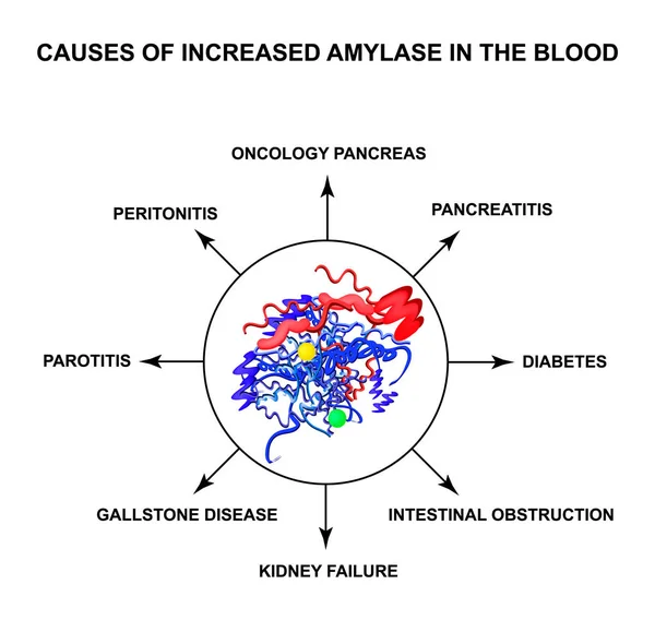
Causes Of Increased Amylase In The Blood. The Enzyme Amylase. Infographics. Vector Illustration On Isolated Background
Vector, 2.12MB, 5000 × 4830 eps
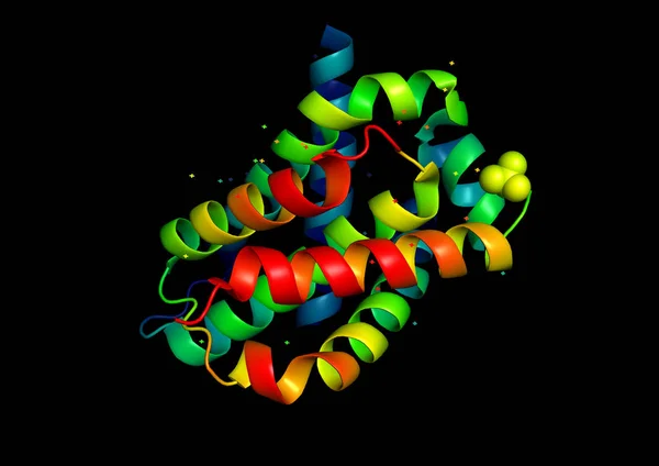
Pepsin Is A Molecular Chemical Formula. Enzyme Of The Stomach. Infographics. Vector Illustration On An Isolated Background.
Vector, 1.47MB, 5000 × 5000 eps
Page 1 >> Next