
Stock image Peptidoglykan

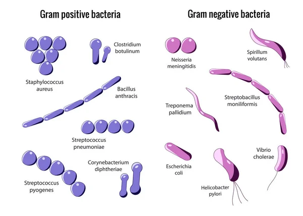
Types Of Bacterial Cell Wall. Gram-negative Bacteria And Gram-negative Bacteria. Comparison, Structure, And Composition. Vector Illustration
Vector, 5.16MB, 5000 × 3688 eps
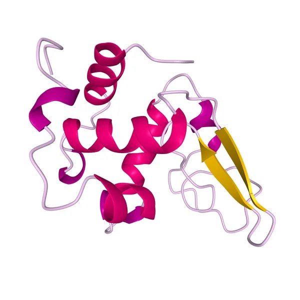
Native Human Lysozyme, 3D Cartoon Model Of The Tertiary Structure With The Elements Of The Secondary Structure Colored, White Background
Image, 1.1MB, 4096 × 4096 jpg
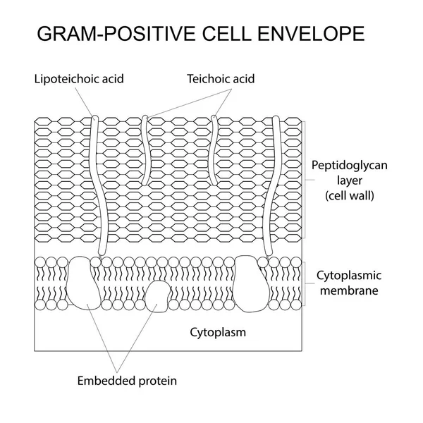
Vector Outlined Illustration Of The Gram-positive Cell Wall. Black And White.
Vector, 5.82MB, 8000 × 8000 eps
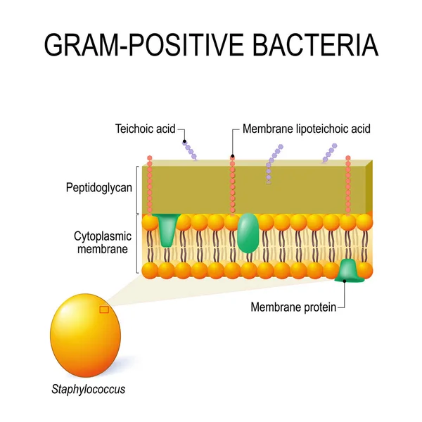
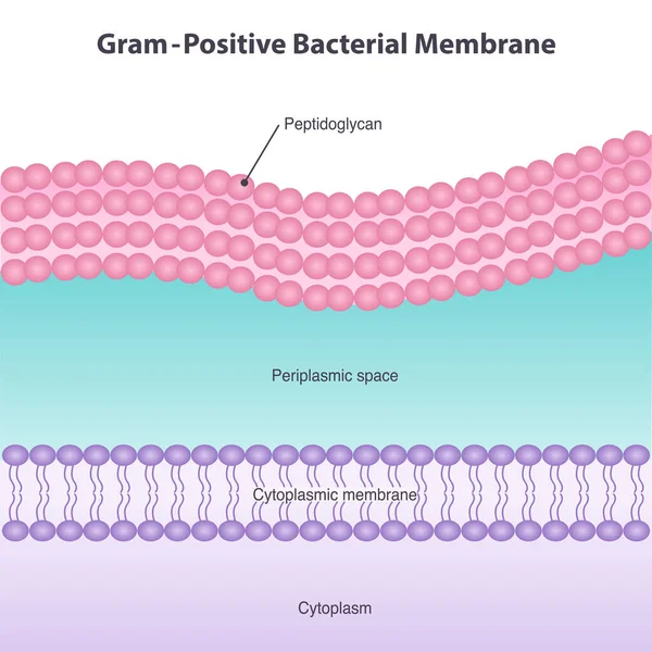
Cell Wall Structure Of Gram-positive Bacteria For Example Staphylococcus. Vector Diagram For Educational, Medical, Biological And Science Use
Vector, 1.74MB, 4095 × 4095 eps
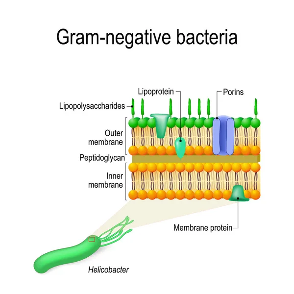
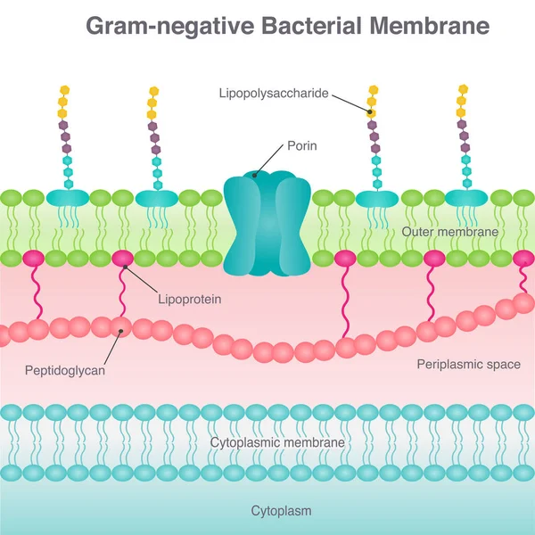
Cell Wall Structure Of Gram-negative Bacteria For Example Helicobacter. Vector Diagram For Educational, Medical, Biological And Science Use
Vector, 2.88MB, 4152 × 4152 eps


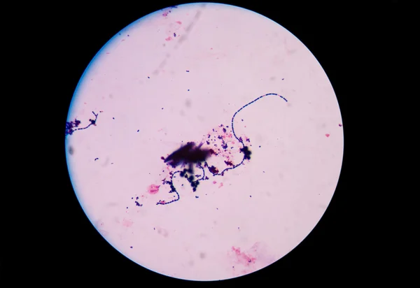
This Differential Staining Procedure Separates Most Bacteria Into Two Groups On The Basis Of Cell Wall Composition, Bacteria Gram Posotive And Gram Negative
Vector, 11.84MB, 2859 × 6420 eps
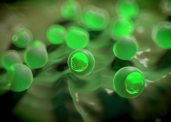
3d Rendering Of Gram Positive Bacteria Have A Thick Peptidoglycan Layer And No Outer Lipid Membrane
Image, 0.45MB, 3840 × 2160 jpg


Gram Positive Versus Negative Cell Wall Structure Differences Outline Diagram
Vector, 7.19MB, 5200 × 3600 eps


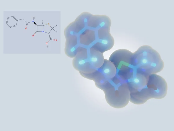
Natural Penicillins Antibiotic Drug Molecule. Benzylpenicillin , Phenoxymethylpenicillin, Almecillin. Structural Chemical Formula
Vector, 0.61MB, 5000 × 5000 eps

Diagram Showing Gram Staining Microbiology Lab Technique Steps - Microbiology Laboratory Using Crystal Violet And Safranin
Vector, 8.54MB, 6001 × 4000 eps

3d Rendering Of Gram-negative Bacteria, Have A Thin Peptidoglycan Cell Wall, Which Is Surrounded By An Outer Membrane Containing Lipopolysaccharide
Image, 0.4MB, 3840 × 2160 jpg

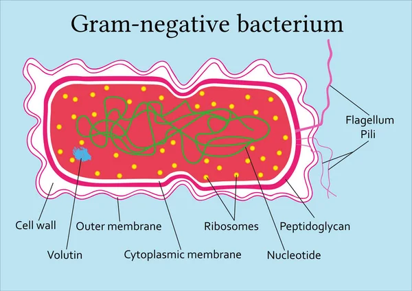
Scientific Designing Of Gram Positive And Gram Negative Bacteria, Illustration.
Image, 0.92MB, 5410 × 3240 jpg

3d Rendering Of Gram-negative Bacteria, Have A Thin Peptidoglycan Cell Wall, Which Is Surrounded By An Outer Membrane Containing Lipopolysaccharide
Image, 0.45MB, 3840 × 2160 jpg

General Formula Of Penicillin PCN Molecule. It Is A Group Of Antibiotics. Structural Chemical Formula And Molecule Model
Vector, 0.37MB, 5000 × 3333 eps

3d Rendering Of Gram Positive Bacteria Have A Thick Peptidoglycan Layer And No Outer Lipid Membrane
Image, 0.54MB, 3840 × 2160 jpg
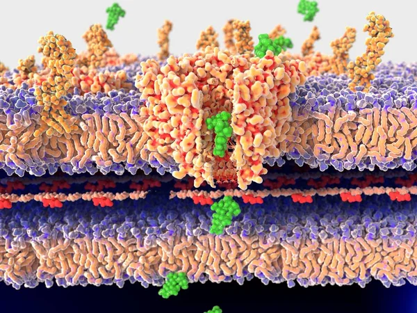
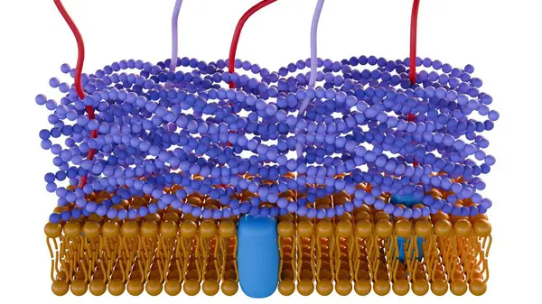
Bacterial Wall Being Passed By Streptomycin Molecules. Structure Of A Bacterial Wall Being Passed By Streptomycin Molecules Through The Channel Protein Porin. 3D Rendering. Illustration
Image, 7.8MB, 8000 × 6000 jpg

3d Rendering Of Gram Positive Bacteria Have A Thick Peptidoglycan Layer And No Outer Lipid Membrane
Image, 0.23MB, 3840 × 2160 jpg

Page 1 >> Next