
Stock image Phagocytes page 2
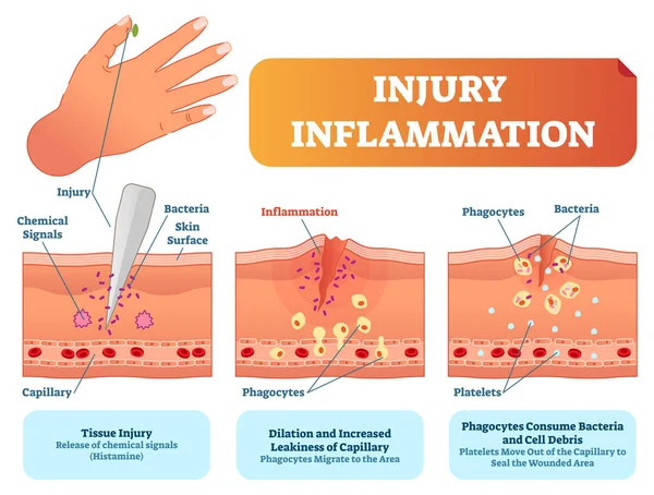
Injury Inflammation Biological Human Body Response Vector Illustration Scheme. Skin Surface Injury Cross Section Poster With Capillary, Phagocytes And Platelets.
Vector, 6.48MB, 5223 × 3957 eps

Illustration Showing The Immune Response To Tattoo Ink (black). During Tattooing, Ink Is Injected Into The Second Skin Layer, Known As The Dermis, And Triggers An Immune Response.
Image, 5.37MB, 7200 × 4050 jpg

Illustration Showing The Immune Response To Tattoo Ink (black). During Tattooing, Ink Is Injected Into The Second Skin Layer, Known As The Dermis, And Triggers An Immune Response. Certain Types Of White Blood Cell Such As Dendritic Cells (blue) And T
Image, 8.16MB, 7200 × 4050 jpg


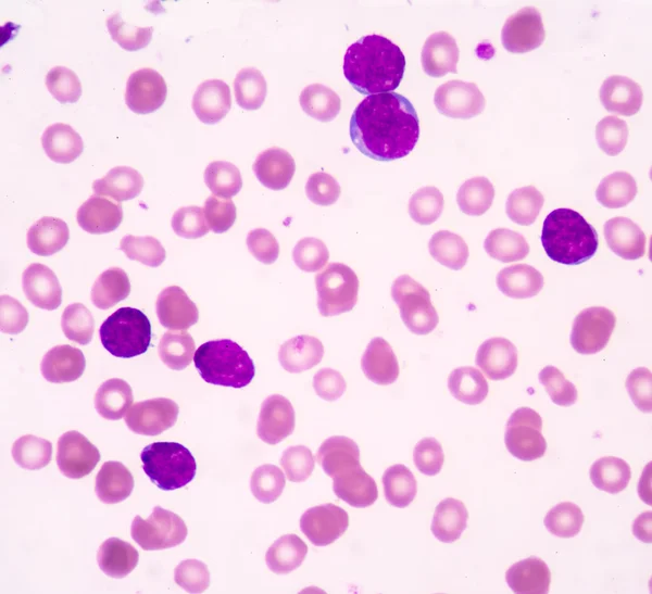

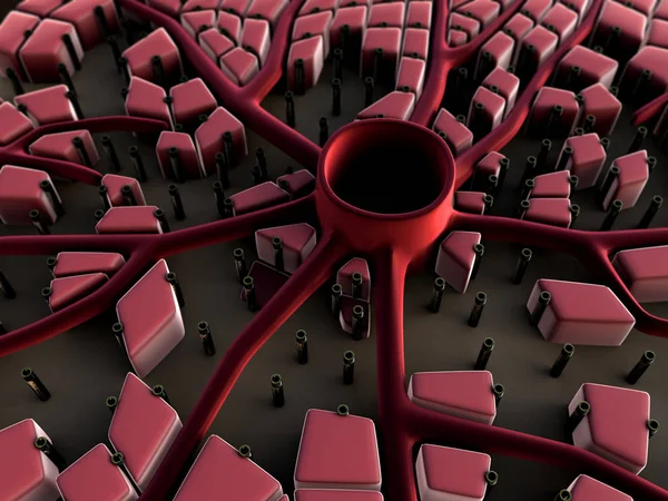
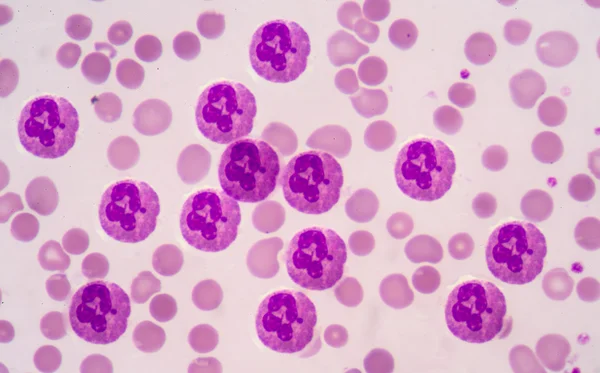


Illustration Showing The Skin Tissue Of An Arm Being Tattooed By A Tattoo Gun (top). Tattoo Guns Work By Piercing The Outer Skin Layer Known As The Epidermis (pink) With Multiple Needles And Depositing Ink Beneath It
Image, 7.48MB, 7200 × 4050 jpg


Coronavirus (COVID-19) Test. Doctor Taking Or DNA Test By Nasal (nose) Swab Probe, Patient Being Tested, Lab Analysis. Cartoon Flat Vector Illustration.
Vector, 0.62MB, 9954 × 6677 eps

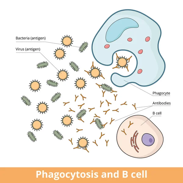
Cooperation Between B Cell And Phagocyte During Immune Responce Caused By Antigens (bacteria Or Virus). B Cell Produces Antibodies That Weaken Antigens And Phagocyte Eliminates Them.
Vector, 7.64MB, 6250 × 6250 eps

Illustration Showing The Immune Response To Tattoo Ink (black). During Tattooing, Ink Is Injected Into The Second Skin Layer, Known As The Dermis, And Triggers An Immune Response.
Image, 5.49MB, 7200 × 4050 jpg

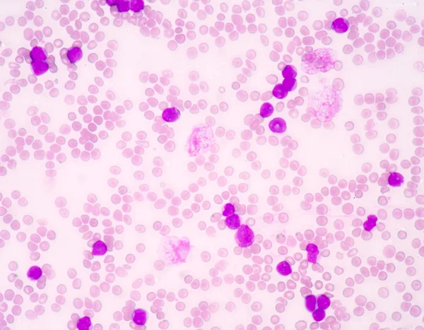

Illustration Showing A Type Of White Blood Cell Known As A Macrophage (purple) Containing Tattoo Ink (black). During Tattooing, Ink Is Injected Into The Second Skin Layer, Known As The Dermis, And Triggers An Immune Response.
Image, 6.29MB, 7200 × 4050 jpg

Illustration Showing The Skin Tissue Of An Arm Being Tattooed By A Tattoo Gun (top). Tattoo Guns Work By Piercing The Outer Skin Layer Known As The Epidermis (horizontal, Centre) With Multiple Needles And Depositing Ink Beneath It
Image, 7.98MB, 7200 × 4050 jpg
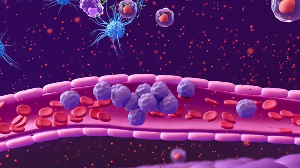
Illustration Showing White Blood Cells (purple) In A Blood Vessel (pink Tube) Moving Towards The Site Of A Bacterial Infection (green, Top).
Image, 6.55MB, 7200 × 4050 jpg


Illustration Showing White Blood Cells (purple) In A Blood Vessel (pink Tube) Moving Towards The Site Of A Bacterial Infection (green, Top). Certain Types Of White Blood Cell, Including Dendritic Cells (blue) And T Helper Cells
Image, 6.57MB, 7200 × 4050 jpg
Previous << Page 2 >> Next