
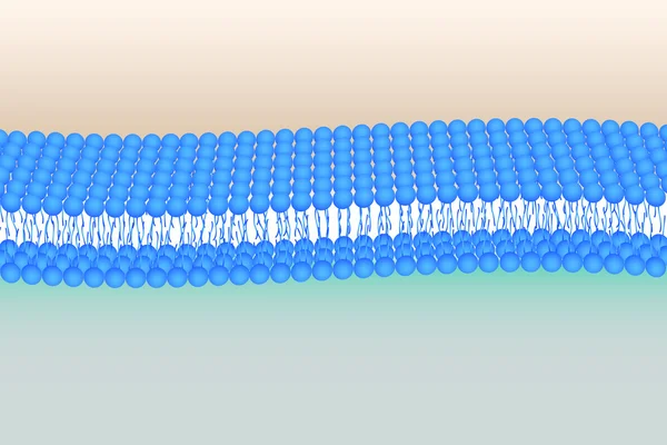
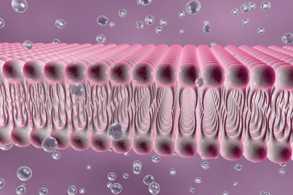
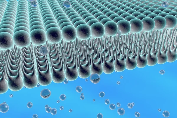
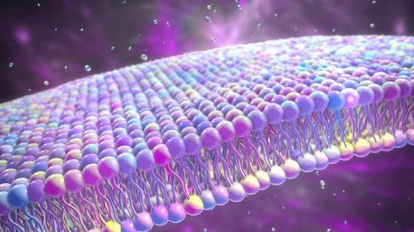
Stock image Phospholipid Bilayer
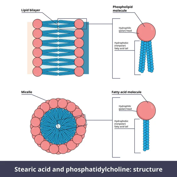
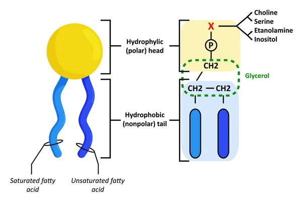
Structure Of Two Lipids. Stearic Acid (fatty Acid) And Phosphatidylcholine (phospholipid) Are Composed Of Chemical Groups That Form Polar Heads (hydrophilic) And Nonpolar Tails" (hydrophobic).
Vector, 10.84MB, 5208 × 5208 eps

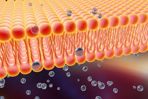
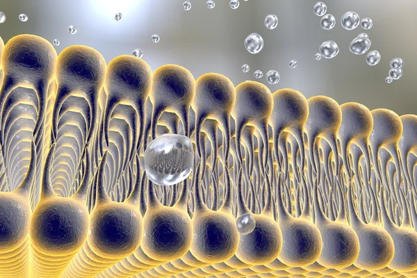
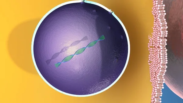
Molecule Passing Through A Protein Channel And Virus Passing Through A Lipid Bilayer Cell Membrane. Cell Transport, 3D Animation.
Image, 10.61MB, 6000 × 4500 jpg
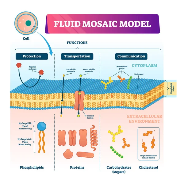
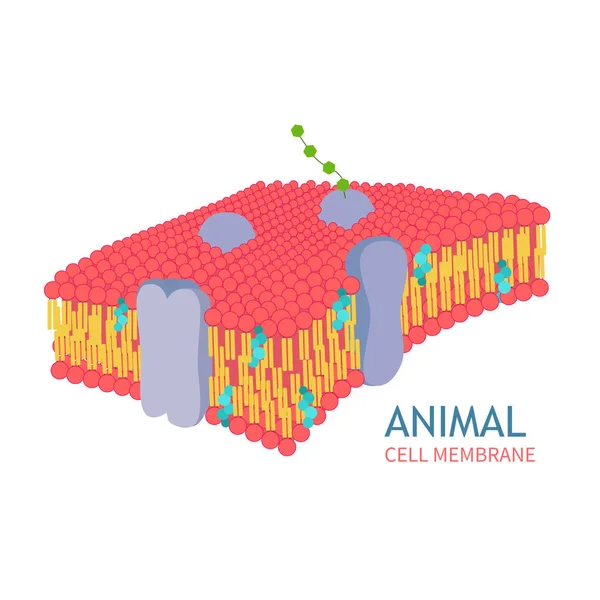
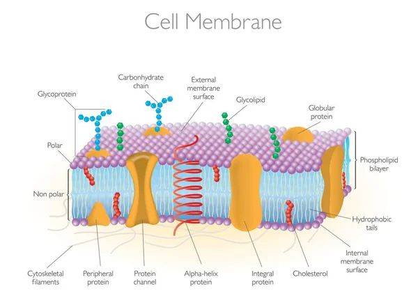
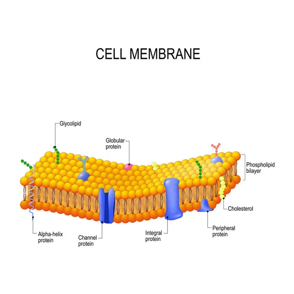
Fluid Mosaic Model Vector Illustration. Cell Membrane Structure Infographic
Vector, 9.21MB, 4000 × 4000 eps
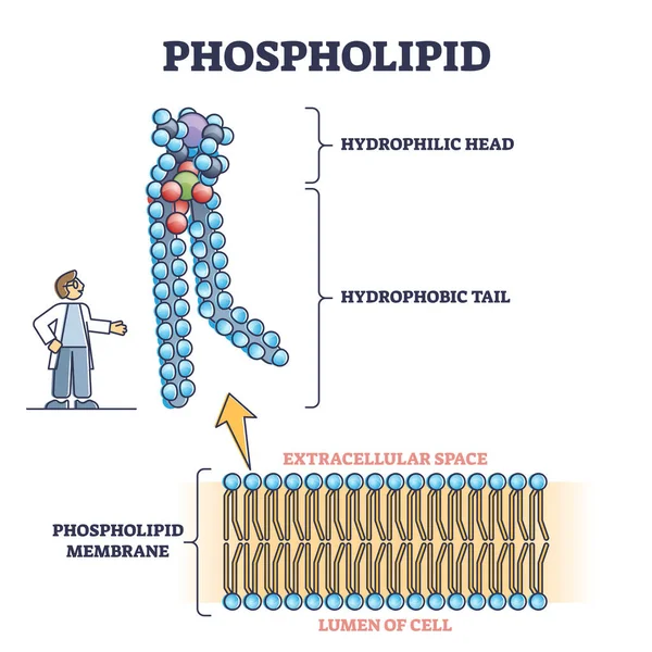
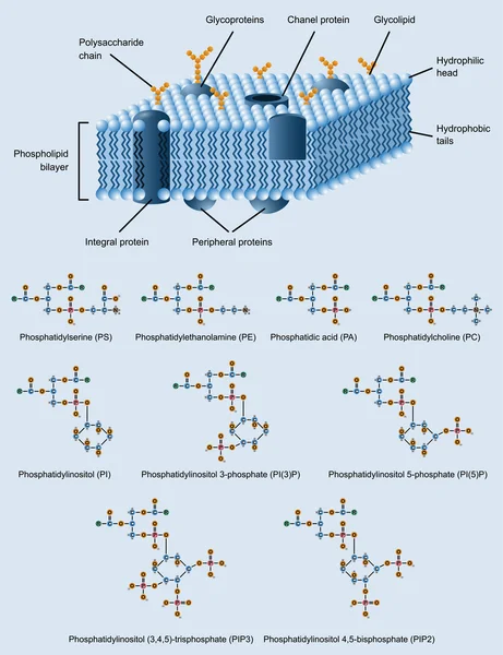
Phospholipid Or Phosphatides Lipids Microscopical Structure Outline Diagram
Vector, 6.22MB, 4000 × 4000 eps
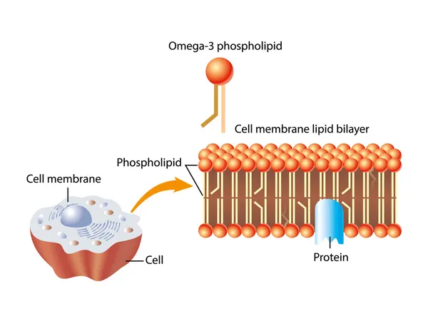
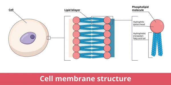
Cell Membrane Structure, That Is Represented By Lipid Bilayer And Its Phosphatidylcholine (a Phospholipid), That Is Composed Of Polar Hydrophilic Head And Nonpolar Hydrophobic Tail.
Vector, 8.5MB, 8333 × 4167 eps
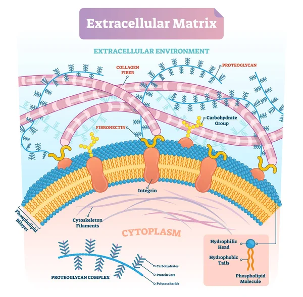
Extracellular Matrix Labeled Infographic Vector Illustration Scheme.
Vector, 9.16MB, 4100 × 4029 eps

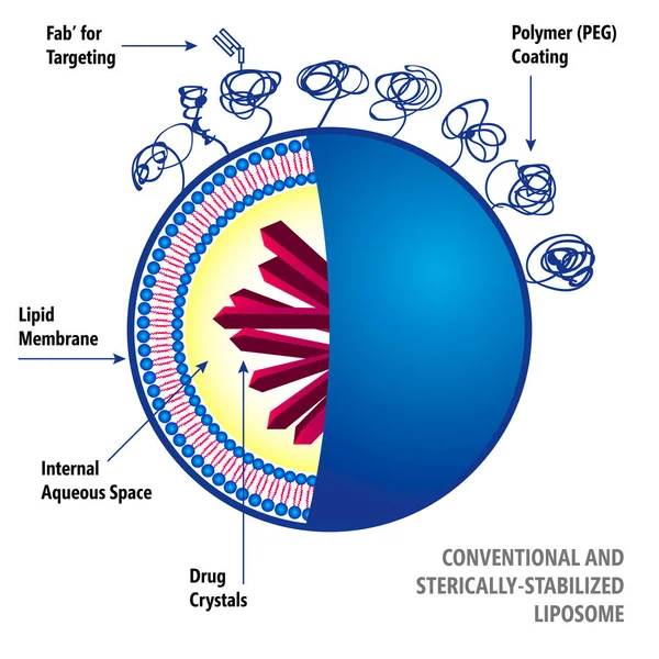


Lipid Bilayer-coated Mesoporous Silica Nanoparticles: Emerging Nanocarriers For Targeted Drug Delivery And 3D-rendered Nanomedicine Release
Image, 0.81MB, 3840 × 2160 jpg

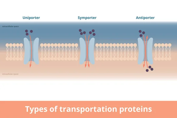
Types Of Cell Membrane Transportation Proteins. Visualization Of Uniporter (one Molecule, One Direction), Symporter (two Molecules, Same Directions), Antiporter (two Molecules, Different Directions).
Vector, 7.12MB, 6250 × 4167 eps
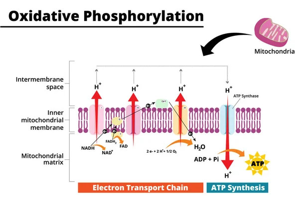
Oxidative Phosphorylation Process. Electron Transport Chain. The Final Step In Cellular Respiration. Vector Illustration. Didatic Illustration.
Vector, 0.73MB, 5000 × 3500 ai

Membrane Proteins. Integral, And Peripheral Membrane Proteins, Single-pass, And Multi-pass Transmembrane Helix, Lipid-anchored Protein. Vector Illustration For Biological, Science And Educational Use
Vector, 4.52MB, 3921 × 3921 eps

A 3D Rendering Of Magnetic Liposomes. These Microscopic Drug Carriers Are Embedded With Nanoparticles, Allowing Magnets To Guide Them To Specific Areas Of The Body.
Image, 0.6MB, 3840 × 2160 jpg

3D Rendering Of Lipid-nucleic Acid Complexes, Known As Lipoplexes, Encompassing Their Formation And Structural Features.
Image, 0.8MB, 3840 × 2160 jpg

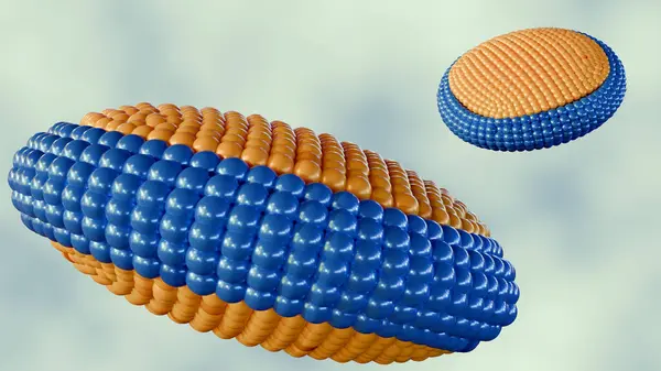
3d Rendering Of Bicelles, They Are Disc-shaped Lipid Assemblies Composed Of Two Different Types Of Lipids: One With A Longer Acyl Chain And The Other With A Shorter Acyl Chain
Image, 1.24MB, 3840 × 2160 jpg

Illustration Of Cross-sections Of Different Types Of Liposome, Categorised By Size And Number Of Phospholipid Bilayers. From Left To Right: Unilamellar (LUV), Oligolamellar (OLV), Multilamellar (MLV). Liposomes Are Artificial Vesicles.
Image, 6.59MB, 9460 × 3700 jpg


Transferosomes Structure Are Vesicular Carrier Systems That Enclosed By A Lipid Bilayer, Together With An Edge Activator 3d Rendering
Image, 0.53MB, 2400 × 2000 jpg
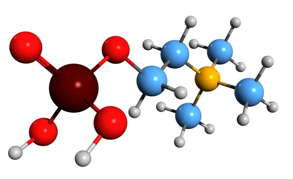
3D Image Of Phosphocholine Skeletal Formula - Molecular Chemical Structure Of Phosphatidylcholine Intermediate Isolated On White Background
Image, 4.39MB, 8070 × 4944 jpg
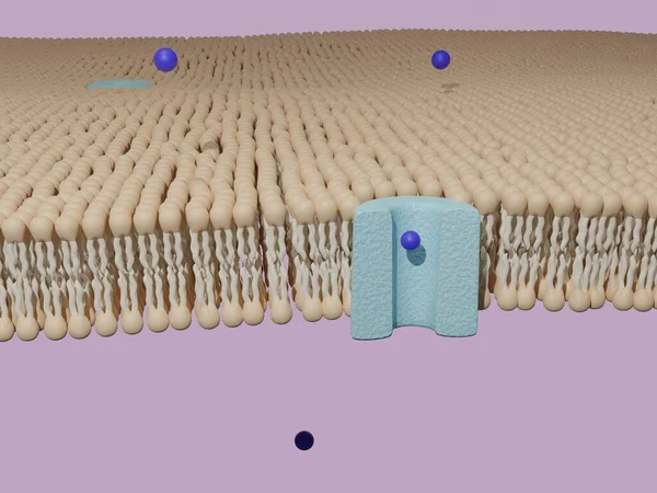
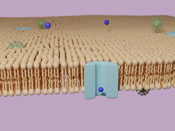
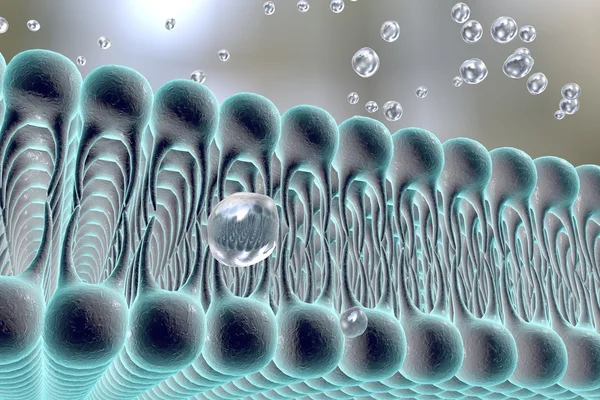
Molecule Passing Through A Protein Channel At A Lipid Bilayer Cell Membrane. Cell Transport, 3D Rendering.
Image, 9.64MB, 6000 × 4500 jpg

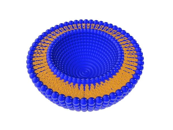
A 3D Rendering Featuring A Half-cut Liposome And Other Liposomes Dispersed On A White Background
Image, 0.6MB, 3840 × 2160 jpg
3d Rendering Of Bicelles, They Are Disc-shaped Lipid Assemblies Composed Of Two Different Types Of Lipids: One With A Longer Acyl Chain And The Other With A Shorter Acyl Chain
Image, 1.19MB, 3840 × 2160 jpg

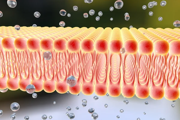
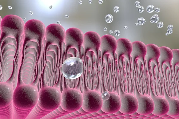
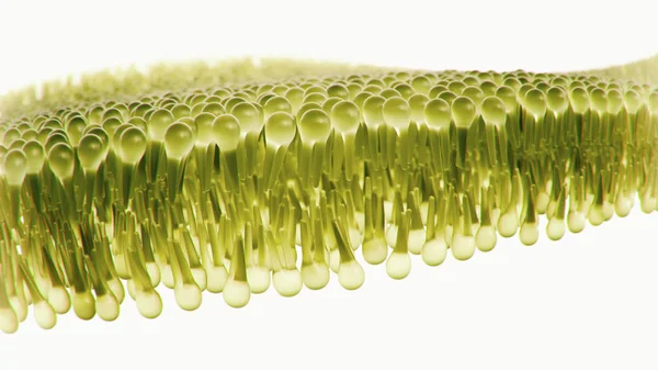
Phospholipid Membrane Structure Cross-section Showing Molecular Structure With Hydrophilic Head And Hydrophobic Tails.
Image, 5.56MB, 6000 × 3375 jpg

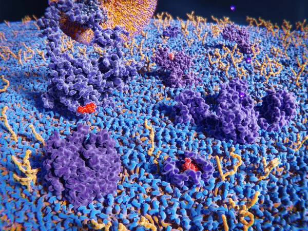
Membrane Proteins (violett), Glycolipids (yellow) And Several Ligands Of The Proteins
Image, 7.89MB, 8000 × 6000 jpg

Page 1 >> Next