photo free
Home Images Vectors Videos

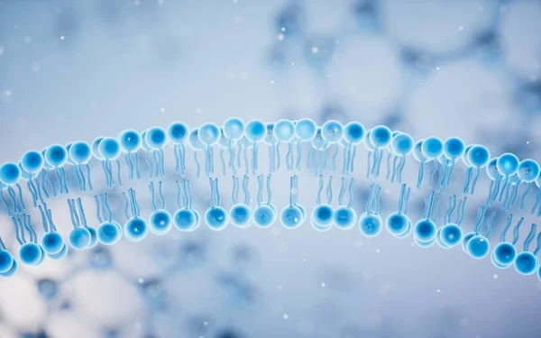
Stock image Phospholipids

stock vector Phospholipid or phosphatides lipids head and tail structure outline diagram

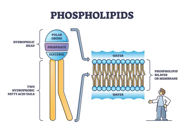
Phospholipid Or Phosphatides Lipids Head And Tail Structure Outline Diagram

Vector, 5.82MB, 5000 × 3472 eps
stock vector Structure of cell membrane

Structure Of Cell Membrane

Vector, 4.12MB, 6131 × 6131 eps
stock vector Plasma membrane

Plasma Membrane

Vector, 4.59MB, 5333 × 4000 eps
stock vector Omega 3 phospholipid

Omega 3 Phospholipid

Vector, 1.45MB, 5000 × 3750 eps
stock image 3d rendering of nanodisc consists of Phospholipids and A stabilizing belt that holds the phospholipids together.

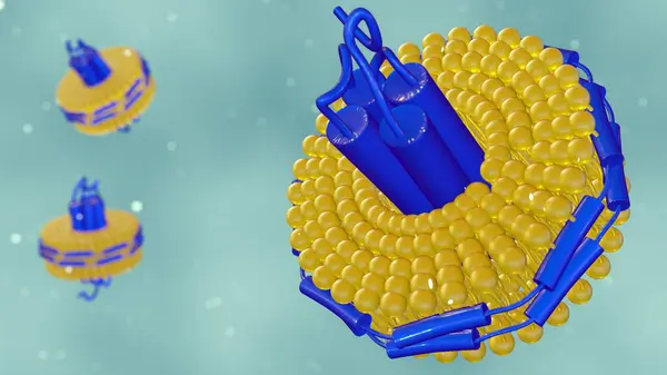
3d Rendering Of Nanodisc Consists Of Phospholipids And A Stabilizing Belt That Holds The Phospholipids Together.

Image, 0.5MB, 3840 × 2160 jpg
stock vector Lipids vector illustration. Triglycerides, waxes and steroids diagram.

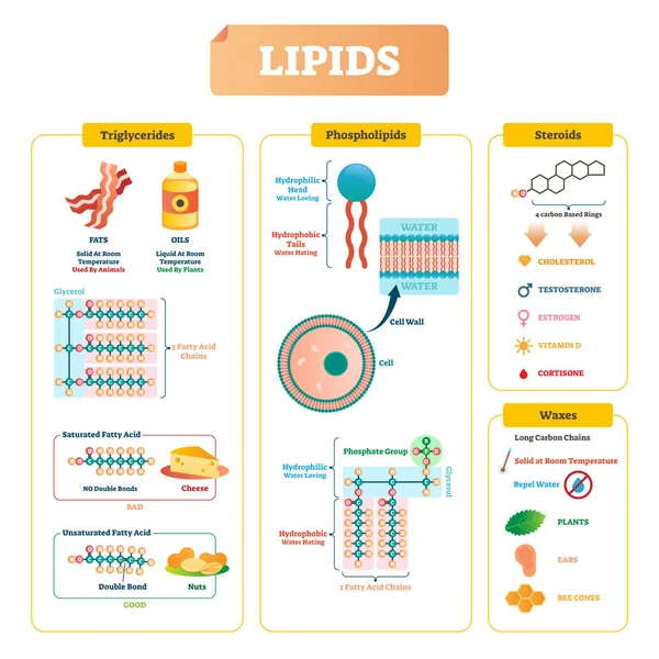
Lipids Vector Illustration. Triglycerides, Waxes And Steroids Diagram.

Vector, 7.45MB, 4000 × 4000 eps
stock image Cell membrane and biology, biological concept, 3d rendering. Computer digital drawing.

Cell Membrane And Biology, Biological Concept, 3d Rendering. Computer Digital Drawing.

Image, 8.51MB, 6000 × 4000 jpg
stock image Interleukin 4 (IL-4) bound to its receptor

Interleukin 4 (IL-4) Bound To Its Receptor

Image, 4.62MB, 8000 × 6000 jpg
stock image High density lipoprotein (HDL) color code: protein ApoA (green), phospholipids (orange with a blue cap), cholesterol (orange with a violet cap). Illustration

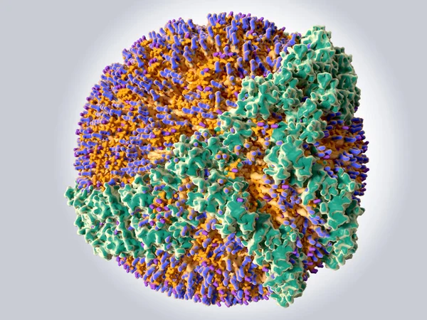
High Density Lipoprotein (HDL) Color Code: Protein ApoA (green), Phospholipids (orange With A Blue Cap), Cholesterol (orange With A Violet Cap). Illustration

Image, 5.76MB, 8000 × 6000 jpg
stock image Cell membrane and biology, biological concept, 3d rendering. Computer digital drawing.

Cell Membrane And Biology, Biological Concept, 3d Rendering. Computer Digital Drawing.

Image, 8.17MB, 6000 × 4000 jpg
stock image Cell membrane and biology, biological concept, 3d rendering. Computer digital drawing.

Cell Membrane And Biology, Biological Concept, 3d Rendering. Computer Digital Drawing.

Image, 8.83MB, 6000 × 4000 jpg
stock image  plasma membrane of a human cell. The plasma membrane is a bilay

Plasma Membrane Of A Human Cell. The Plasma Membrane Is A Bilay

Image, 4.96MB, 5250 × 3000 jpg
stock image Soy lecithin granules

Soy Lecithin Granules

Image, 5.49MB, 3168 × 4752 jpg
stock image Soy lecithin granules

Soy Lecithin Granules

Image, 7.22MB, 4288 × 2848 jpg
stock image Prague, Czech Republic - July 9 2024: ESSENTIALE capsules with PHOSPHOLIPIDS active substance by SANOFI, used for liver health and function improvement.

Prague, Czech Republic - July 9 2024: ESSENTIALE Capsules With PHOSPHOLIPIDS Active Substance By SANOFI, Used For Liver Health And Function Improvement.

Image, 2.49MB, 4032 × 3024 jpg
stock image A 3D rendering of a lamellar phase and an inverse hexagonal phase, arranged side by side, each featuring a lipid monolayer on a hexagonal lattice.

A 3D Rendering Of A Lamellar Phase And An Inverse Hexagonal Phase, Arranged Side By Side, Each Featuring A Lipid Monolayer On A Hexagonal Lattice.

Image, 1.85MB, 3840 × 2160 jpg
stock image A 3D rendering of a lamellar phase and an inverse hexagonal phase, arranged side by side, each featuring a lipid monolayer on a hexagonal lattice.

A 3D Rendering Of A Lamellar Phase And An Inverse Hexagonal Phase, Arranged Side By Side, Each Featuring A Lipid Monolayer On A Hexagonal Lattice.

Image, 2.23MB, 3840 × 2160 jpg
stock image Illustration of cross-sections of different types of liposome, categorised by size and number of phospholipid bilayers. From left to right: unilamellar (LUV), oligolamellar (OLV), multilamellar (MLV). Liposomes are artificial vesicles.

Illustration Of Cross-sections Of Different Types Of Liposome, Categorised By Size And Number Of Phospholipid Bilayers. From Left To Right: Unilamellar (LUV), Oligolamellar (OLV), Multilamellar (MLV). Liposomes Are Artificial Vesicles.

Image, 6.59MB, 9460 × 3700 jpg
stock image Plasma Membrane Of A Cell

Plasma Membrane Of A Cell

Image, 7.16MB, 6000 × 3000 jpg
stock image Rice bran oil in bottle glass with rice paddy on white

Rice Bran Oil In Bottle Glass With Rice Paddy On White

Image, 4.74MB, 4061 × 2692 jpg
stock image 3d rendering of compromised liposome releasing its contents. Magnified image of a liposome, showing leakage of its encapsulated material.

3d Rendering Of Compromised Liposome Releasing Its Contents. Magnified Image Of A Liposome, Showing Leakage Of Its Encapsulated Material.

Image, 0.65MB, 3840 × 2160 jpg
stock image A 3D rendering featuring a half-cut liposome and other liposomes dispersed on a white background

A 3D Rendering Featuring A Half-cut Liposome And Other Liposomes Dispersed On A White Background

Image, 0.6MB, 3840 × 2160 jpg
stock image Lecithin

Lecithin

Image, 9.66MB, 6016 × 4016 jpg
stock image Phospholipid coated nanobubble for drug delivery 3d rendering

Phospholipid Coated Nanobubble For Drug Delivery 3d Rendering

Image, 0.27MB, 2400 × 2000 jpg
stock image Cell membrane with blue background, 3d rendering. Computer digital drawing.

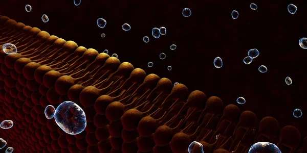
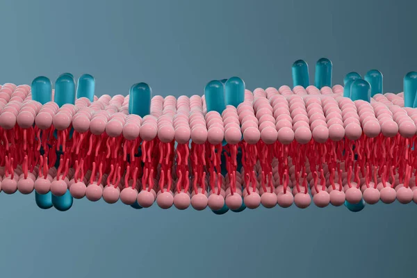
Cell Membrane With Blue Background, 3d Rendering. Computer Digital Drawing.

Image, 14.13MB, 8000 × 5000 jpg
stock image Chylomicrons or ultra low-density lipoproteins or ULDL in the blood vessel flow with red blood cells 3d rendering

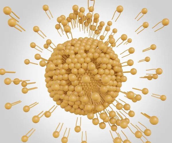
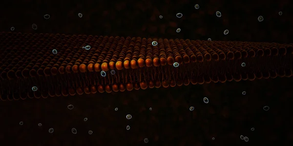
Chylomicrons Or Ultra Low-density Lipoproteins Or ULDL In The Blood Vessel Flow With Red Blood Cells 3d Rendering

Image, 0.42MB, 2400 × 2000 jpg
stock image The liposomes can burst or be broken down to release nanodrugs or nanomedicine 3d rendering

The Liposomes Can Burst Or Be Broken Down To Release Nanodrugs Or Nanomedicine 3d Rendering

Image, 0.36MB, 2400 × 2000 jpg
stock image A 3d rendering of inverse hexagonal HIIc phase consists of DNA rods coated with a lipid monolayer arranged on a hexagonal lattice.

A 3d Rendering Of Inverse Hexagonal HIIc Phase Consists Of DNA Rods Coated With A Lipid Monolayer Arranged On A Hexagonal Lattice.

Image, 1.02MB, 3840 × 2160 jpg
stock image dried fruits over isolated background

Dried Fruits Over Isolated Background

Image, 2.55MB, 3948 × 2494 jpg
stock image Low density lipoprotein (LDL) color code: protein ApoB 100 (blue), phospholipids (orange with a blue cap), cholesterol (orange with a violet cap). Illustration

Low Density Lipoprotein (LDL) Color Code: Protein ApoB 100 (blue), Phospholipids (orange With A Blue Cap), Cholesterol (orange With A Violet Cap). Illustration

Image, 4.91MB, 6000 × 4500 jpg
stock image Soya Lecithin Granules background texture. Vitamin and dietary supplements. Healthy nutrition concept

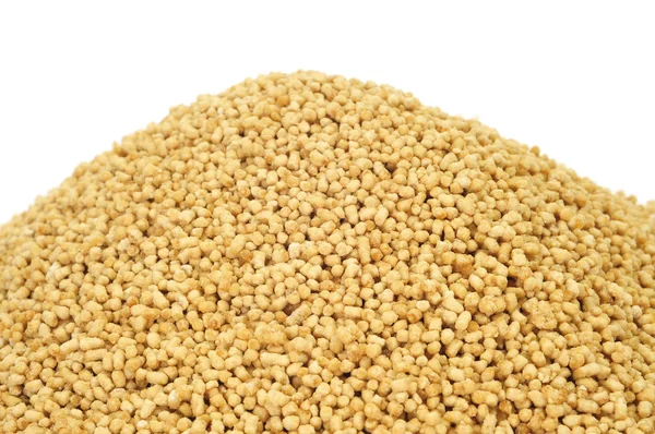
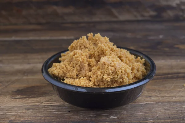
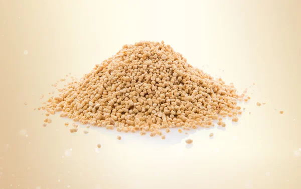
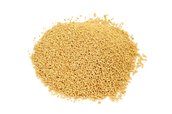
Soya Lecithin Granules Background Texture. Vitamin And Dietary Supplements. Healthy Nutrition Concept

Image, 6.65MB, 3001 × 2001 jpg
stock image Cell membrane and biology, biological concept, 3d rendering. Computer digital drawing.

Cell Membrane And Biology, Biological Concept, 3d Rendering. Computer Digital Drawing.

Image, 8.25MB, 6000 × 4000 jpg
stock image Illustration of different phospholipid arrangements including (from left to right) micelle, liposome, lipid bilayer. Phospholipids have a hydrophilic head and hydrophobic tails.

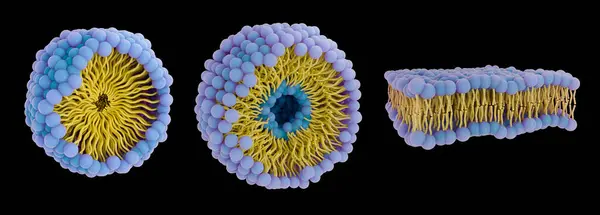
Illustration Of Different Phospholipid Arrangements Including (from Left To Right) Micelle, Liposome, Lipid Bilayer. Phospholipids Have A Hydrophilic Head And Hydrophobic Tails.

Image, 4.82MB, 10000 × 3588 jpg
stock image Illustration of a cross-section of a liposome containing RNA. Liposomes are artificial vesicles enclosed by a lipid bilayer. Liposome lipid bilayers are most commonly formed of phospholipids, which have a hydrophilic head (blue) and hydrophobic tails

Illustration Of A Cross-section Of A Liposome Containing RNA. Liposomes Are Artificial Vesicles Enclosed By A Lipid Bilayer. Liposome Lipid Bilayers Are Most Commonly Formed Of Phospholipids, Which Have A Hydrophilic Head (blue) And Hydrophobic Tails

Image, 2.83MB, 7000 × 3500 jpg
stock image 3d rendering of hexagonal phase consists of DNA rods between rodlike lipid micelles arranged on a hexagonal lattice

3d Rendering Of Hexagonal Phase Consists Of DNA Rods Between Rodlike Lipid Micelles Arranged On A Hexagonal Lattice

Image, 0.69MB, 3840 × 2160 jpg
stock image Plasma Membrane Of A Cell

Plasma Membrane Of A Cell

Image, 9.51MB, 6000 × 3000 jpg
stock image Plasma Membrane Of A Cell

Plasma Membrane Of A Cell

Image, 5.55MB, 6000 × 3000 jpg
stock image The angiotensin converting enzyme 2 (ACE2, blue) in complex with the amino acid transporter BOAT 1 (violet). ACE2 is involved in control of blood pressure and is the target for the SARS-CoV-2 virus to infect human cells.

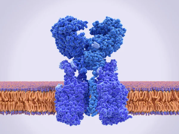
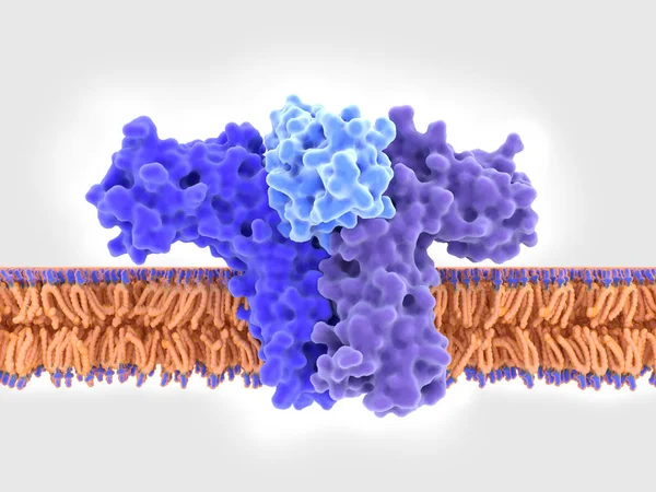
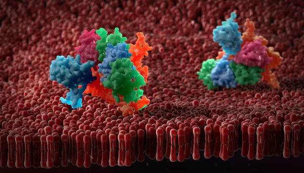
The Angiotensin Converting Enzyme 2 (ACE2, Blue) In Complex With The Amino Acid Transporter BOAT 1 (violet). ACE2 Is Involved In Control Of Blood Pressure And Is The Target For The SARS-CoV-2 Virus To Infect Human Cells.

Image, 4.5MB, 8000 × 6000 jpg
stock image Lipid nanoparticle mRNA vaccine, a type of vaccine used against Covid-19 and influenza. 3D illustration showing cross-section of a lipid nanoparticle carrying mRNA of the virus (orange).

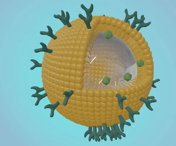
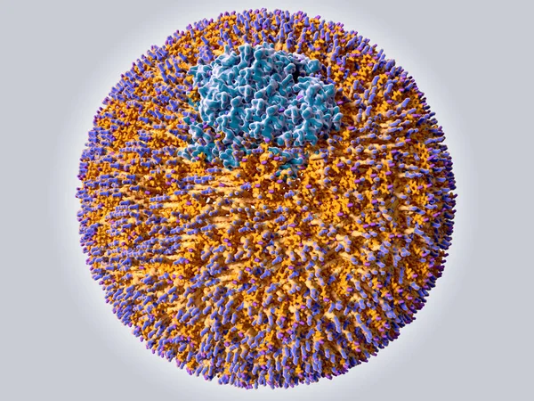
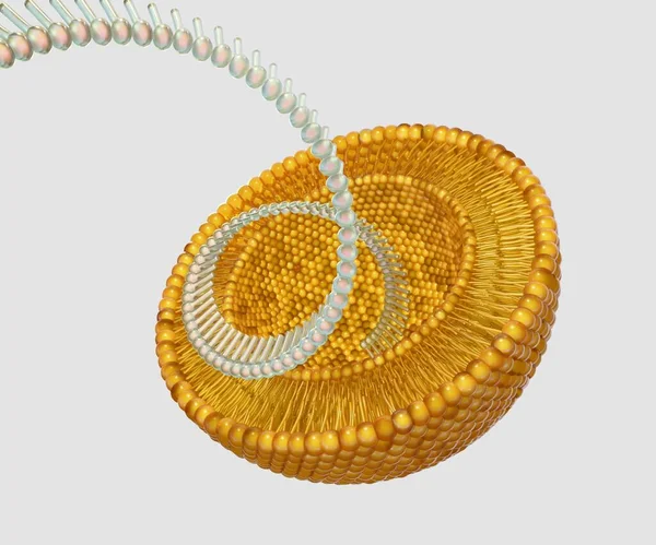
Lipid Nanoparticle MRNA Vaccine, A Type Of Vaccine Used Against Covid-19 And Influenza. 3D Illustration Showing Cross-section Of A Lipid Nanoparticle Carrying MRNA Of The Virus (orange).

Image, 8.8MB, 4492 × 4492 jpg
stock image The liposomes can burst or be broken down to release nanodrugs or nanomedicine 3d rendering

The Liposomes Can Burst Or Be Broken Down To Release Nanodrugs Or Nanomedicine 3d Rendering

Image, 0.61MB, 2400 × 2000 jpg
stock image Lecithin

Lecithin

Image, 9.77MB, 6016 × 4016 jpg
stock image LDL particles in the blood stream Low-density lipoprotein (LDL) particles transport the water insoluble lipids in blood plasma from the liver to other organs and tissues. Illustration

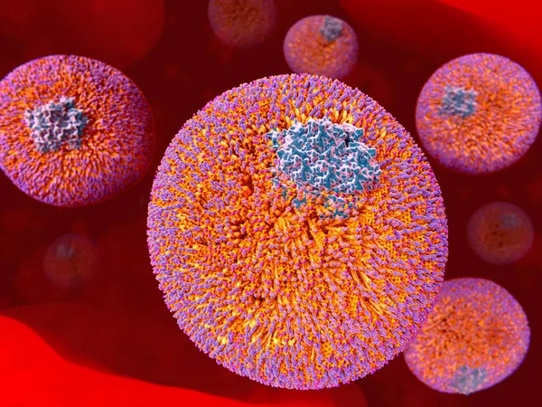
LDL Particles In The Blood Stream Low-density Lipoprotein (LDL) Particles Transport The Water Insoluble Lipids In Blood Plasma From The Liver To Other Organs And Tissues. Illustration

Image, 8.07MB, 8000 × 6000 jpg
stock image A 3D rendering featuring a half-cut liposome and other liposomes dispersed on a white background

A 3D Rendering Featuring A Half-cut Liposome And Other Liposomes Dispersed On A White Background

Image, 0.48MB, 3840 × 2160 jpg
stock vector Fluid mosaic model with cell membrane anatomical structure outline diagram. Labeled educational scheme with glycoprotein, integral protein, glycolipid and phospholipid vector illustration.

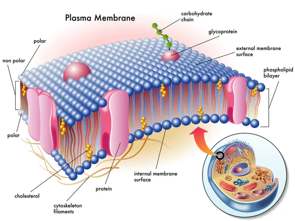
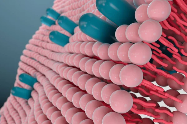
Fluid Mosaic Model With Cell Membrane Anatomical Structure Outline Diagram. Labeled Educational Scheme With Glycoprotein, Integral Protein, Glycolipid And Phospholipid Vector Illustration.

Vector, 6.23MB, 5000 × 3750 eps
stock image 3d rendering of hexagonal lipid phase. In this phase, long, tubular aggregates form in a hexagonal pattern

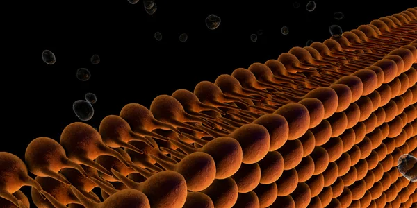
3d Rendering Of Hexagonal Lipid Phase. In This Phase, Long, Tubular Aggregates Form In A Hexagonal Pattern

Image, 0.51MB, 3840 × 2160 jpg
stock image 3d rendering of Liposomes within liposomes are known as multivesicular liposomes or nested liposomes.

3d Rendering Of Liposomes Within Liposomes Are Known As Multivesicular Liposomes Or Nested Liposomes.

Image, 0.81MB, 3840 × 2160 jpg
stock image Plasma Membrane Of A Cell

Plasma Membrane Of A Cell

Image, 4.34MB, 4088 × 3000 jpg
stock image Soya Lecithin Granules and plastic spoon, macro photo. Vitamin and dietary supplements. Healthy nutrition concept

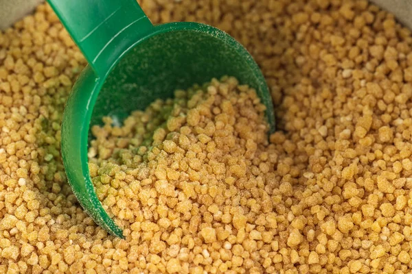
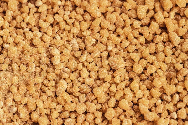
Soya Lecithin Granules And Plastic Spoon, Macro Photo. Vitamin And Dietary Supplements. Healthy Nutrition Concept

Image, 5.13MB, 3001 × 2001 jpg
stock image liposomes as nanocarriers for small interference RNA (siRNA) delivery 3d rendering

Liposomes As Nanocarriers For Small Interference RNA (siRNA) Delivery 3d Rendering

Image, 0.2MB, 2400 × 2000 jpg
stock image Soy lecithin granules

Soy Lecithin Granules

Image, 5.27MB, 4288 × 2848 jpg
Page 1 >> Next