
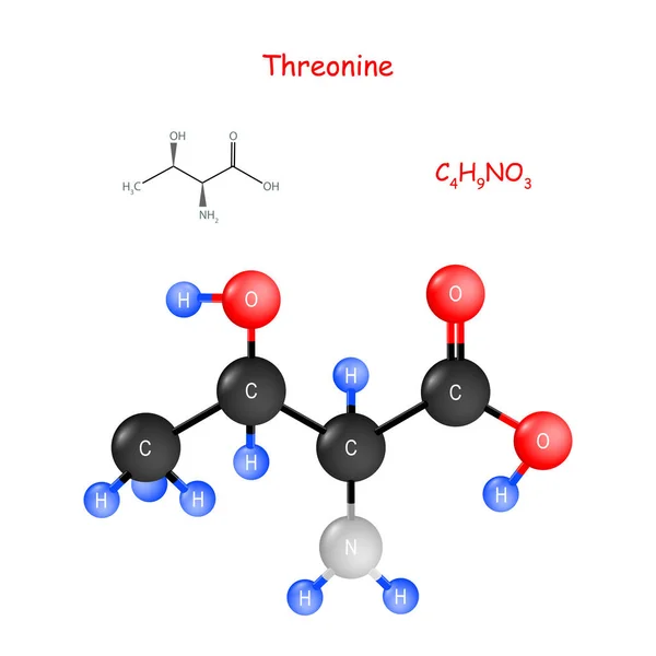
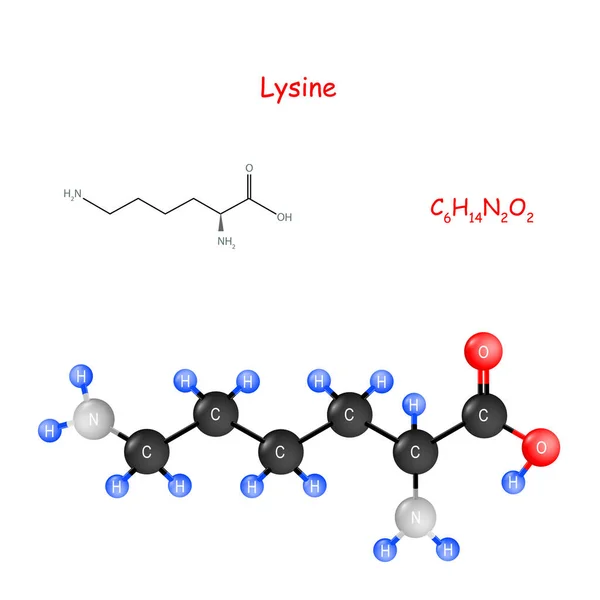
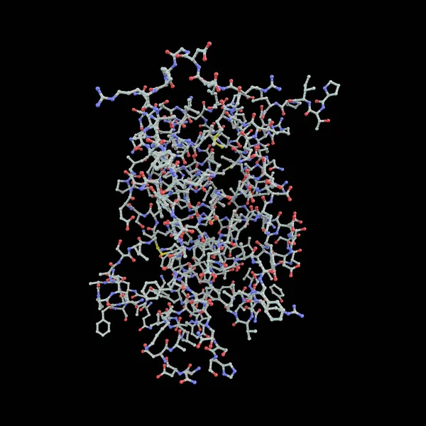
Stock image Polypeptide

Protein Structure Levels From Amino Acid To Complex Molecule Outline Diagram
Vector, 6.02MB, 5000 × 3333 eps

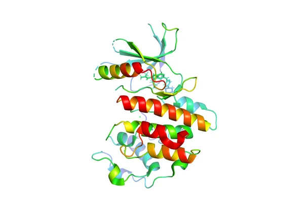
Thioredoxin Antioxidant Enzyme. 3D Illustration. Cartoon & Wireframe Representation. N-term To C-term Gradient Coloring.
Image, 9.9MB, 8000 × 7207 jpg

Several Vials With Soluble Protease Proteins For Activation Of The Coronavirus Severe Acute Respiratory Syndrome (SARS) Trypsin-like Protein In Human Respiratory Tract In A Hospital, Spain
Image, 7.23MB, 5184 × 3600 jpg

Human Nucleosome With CpG Methylated (red) DNA Showed, 3D Cartoon Model Isolated, Black Background
Image, 2.26MB, 6000 × 4056 jpg

Interferon Alpha 2a (IFNA2) Molecule, 3D Rendering. Pegylated Analogs Of This Cytokine Are Used To Treat Hepatitis B And C Infections.
Image, 11.43MB, 6450 × 8000 jpg
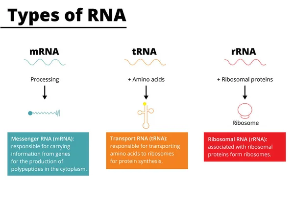
Types Of RNA: Messenger RNA (mRNA), Transport RNA (tRNA), Ribosomal RNA (rRNA). Vector Illustration.
Vector, 0.73MB, 5000 × 3500 ai
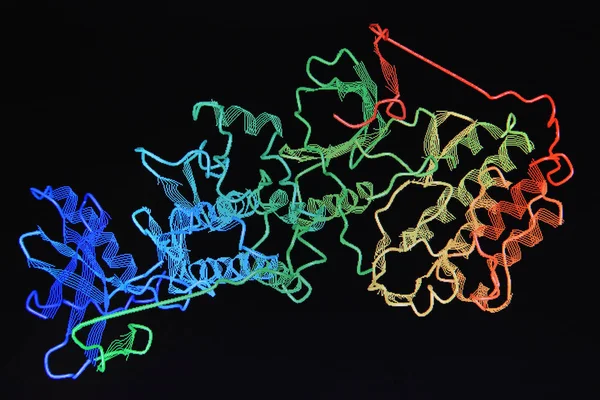
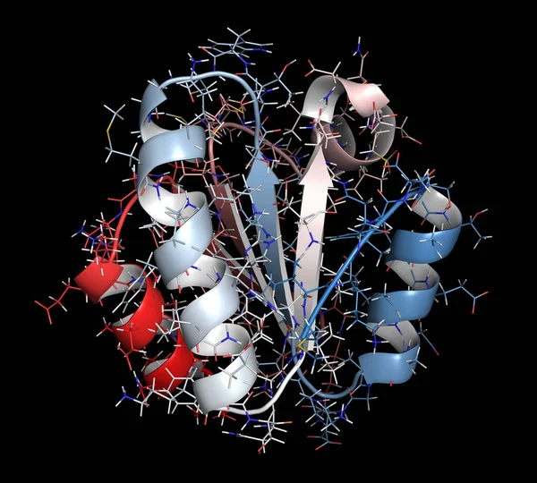
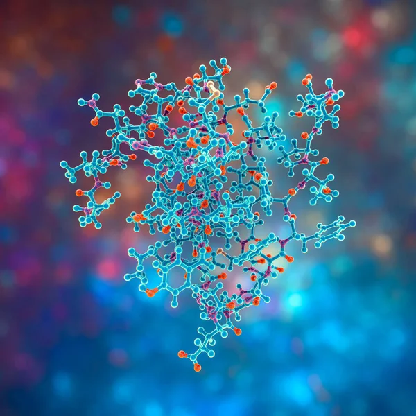
Three-dimensional Crystal Structure Of Protein Molecule, Tumor Growth Marker. 3D Model Of A Biopolymer Is A Peptide.
Image, 2.1MB, 3640 × 2453 jpg
Three-dimensional Crystal Structure Of Protein Molecule, Tumor Growth Marker. 3D Model Of A Biopolymer Is A Peptide.
Image, 2.13MB, 3200 × 2500 jpg
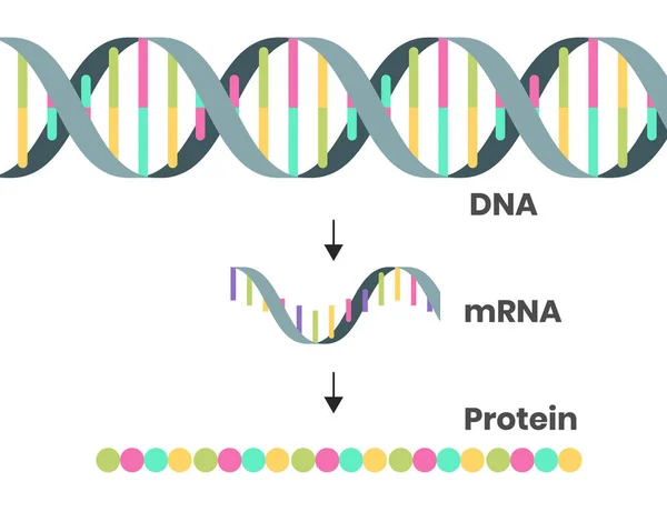
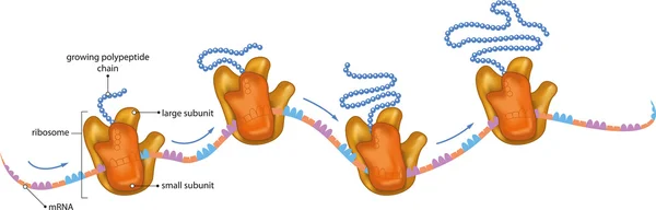
Protein Syntesis Schematic Illustration. Vector Illustration Of The DNA, MRNA And Polypeptide Chain
Vector, 0.96MB, 5102 × 4000 eps
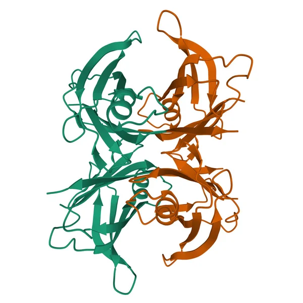
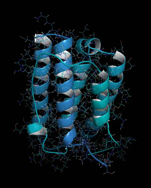
Wild Type Human Transthyretin (TTR), A 3D Ribbon Model Of The Homodimer Isolated, White Background
Image, 2.97MB, 4096 × 4096 jpg

Tay-Sachs Disease, 3D Illustration. A Genetic Disorder That Progressively Destroys Brain Neurons, Is Caused By A Mutation In The HEXA Gene Of Chromosome 15 Leading To Deficiency Of Hexosaminidase A
Image, 24.4MB, 14000 × 8436 jpg
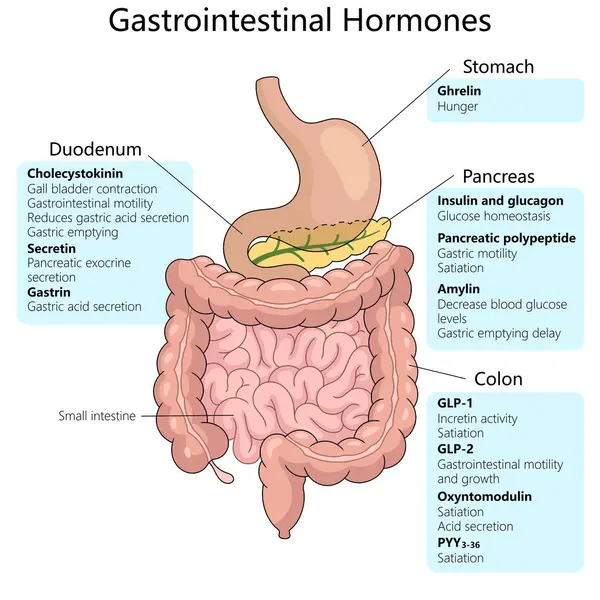
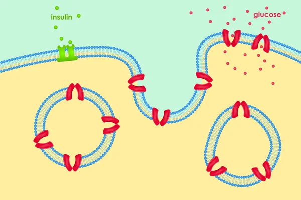
Gastrointestinal Hormones, Including Their Origins In The Stomach, Duodenum, Pancreas, And Colon, And Their Specific Functions Schematic Raster Illustration. Medical Science Educational Illustration
Image, 5.2MB, 6000 × 6000 jpg
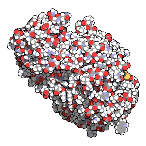
Human Insulin Hormone Molecule, 3D Illustration. Drug In Diabetes Treament
Image, 14.23MB, 6000 × 6000 jpg
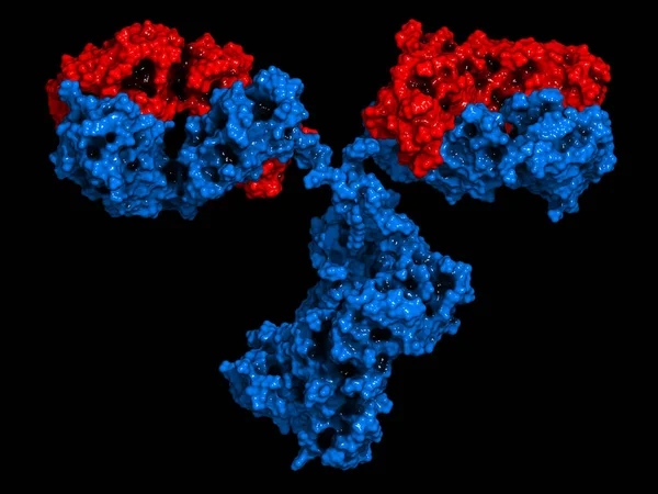
IgG2a Monoclonal Antibody (immunoglobulin). Many Biotech Drugs Are Antibodies. 3D Render. Molecular Surface Model.
Image, 11.96MB, 8000 × 6000 jpg
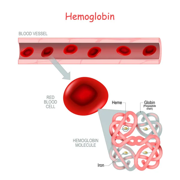
Structure Of The Hemoglobin Molecule With Heme (Iron And Oxygen Molecule) And Polypeptide Chain (Globin). Blood Vessel And Close-up Of Red Blood Cell. Showing Alpha And Beta Chains, Heme Groups And Iron Atoms. Vector Medical Icon.
Vector, 10.3MB, 4444 × 4444 eps

Ferritin Round Droplets Scattered Randomly. Beauty Treatment And Nutrition Skin Care. Medical And Scientific Background. Healthy Life Concept.
Vector, 9.01MB, 4000 × 4000 eps

Ferritin Round Droplets Scattered Randomly. Beauty Treatment And Nutrition Skin Care. Essential Vitamins Vector Illustration. Wellness Concept.
Vector, 19.6MB, 4000 × 4000 eps


A 3D Rendering Of A Polymer Chain. The Chain Is Made Up Of Individual Monomers, Which Are Linked Together By Chemical Bonds
Image, 0.18MB, 3840 × 2160 jpg

3d Rendering Of Scattered Gold Antibody Molecules In The Black Background
Image, 0.36MB, 3840 × 2160 jpg

New York, USA - 1 August 2024: PolyPeptide Group Logo On Phone Screen, Company Icon On Display.
Image, 10.97MB, 6240 × 4105 jpg

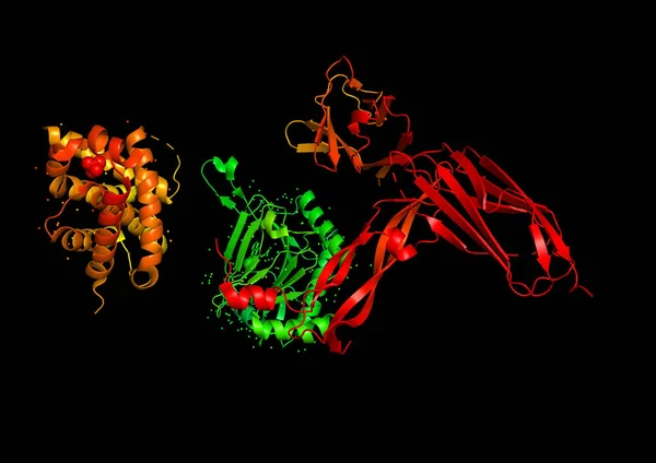
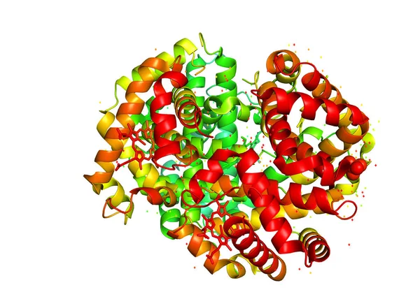
3D Rendering. The Structure Of The Protein Molecule, An Activator Of Angiogenesis. X-ray Crystalline Model Of One Of The Subunits Of The Hif1A Molecule.
Image, 3.98MB, 5531 × 3915 jpg
Amylase (human Pancreatic Alpha-amylase) Protein. Digestive Enzyme, Responsible For The Hydrolysis Of Starch Into Sugars. 3D Illustration.
Image, 4.69MB, 4096 × 4096 jpg

Types Of Protein Structure. Proteins Are Biological Polymers Composed Of Amino Acids.
Image, 5.33MB, 8840 × 11811 jpg

Structure Of Human Activin A Homodimer, 3D Cartoon Model, White Background
Image, 1.88MB, 6000 × 4000 jpg
Human Growth Hormone Molecule (hGH, Somatotropin), 3D Illustration. Natural Hormone That Is Used Both As A Medicine And As A Doping Agent.
Image, 5.53MB, 6000 × 6000 jpg
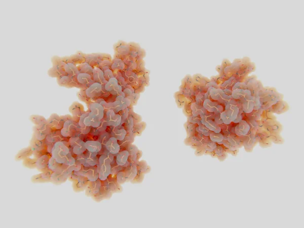
The Crystal Structure Of The Tumor Marker Protein. The 3D Model Of The Biological Macromolecule.
Image, 1.64MB, 4000 × 2933 jpg

The Crystal Structure Of The Tumor Marker Protein. The 3D Model Of The Biological Macromolecule.
Image, 2.61MB, 4000 × 2933 jpg

The Crystal Structure Of The Tumor Marker Protein. The 3D Model Of The Biological Macromolecule.
Image, 1.21MB, 4000 × 2667 jpg

Scientist Holds Vial With Catalase Enzyme That Helps Regulate Cytokine Production, Protects Oxidative Injury And Suppresses SARS-CoV-2 Replication, Conceptual Image
Image, 10.02MB, 5184 × 3521 jpg
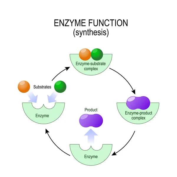

Digestion Of Protein. Breaking The Complex Molecule First Into Peptides Then Into Individual Amino Acids. The Pepsins Are Enzymes Secreted By The Stomach That Breaks Down Proteins. Vector Illustration
Vector, 5.26MB, 6000 × 3321 eps

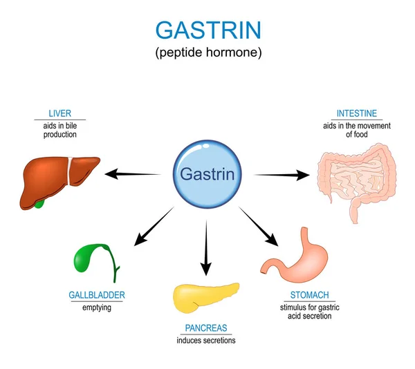
Gastrin Hormone Function. Gastrointestinal Hormone That Affects Gastric Acid Secretion In Stomach, Aids In Bile Production In Liver, Pancreas Induces Secretions, Aids In The Movement Of Food In Intestine. Vector Illustration, Medical Poster
Vector, 1.58MB, 4444 × 3963 eps
Page 1 >> Next