
Stock image Porins

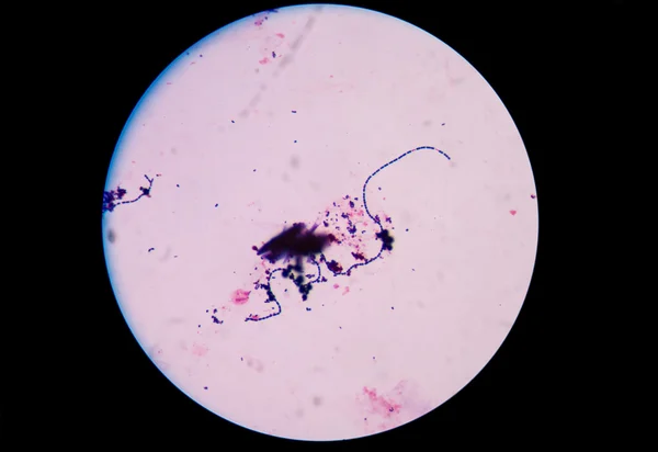
This Differential Staining Procedure Separates Most Bacteria Into Two Groups On The Basis Of Cell Wall Composition, Bacteria Gram Posotive And Gram Negative
Vector, 11.84MB, 2859 × 6420 eps

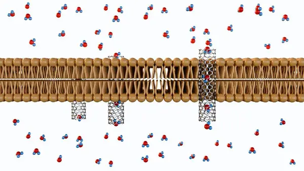
3d Rendering Of Molecules Passing Through Carbon Nanotube Porins On Lipid Bilayer Membrane
Image, 0.81MB, 3840 × 2160 jpg

3d Rendering Of Molecules Passing Through Carbon Nanotube Porins On Lipid Bilayer Membrane
Image, 0.57MB, 3840 × 2160 jpg

3D Rendering Of Carbon Nanotube Porins, Short Pieces Of Carbon Nanotubes Capable Of Self-inserting Into A Lipid Bilayer, Model Of Biological Membrane Channels.
Image, 0.81MB, 3840 × 2160 jpg


3D Rendering Of Carbon Nanotube Porins, Short Pieces Of Carbon Nanotubes Capable Of Self-inserting Into A Lipid Bilayer, Model Of Biological Membrane Channels.
Image, 0.43MB, 3840 × 2160 jpg

3D Rendering Of Carbon Nanotube Porins, Short Pieces Of Carbon Nanotubes Capable Of Self-inserting Into A Lipid Bilayer, Model Of Biological Membrane Channels.
Image, 0.69MB, 3840 × 2160 jpg

3d Rendering Of Molecules Passing Through Carbon Nanotube Porins On Lipid Bilayer Membrane
Image, 0.6MB, 3840 × 2160 jpg
3d Rendering Of Molecules Passing Through Carbon Nanotube Porins On Lipid Bilayer Membrane
Image, 0.34MB, 3840 × 2160 jpg
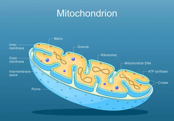
Mitochondria Structure. Anatomy Of Motochondrion. Cross Section Of Motochondrion. Close-up Of Ribosomes, ATP Synthase, Cristae, Granule, Porins, Matrix. Isometric Flat Vector Illustration
Vector, 2.61MB, 5000 × 3500 eps

3D Rendering Of Carbon Nanotube Porins, Short Pieces Of Carbon Nanotubes Capable Of Self-inserting Into A Lipid Bilayer, Model Of Biological Membrane Channels.
Image, 0.79MB, 3840 × 2160 jpg

3D Rendering Of Carbon Nanotube Porins, Short Pieces Of Carbon Nanotubes Capable Of Self-inserting Into A Lipid Bilayer, Model Of Biological Membrane Channels.
Image, 0.95MB, 3840 × 2160 jpg

3d Rendering Of Molecules Passing Through Carbon Nanotube Porins On Lipid Bilayer Membrane
Image, 0.67MB, 3840 × 2160 jpg
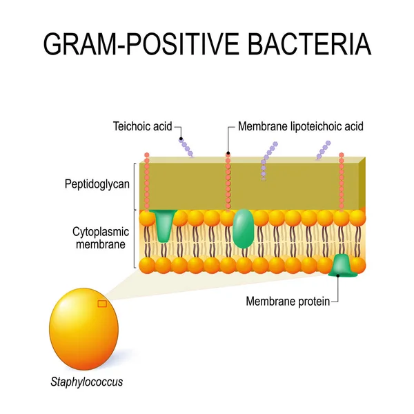
Cell Wall Structure Of Gram-positive Bacteria For Example Staphylococcus. Vector Diagram For Educational, Medical, Biological And Science Use
Vector, 1.74MB, 4095 × 4095 eps

3D Rendering Of Carbon Nanotube Porins, Short Pieces Of Carbon Nanotubes Capable Of Self-inserting Into A Lipid Bilayer, Model Of Biological Membrane Channels.
Image, 0.4MB, 3840 × 2160 jpg

3d Rendering Of Molecules Passing Through Carbon Nanotube Porins On Lipid Bilayer Membrane
Image, 0.8MB, 3840 × 2160 jpg
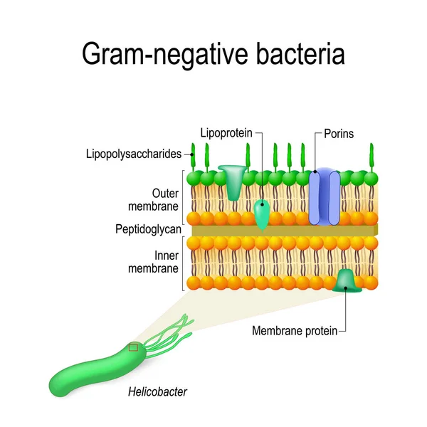
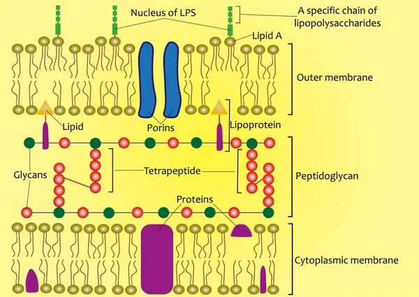
Cell Wall Structure Of Gram-negative Bacteria For Example Helicobacter. Vector Diagram For Educational, Medical, Biological And Science Use
Vector, 2.88MB, 4152 × 4152 eps
Page 1 >> Next