photo free
Home Images Vectors Videos

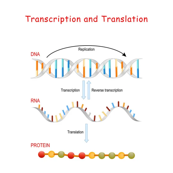
Stock image Protein Structure

stock vector Molecular structure of the protein

Molecular Structure Of The Protein

Vector, 0.62MB, 4000 × 5333 eps
stock vector Protein structure vector illustration. Labeled amino acid chain molecules.

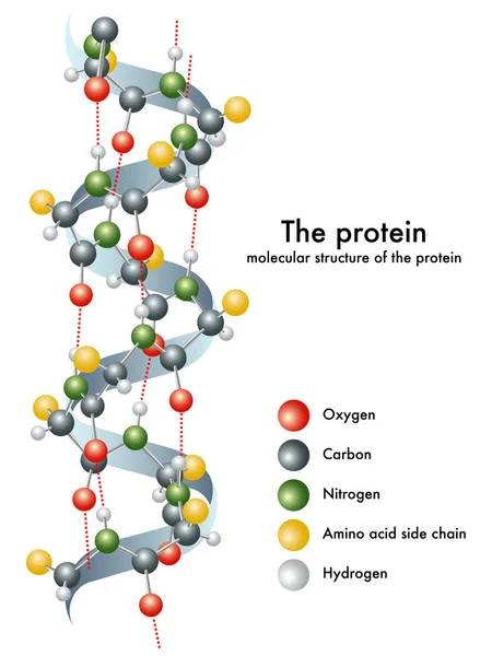
Protein Structure Vector Illustration. Labeled Amino Acid Chain Molecules.

Vector, 6.85MB, 4500 × 4500 eps
stock image Leptin, the human obesity protein

Leptin, The Human Obesity Protein

Image, 4.33MB, 5021 × 4323 jpg
stock vector Protein structure. From Linear sequence of amino acids, alpha helices and Linear sequence to Polypeptide chain and Multiple protein subunits in one complex. Vector diagram for scientific, medical, and educational use. poster for infographics

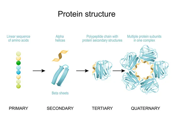
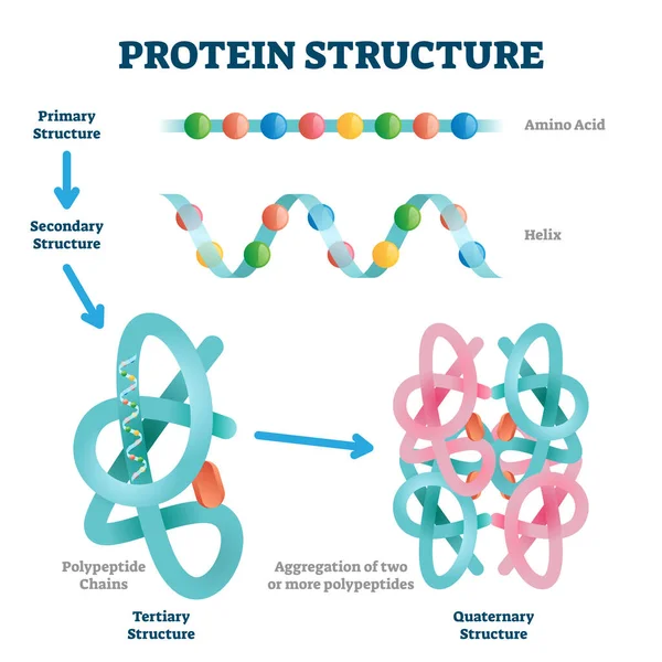
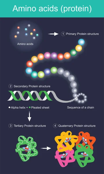
Protein Structure. From Linear Sequence Of Amino Acids, Alpha Helices And Linear Sequence To Polypeptide Chain And Multiple Protein Subunits In One Complex. Vector Diagram For Scientific, Medical, And Educational Use. Poster For Infographics

Vector, 2.27MB, 5000 × 3483 eps
stock image Structure of the human growth hormone, 3D cartoon model. white background

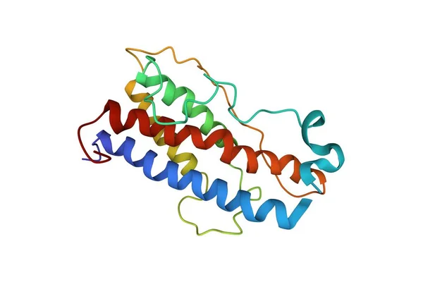
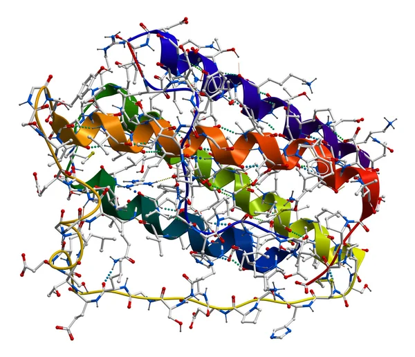
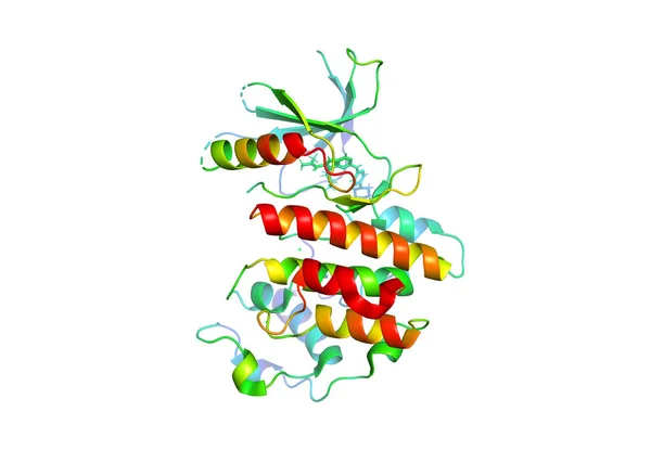
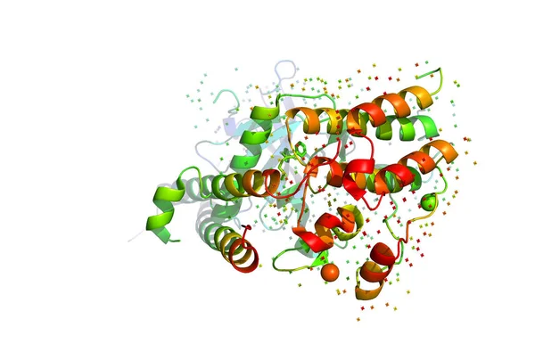
Structure Of The Human Growth Hormone, 3D Cartoon Model. White Background

Image, 1.74MB, 6000 × 4000 jpg
stock vector Structure of protein molecules

Structure Of Protein Molecules

Vector, 1.67MB, 4250 × 4250 eps
stock image Molecular structure of pancratic lipase

Molecular Structure Of Pancratic Lipase

Image, 0.57MB, 2590 × 1600 jpg
stock vector When we eat food protein The body will digest those proteins into amino acids before being absorbed into the bloodstream.

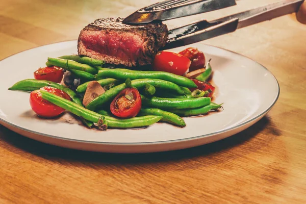
When We Eat Food Protein The Body Will Digest Those Proteins Into Amino Acids Before Being Absorbed Into The Bloodstream.

Vector, 11.61MB, 4200 × 7000 eps
stock image Amino acid cysteine molecular structure

Amino Acid Cysteine Molecular Structure

Image, 2.35MB, 8765 × 6790 jpg
stock vector DNA Replication, Protein synthesis, Transcription and translatio

DNA Replication, Protein Synthesis, Transcription And Translatio

Vector, 2.85MB, 4444 × 4444 eps
stock image DNA polymerase I. An enzyme that participates in the DNA replica

DNA Polymerase I. An Enzyme That Participates In The DNA Replica

Image, 6.74MB, 5048 × 4169 jpg
stock image The crystal structure of the tumor marker protein. The 3D model of the biological macromolecule.

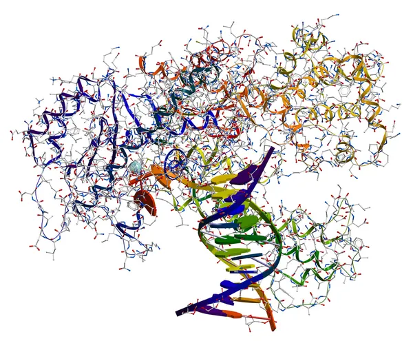
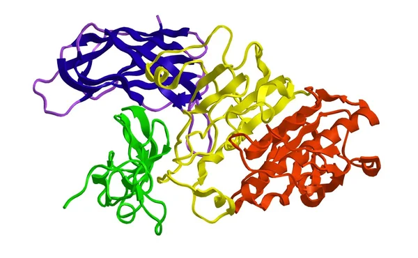
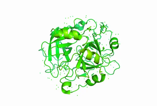
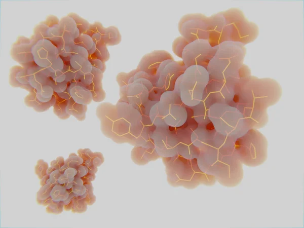
The Crystal Structure Of The Tumor Marker Protein. The 3D Model Of The Biological Macromolecule.

Image, 1.64MB, 4000 × 2933 jpg
stock image 3D model of a protein molecule.

3D Model Of A Protein Molecule.

Image, 2.35MB, 3640 × 2453 jpg
stock image 3D model of a protein molecule.

3D Model Of A Protein Molecule.

Image, 1.55MB, 3171 × 2137 jpg
stock image Adenosine triphosphate (ATP) molecule isolated on white

Adenosine Triphosphate (ATP) Molecule Isolated On White

Image, 3.2MB, 6000 × 5139 jpg
stock vector Detailed vector illustration of round membrane cells

Detailed Vector Illustration Of Round Membrane Cells

Vector, 13.66MB, 4000 × 4000 eps
stock image Hemoglobin molecule isolated on a white background

Hemoglobin Molecule Isolated On A White Background

Image, 4.59MB, 5359 × 4656 jpg
stock vector Skin aging

Skin Aging

Vector, 4.05MB, 6678 × 3643 eps
stock image Gliadin molecule, component of gluten

Gliadin Molecule, Component Of Gluten

Image, 2.53MB, 5988 × 4101 jpg
stock vector The general formula for an Amino Acid.

The General Formula For An Amino Acid.

Vector, 5.27MB, 12500 × 8333 eps
stock vector The structure of Serine. Serine is an amino acid that has a side chain consisting of a hydroxymethyl group.

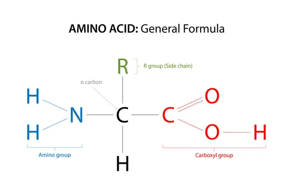
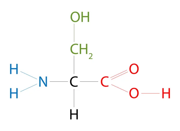
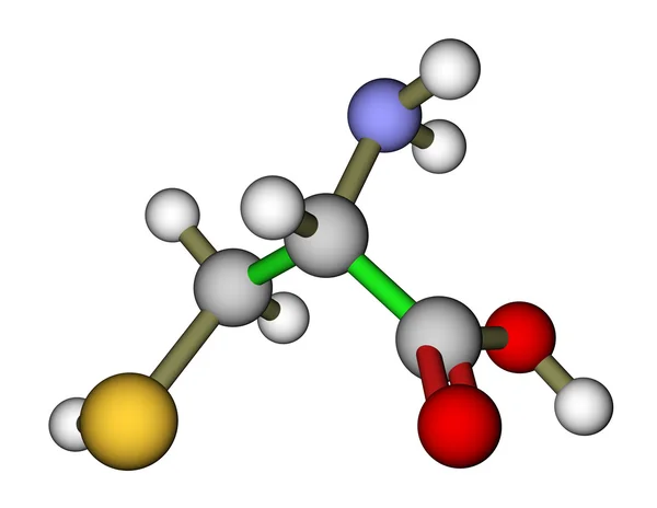
The Structure Of Serine. Serine Is An Amino Acid That Has A Side Chain Consisting Of A Hydroxymethyl Group.

Vector, 5.21MB, 11458 × 8333 eps
stock image Three-dimensional model of Alpha-Amylase - 3d render

Three-dimensional Model Of Alpha-Amylase - 3d Render

Image, 9.06MB, 5200 × 3100 jpg
stock vector Ribosomes

Ribosomes

Vector, 1.78MB, 5351 × 1715 eps
stock image Insulin molecules

Insulin Molecules

Image, 2.16MB, 8000 × 6000 jpg
stock image Three-dimensional model of Catalase - 3d render

Three-dimensional Model Of Catalase - 3d Render

Image, 6.84MB, 3592 × 4943 jpg
stock image Metal pliers serving cooked Steak

Metal Pliers Serving Cooked Steak

Image, 4.26MB, 3674 × 2449 jpg
stock vector Antibody

Antibody

Vector, 0.7MB, 5577 × 3500 eps
stock image Proteomics and Functional Genomics - The Large-scale Study of Proteins in Living Organisms - A Protein Isolated on Blue Tech Background - Conceptual Illustration with Copy Space

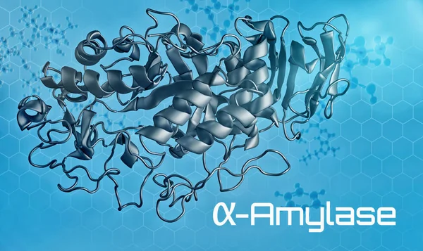
Proteomics And Functional Genomics - The Large-scale Study Of Proteins In Living Organisms - A Protein Isolated On Blue Tech Background - Conceptual Illustration With Copy Space

Image, 10.29MB, 9584 × 5298 jpg
stock image Proteomics and Protein Folding Prediction Through Computational Means - The Study of the Function and Structure of Proteins Within Living Organisms - Conceptual Illustration

Proteomics And Protein Folding Prediction Through Computational Means - The Study Of The Function And Structure Of Proteins Within Living Organisms - Conceptual Illustration

Image, 10.62MB, 6500 × 3656 jpg
stock vector Protein structure - molecule 3D icon

Protein Structure - Molecule 3D Icon

Vector, 5.3MB, 5834 × 5835 eps
stock image Collection of different covid variants

Collection Of Different Covid Variants

Image, 6.57MB, 3840 × 2160 jpg
stock image resting, activated, and inactivated states of an ion channel in the plasma membrane with ion flow structure diagram hand drawn schematic raster illustration. Medical science educational illustration

Resting, Activated, And Inactivated States Of An Ion Channel In The Plasma Membrane With Ion Flow Structure Diagram Hand Drawn Schematic Raster Illustration. Medical Science Educational Illustration

Image, 3.93MB, 6000 × 4500 jpg
stock image Inner Life of Proteins

Inner Life Of Proteins

Image, 5.58MB, 3600 × 2700 jpg
stock vector Illustration of collagen sequence, molecule, and fiber

Illustration Of Collagen Sequence, Molecule, And Fiber

Vector, 0.68MB, 3662 × 6449 eps
stock vector Collagen is a fibrillar protein that forms the basis of the connective tissue of the whole organism, providing its strength and elasticity.

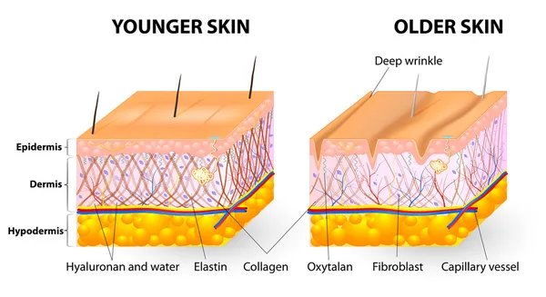
Collagen Is A Fibrillar Protein That Forms The Basis Of The Connective Tissue Of The Whole Organism, Providing Its Strength And Elasticity.

Vector, 1.73MB, 10833 × 5000 eps
stock vector Normal protein and prion diseases. Vector scheme

Normal Protein And Prion Diseases. Vector Scheme

Vector, 0.42MB, 5041 × 4265 eps
stock vector dna background

Dna Background

Vector, 0.29MB, 4167 × 4167 eps
stock image Molecular structure of Glutamic Acid isolated on white background

Molecular Structure Of Glutamic Acid Isolated On White Background

Image, 2.77MB, 5432 × 5519 jpg
stock vector Protein Digestion. Enzymes (proteases and peptidases) are digestion breaks the protein into smaller peptide chains and into single amino acids, which are absorbed into the blood.

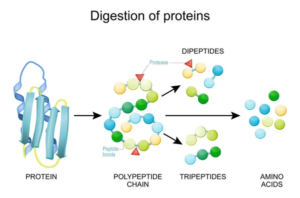
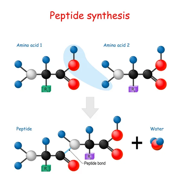
Protein Digestion. Enzymes (proteases And Peptidases) Are Digestion Breaks The Protein Into Smaller Peptide Chains And Into Single Amino Acids, Which Are Absorbed Into The Blood.

Vector, 2.17MB, 5000 × 3297 eps
stock image Nutrition and diet therapy concept

Nutrition And Diet Therapy Concept

Image, 6.76MB, 4000 × 4000 jpg
stock vector How enzymes work. Fermentation and digestion food

How Enzymes Work. Fermentation And Digestion Food

Vector, 0.75MB, 4779 × 3362 eps
stock vector Peptide synthesis.

Peptide Synthesis.

Vector, 4.57MB, 4444 × 4444 eps
stock vector Structure of hemoglobin. Red blood cell, hemoglobin molecule, and structural formula of a Heme b-group. Vector illustration

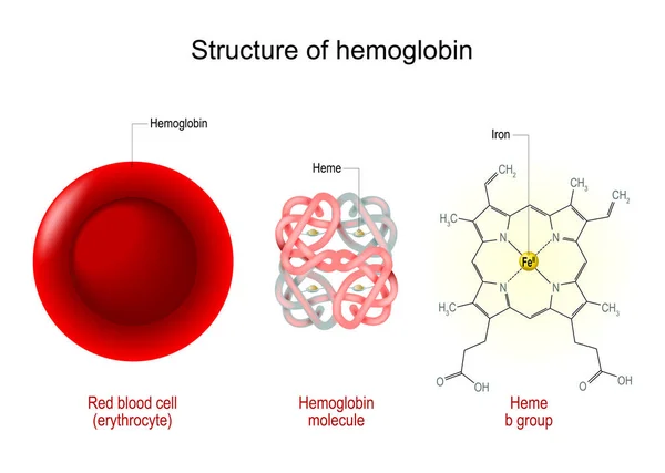
Structure Of Hemoglobin. Red Blood Cell, Hemoglobin Molecule, And Structural Formula Of A Heme B-group. Vector Illustration

Vector, 11.36MB, 5000 × 3562 eps
stock vector Enzymes breaking down food into nutrients. Digestive systems work vector illustrative infographics

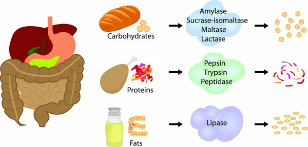
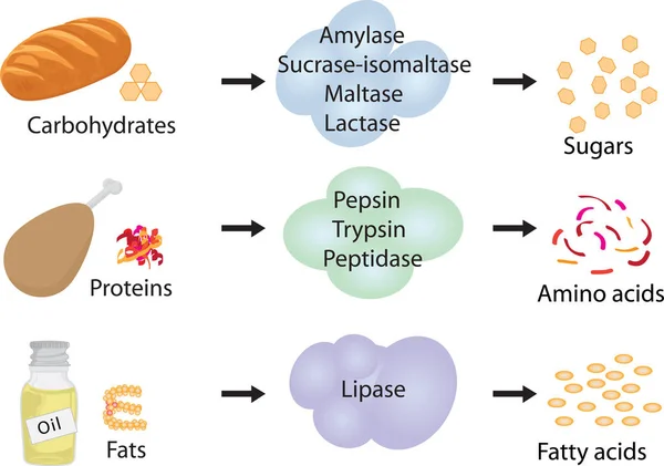
Enzymes Breaking Down Food Into Nutrients. Digestive Systems Work Vector Illustrative Infographics

Vector, 0.79MB, 5696 × 2731 eps
stock image Lysine (symbol Lys or K) is an amino acid that is used in the biosynthesis of proteins. Formula: C6H14N2O2. 3D illustration. Chemical structure model: Ball and Stick + Space-Filling. White background

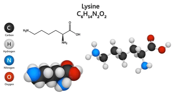
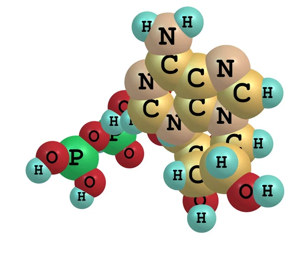
Lysine (symbol Lys Or K) Is An Amino Acid That Is Used In The Biosynthesis Of Proteins. Formula: C6H14N2O2. 3D Illustration. Chemical Structure Model: Ball And Stick + Space-Filling. White Background

Image, 1.07MB, 3840 × 2160 jpg
stock vector Healthy food plate guide concept. Vector flat modern illustration. Infographic of recomendation nutrition plan with percent labels. Colorful meat, fruit, vegetables and grains icon set.

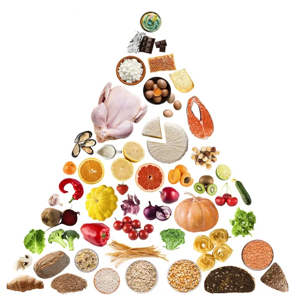
Healthy Food Plate Guide Concept. Vector Flat Modern Illustration. Infographic Of Recomendation Nutrition Plan With Percent Labels. Colorful Meat, Fruit, Vegetables And Grains Icon Set.

Vector, 0.24MB, 4000 × 4000 ai
stock image 3D image of tyrosine skeletal formula - molecular chemical structure of 4-hydroxyphenylalanine isolated on white background

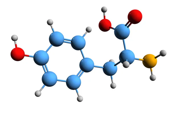
3D Image Of Tyrosine Skeletal Formula - Molecular Chemical Structure Of 4-hydroxyphenylalanine Isolated On White Background

Image, 2.01MB, 5500 × 3630 jpg
stock vector Arginine (Arg, R) amino acid molecule. (Chemical formula C6H14N4O2) it is used in the biosynthesis of proteins. Ball-and-stick model, space-filling model and skeletal formula. Layered vector illustration

Arginine (Arg, R) Amino Acid Molecule. (Chemical Formula C6H14N4O2) It Is Used In The Biosynthesis Of Proteins. Ball-and-stick Model, Space-filling Model And Skeletal Formula. Layered Vector Illustration

Vector, 5.64MB, 5209 × 3334 eps
stock vector The structure of the ribosome. Infographics. Vector illustration on isolated background

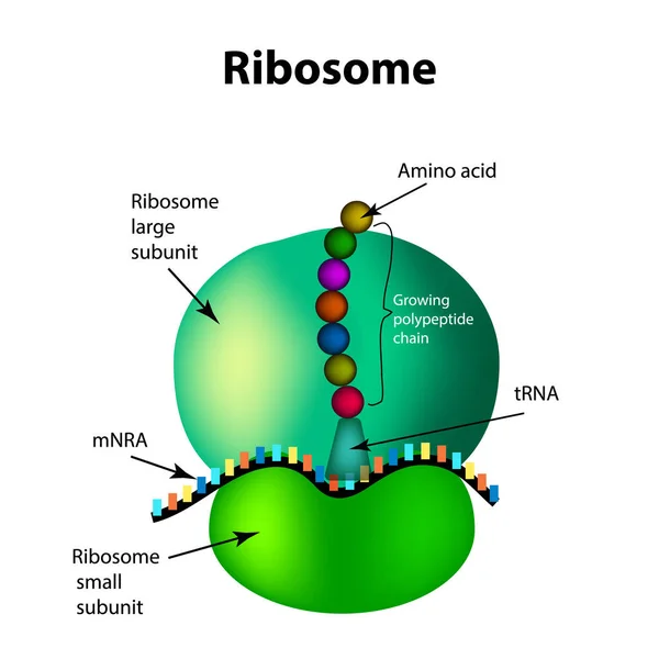
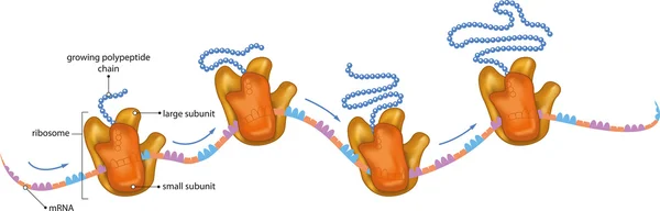
The Structure Of The Ribosome. Infographics. Vector Illustration On Isolated Background

Vector, 2.01MB, 5000 × 5000 eps
stock vector Genetic mutation. Normal and mutated genes that synthesis normal and Abnormal proteins. Vector poster for education and science.

Genetic Mutation. Normal And Mutated Genes That Synthesis Normal And Abnormal Proteins. Vector Poster For Education And Science.

Vector, 0.83MB, 4444 × 4444 eps
Page 1 >> Next