
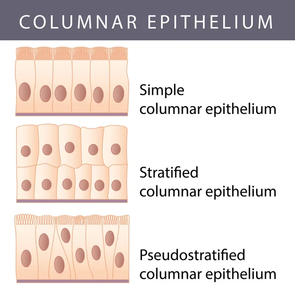
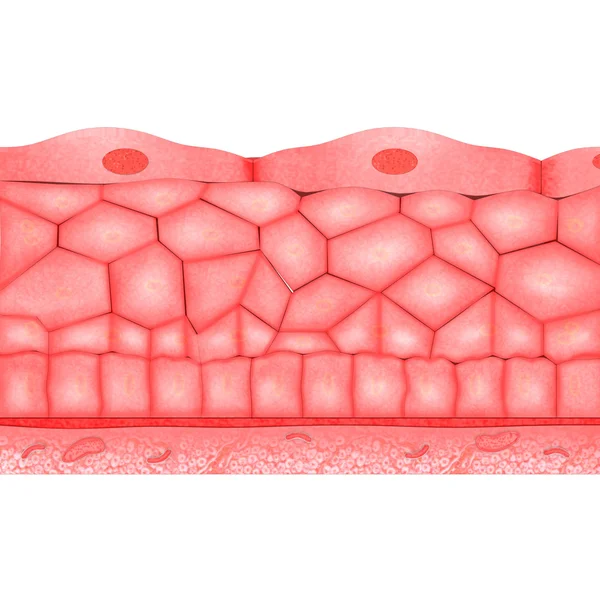
Stock image Pseudostratified
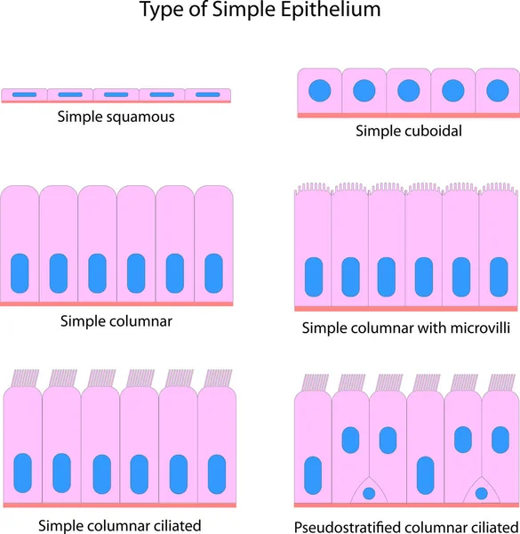
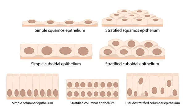
Cells Of Epithelial Tissue: Squamous (flattened And Thin), Cuboidal (boxy, As Wide As It Is Tall), Columnar (rectangular, Taller Than It Is Wide), Pseudostratified.
Vector, 6.24MB, 5209 × 3125 eps
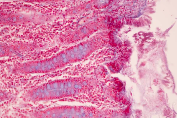
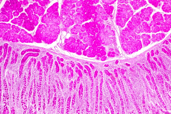
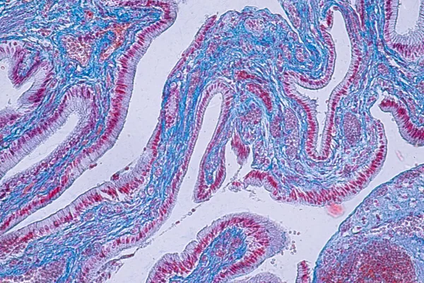
Tissue Of Small Intestine (Duodenum), Large Intestine Human And Stomach Human Under The Microscope In Lab.
Image, 17.29MB, 8192 × 5461 jpg
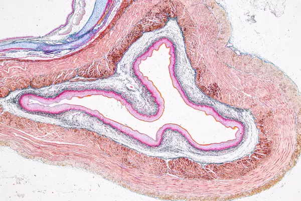
Tissue Of Small Intestine (Duodenum) And Vermiform Appendix Human Under The Microscope In Lab.
Image, 22.69MB, 6000 × 4000 jpg

Tissue Of Small Intestine (Duodenum) And Vermiform Appendix Human Under The Microscope In Lab.
Image, 20.31MB, 6000 × 4000 jpg

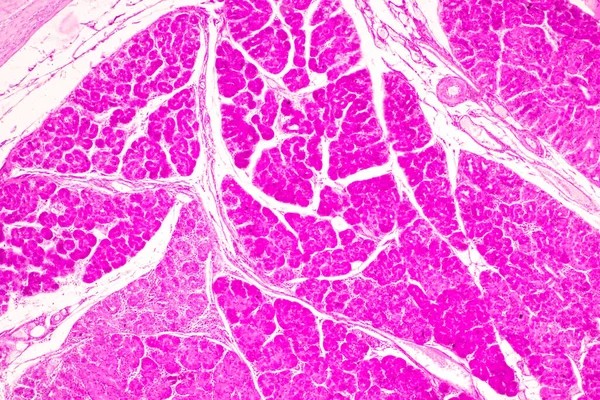
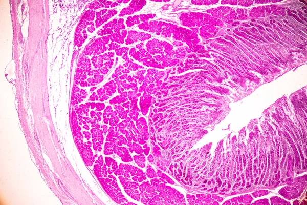
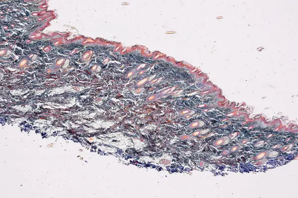
Pathology And Histology Tissue Of Mouse, Rabbit, Cat And Cow Under Microscope.
Image, 32.75MB, 6000 × 4000 jpg

Pathology And Histology Tissue Of Mouse, Rabbit, Cat And Cow Under Microscope.
Image, 6.85MB, 2667 × 4000 jpg
Pathology And Histology Tissue Of Mouse, Rabbit, Cat And Cow Under Microscope.
Image, 18.37MB, 6000 × 4000 jpg

Pathology And Histology Tissue Of Mouse, Rabbit, Cat And Cow Under Microscope.
Image, 8.03MB, 6000 × 3245 jpg

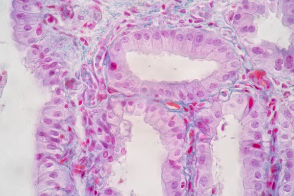
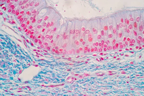
Characteristics Of Columnar Epithellum Cell (Cell Structure) Of Human Under Microscope View For Education In Laboratory.
Image, 16.38MB, 6720 × 4480 jpg
Tissue Of Small Intestine (Duodenum) And Vermiform Appendix Human Under The Microscope In Lab.
Image, 21.54MB, 6000 × 4000 jpg
Tissue Of Small Intestine (Duodenum) And Vermiform Appendix Human Under The Microscope In Lab.
Image, 18.99MB, 6000 × 4000 jpg

Pathology And Histology Tissue Of Mouse, Rabbit, Cat And Cow Under Microscope.
Image, 34.2MB, 6000 × 4000 jpg

Pathology And Histology Tissue Of Mouse, Rabbit, Cat And Cow Under Microscope.
Image, 18.99MB, 6000 × 4000 jpg
Characteristics Of Columnar Epithellum Cell (Cell Structure) Of Human Under Microscope View For Education In Laboratory.
Image, 17.03MB, 6720 × 4480 jpg

Pathology And Histology Tissue Of Mouse, Rabbit, Cat And Cow Under Microscope.
Image, 34.94MB, 6000 × 4000 jpg

Pathology And Histology Tissue Of Mouse, Rabbit, Cat And Cow Under Microscope.
Image, 10.76MB, 6000 × 4000 jpg

Pathology And Histology Tissue Of Mouse, Rabbit, Cat And Cow Under Microscope.
Image, 16.84MB, 6000 × 4000 jpg
Characteristics Of Columnar Epithellum Cell (Cell Structure) Of Human Under Microscope View For Education In Laboratory.
Image, 17.31MB, 6720 × 4480 jpg


Tissue Of Small Intestine (Duodenum) And Vermiform Appendix Human Under The Microscope In Lab.
Image, 17.16MB, 6000 × 4000 jpg
Characteristics Of Columnar Epithellum Cell (Cell Structure) Of Human Under Microscope View For Education In Laboratory.
Image, 16.28MB, 6720 × 4480 jpg

Pathology And Histology Tissue Of Mouse, Rabbit, Cat And Cow Under Microscope.
Image, 20.89MB, 6000 × 4000 jpg
Pathology And Histology Tissue Of Mouse, Rabbit, Cat And Cow Under Microscope.
Image, 18.02MB, 6000 × 4000 jpg


Pathology And Histology Tissue Of Mouse, Rabbit, Cat And Cow Under Microscope.
Image, 29.99MB, 6000 × 4000 jpg


Pathology And Histology Tissue Of Mouse, Rabbit, Cat And Cow Under Microscope.
Image, 13.68MB, 6000 × 4000 jpg

Pathology And Histology Tissue Of Mouse, Rabbit, Cat And Cow Under Microscope.
Image, 20.61MB, 6000 × 4000 jpg
Characteristics Of Columnar Epithellum Cell (Cell Structure) Of Human Under Microscope View For Education In Laboratory.
Image, 18.67MB, 6720 × 4480 jpg
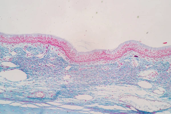
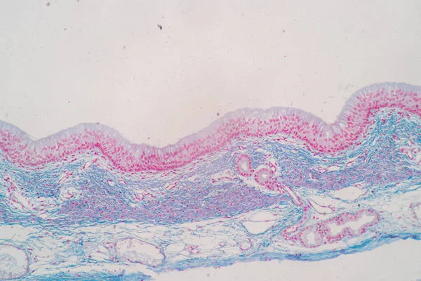
Cross Section Of Ciliated Epithelium Under The Microscope For Education Histology. Human Tissue.
Image, 14.75MB, 5168 × 3448 jpg
Epithelium. Squamous, Cubic, Ciliated, Glandular. Set. Infographics. Vector Illustration.
Vector, 1.56MB, 5000 × 5093 eps
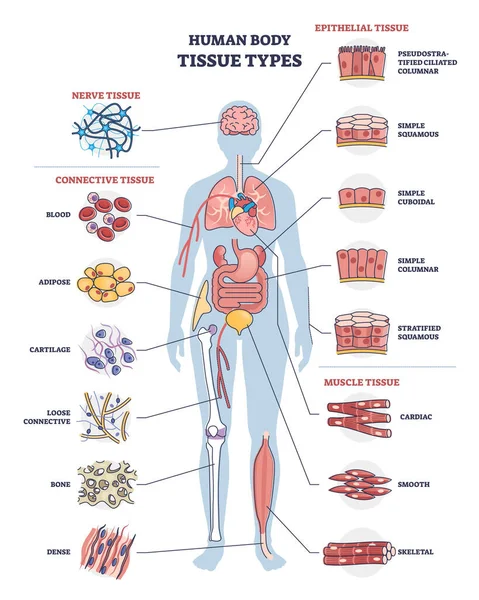
Human Body Tissue Types With Nerve, Connective And Epithelial Outline Diagram
Vector, 7.23MB, 3840 × 4800 eps

The Structure Of The Glandular Epithelium. Infographics. Vector Illustration On Isolated Background
Vector, 0.78MB, 5000 × 5000 eps
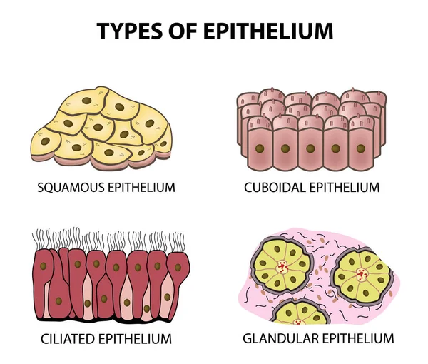
Types Of Epithelium. Squamous, Cubic, Ciliated, Glandular. Set. Infographics. Vector Illustration On Isolated Background
Vector, 2.27MB, 5000 × 4253 eps

Pathology And Histology Tissue Of Mouse, Rabbit, Cat And Cow Under Microscope.
Image, 32.96MB, 6000 × 4000 jpg


Pathology And Histology Tissue Of Mouse, Rabbit, Cat And Cow Under Microscope.
Image, 24.96MB, 6000 × 4000 jpg

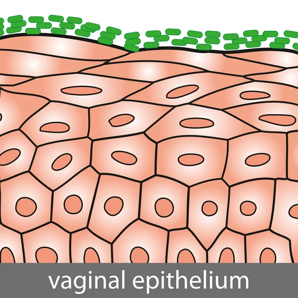
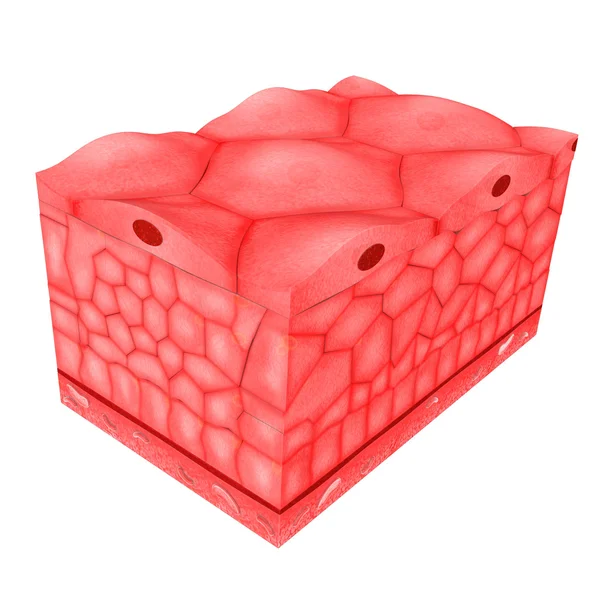
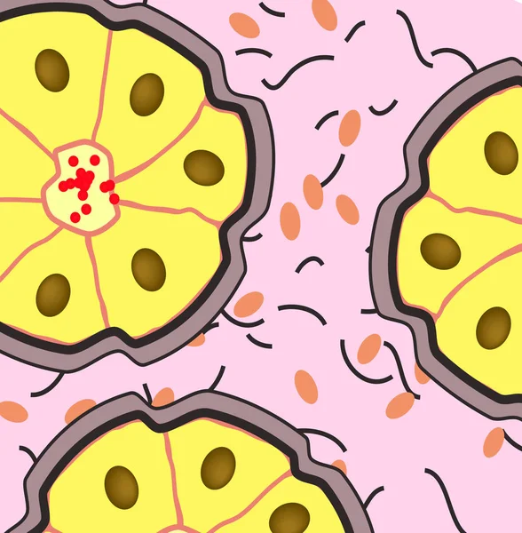
Pseudostratified Epithelium Is A Type Of Epithelium That, Though Comprising Only A Single Layer Of Cells.
Image, 11.24MB, 5840 × 3893 jpg

Pathology And Histology Tissue Of Mouse, Rabbit, Cat And Cow Under Microscope.
Image, 23.2MB, 6000 × 4000 jpg

Pathology And Histology Tissue Of Mouse, Rabbit, Cat And Cow Under Microscope.
Image, 30.02MB, 6000 × 4000 jpg

Pathology And Histology Tissue Of Mouse, Rabbit, Cat And Cow Under Microscope.
Image, 29.48MB, 6000 × 4000 jpg
Page 1 >> Next