
Stock image Pseudounipolar


High Magnification Micrograph Of Pseudounipolar Neurons Of A Dorsal Root Ganglion Stained With The Cajal's Formol-uranium Silver Method That Demonstrates The Golgi Apparatus. It Appears As A Brown Network Located In The Neuron Cell Body Around The Nu
Image, 5.13MB, 3840 × 3072 jpg
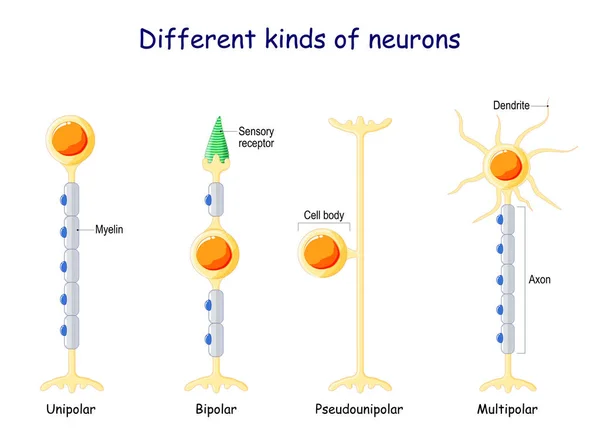
Different Kinds Of Neurons. Neuron Types: Unipolar, Bipolar, Multipolar, And Pseudounipolar Neuron. Vector Illustration.
Vector, 2.32MB, 5000 × 3665 eps

Types Of Neurons- Multipolar, Pseudounipolar, Bipolar - Structure Anatomy Colorful Illustration.
Vector, 3.94MB, 5000 × 5000 eps
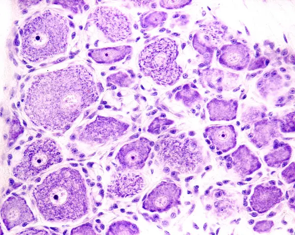
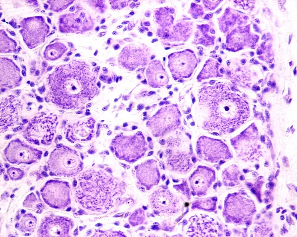
Light Microscope Micrograph Showing Neurons In A Dorsal Root Ganglion Stained With Cresyl Violet. They Are Pseudounipolar Neurons Of Rounded Soma Showing Small, Thin Nissl Bodies Characteristic Of Sensitive Neurons And A Large Nucleus With A Prominen
Image, 10.74MB, 3840 × 3072 jpg
Light Microscope Micrograph Showing Neurons In A Dorsal Root Ganglion Stained With Cresyl Violet. They Are Pseudounipolar Neurons Of Rounded Soma Showing Small, Thin Nissl Bodies Characteristic Of Sensitive Neurons And A Large Nucleus With A Prominen
Image, 9.97MB, 3840 × 3072 jpg

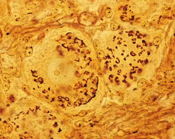
Light Micrograph Showing The Golgi Apparatus In Neurons Of Dorsal Root Ganglion. Cajal's Formol-uranium Silver Method. The Golgi Apparatus Is Distributed Throughout The Cell Body Cytoplasm Around The Nucleus.
Image, 15.85MB, 3840 × 3072 jpg

Low Magnification Light Microscope Micrograph Of A Dorsal Root Ganglion Stained With The Cajal's Formol-uranium Silver Method That Demonstrates The Golgi Apparatus In The Dorsal Root Ganglion Neurons. The Golgi Apparatus Is Distributed Throughout The
Image, 17.72MB, 3840 × 3072 jpg

High Magnification Micrograph Of Pseudounipolar Neurons Of A Dorsal Root Ganglion Stained With The Cajal's Formol-uranium Silver Method That Demonstrates The Golgi Apparatus. It Appears As A Brown Network Located In The Neuron Cell Body Around The Nu
Image, 9.6MB, 3840 × 3072 jpg


High Magnification Micrograph Of Pseudounipolar Neurons Of A Dorsal Root Ganglion Stained With The Cajal's Formol-uranium Silver Method That Demonstrates The Golgi Apparatus. It Appears As A Brown Network Located In The Neuron Cell Body Around The Nu
Image, 9.14MB, 3840 × 3072 jpg
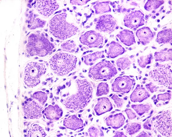
Light Microscope Micrograph Showing Neurons In A Dorsal Root Ganglion Stained With Cresyl Violet. They Are Pseudounipolar Neurons Of Rounded Soma Showing Small, Thin Nissl Bodies Characteristic Of Sensitive Neurons And A Large Nucleus With A Prominen
Image, 12.15MB, 3840 × 3072 jpg

High Magnification Micrograph Of Pseudounipolar Neurons Of A Dorsal Root Ganglion Stained With The Cajal's Formol-uranium Silver Method That Demonstrates The Golgi Apparatus. It Appears As A Brown Network Located In The Neuron Cell Body Around The Nu
Image, 10.8MB, 3840 × 3072 jpg
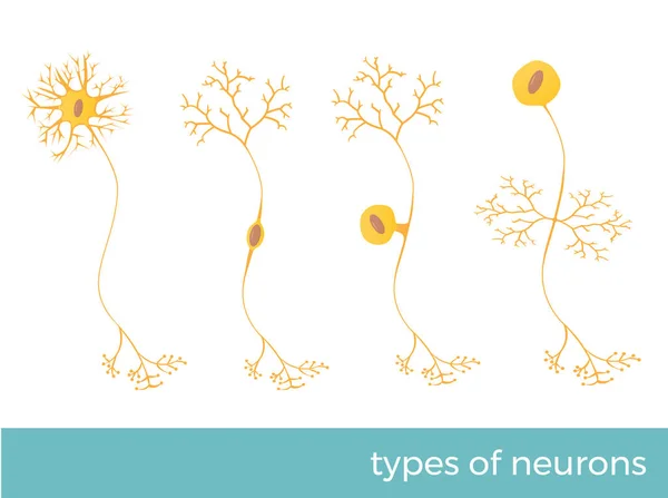
Types Of Neurons - Part Of Human's Central Nervous System. Vector Format Illustration.
Vector, 5.2MB, 8041 × 6000 eps

High Magnification Micrograph Of Pseudounipolar Neurons Of A Dorsal Root Ganglion Stained With The Cajal's Formol-uranium Silver Method That Demonstrates The Golgi Apparatus. It Appears As A Brown Network Located In The Neuron Cell Body Around The Nu
Image, 9.79MB, 3840 × 3072 jpg

High Magnification Micrograph Of Pseudounipolar Neurons Of A Dorsal Root Ganglion Stained With The Cajal's Formol-uranium Silver Method That Demonstrates The Golgi Apparatus. It Appears As A Brown Network Located In The Neuron Cell Body Around The Nu
Image, 9.43MB, 3840 × 3072 jpg
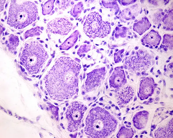
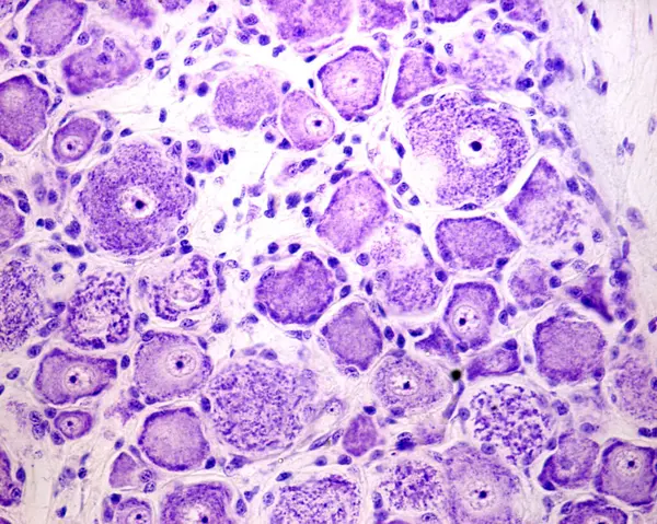
Light Microscope Micrograph Showing Neurons In A Dorsal Root Ganglion Stained With Cresyl Violet. They Are Pseudounipolar Neurons Of Rounded Soma Showing Small, Thin Nissl Bodies Characteristic Of Sensitive Neurons And A Large Nucleus With A Prominen
Image, 11.59MB, 3840 × 3072 jpg

High Magnification Micrograph Of Pseudounipolar Neurons Of A Dorsal Root Ganglion Stained With The Cajal's Formol-uranium Silver Method That Demonstrates The Golgi Apparatus. It Appears As A Brown Network Located In The Neuron Cell Body Around The Nu
Image, 10.98MB, 3840 × 3072 jpg
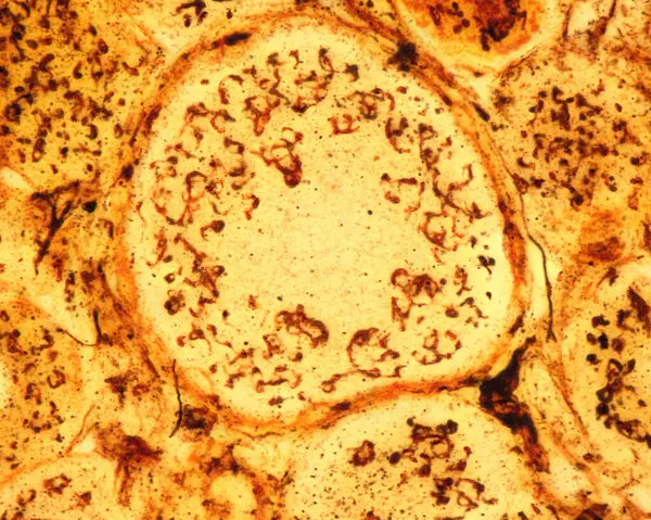
High Magnification Micrograph Of Pseudounipolar Neurons Of A Dorsal Root Ganglion Stained With The Cajal's Formol-uranium Silver Method That Demonstrates The Golgi Apparatus. It Appears As A Brown Network Located In The Neuron Cell Body Around The Nu
Image, 8.79MB, 3840 × 3072 jpg
High Magnification Micrograph Of Pseudounipolar Neurons Of A Dorsal Root Ganglion Stained With The Cajal's Formol-uranium Silver Method That Demonstrates The Golgi Apparatus. It Appears As A Brown Network Located In The Neuron Cell Body Around The Nu
Image, 9.35MB, 3840 × 3072 jpg

High Magnification Micrograph Of Pseudounipolar Neurons Of A Dorsal Root Ganglion Stained With The Cajal's Formol-uranium Silver Method That Demonstrates The Golgi Apparatus. It Appears As A Brown Network Located In The Neuron Cell Body Around The Nu
Image, 5.39MB, 3840 × 3072 jpg
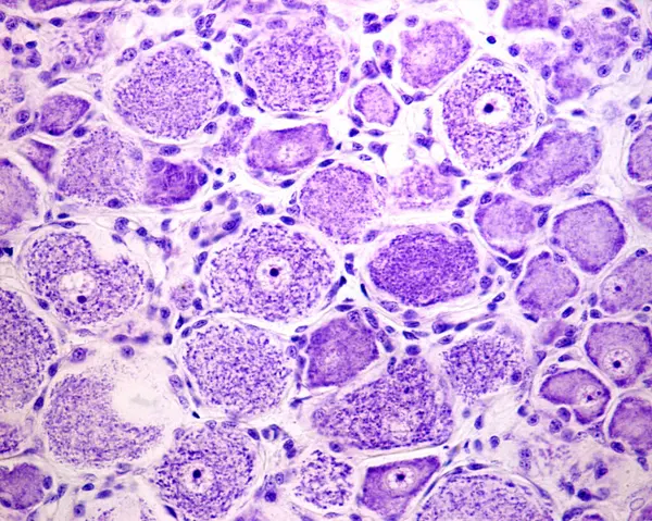
Light Microscope Micrograph Showing Neurons In A Dorsal Root Ganglion Stained With Cresyl Violet. They Are Pseudounipolar Neurons Of Rounded Soma Showing Small, Thin Nissl Bodies Characteristic Of Sensitive Neurons And A Large Nucleus With A Prominen
Image, 10.51MB, 3840 × 3072 jpg
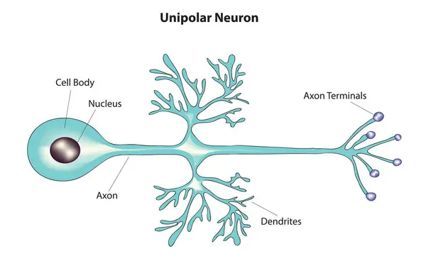
Unipolar Neuron, Unipolar Neuron Anatomy, Unipolar Neuron Structure, Unipolar Neuron Diagram
Vector, 8.58MB, 5121 × 3229 eps

Light Micrograph Showing The Golgi Apparatus In Neurons Of Dorsal Root Ganglion. Cajal's Formol-uranium Silver Method. The Golgi Apparatus Is Distributed Throughout The Cell Body Cytoplasm Around The Nucleus.
Image, 9.83MB, 3840 × 3072 jpg
Light Microscope Micrograph Showing Neurons In A Dorsal Root Ganglion Stained With Cresyl Violet. They Are Pseudounipolar Neurons Of Rounded Soma Showing Small, Thin Nissl Bodies Characteristic Of Sensitive Neurons And A Large Nucleus With A Prominen
Image, 11.94MB, 3840 × 3072 jpg

High Magnification Micrograph Of Pseudounipolar Neurons Of A Dorsal Root Ganglion Stained With The Cajal's Formol-uranium Silver Method That Demonstrates The Golgi Apparatus. It Appears As A Brown Network Located In The Neuron Cell Body Around The Nu
Image, 8.53MB, 3840 × 3072 jpg
Page 1 >> Next