
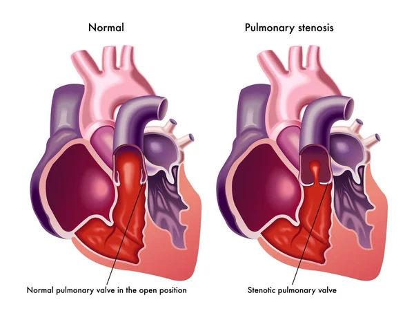
Stock image Pulmonary Stenosis

CTA Coronary Artery 3D Rendering Image Compare With Axial And Vertical Long Axis Plane From The Screen For Finding Coronary Artery Disease.
Image, 1.83MB, 3984 × 2596 jpg


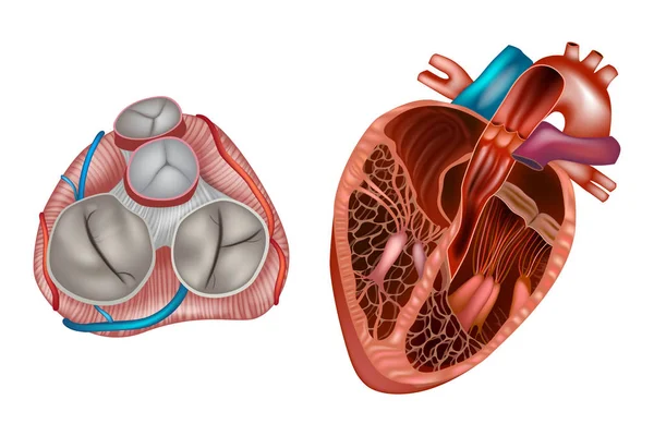
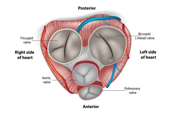
Heart Valves Anatomy. Mitral Valve, Pulmonary Valve, Aortic Valve And The Tricuspid Valve.
Vector, 11.68MB, 5000 × 3334 eps

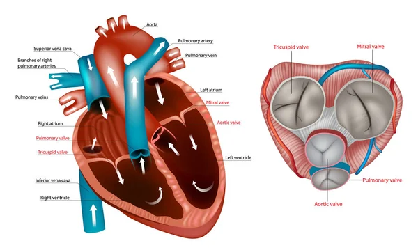
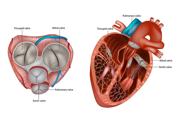
Structure Of The Heart Valves Anatomy. Mitral Valve, Pulmonary Valve, Aortic Valve And The Tricuspid Valve. Anterior Cut-away View Of Heart And Normal Circulation Showing Valves
Vector, 5.17MB, 6000 × 3600 eps

CTA Coronary Artery 3D Rendering Image For Finding Coronary Artery Disease On Blurred Screen Background.
Image, 2.12MB, 3736 × 2385 jpg
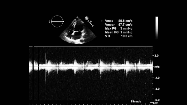
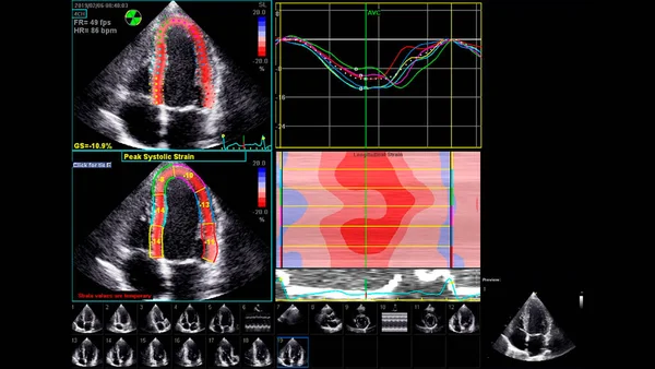
Image Of The Heart In Gray-scale Mode During Transesophageal Ultrasound.
Image, 1.9MB, 4000 × 2252 jpg

Image Of The Heart In Gray-scale Mode During Transesophageal Ultrasound.
Image, 2.34MB, 4000 × 2251 jpg

Angioplasty (or Balloon Angioplasty) Is An Endovascular Procedure To Widen Narrowed Or Obstructed Arteries Or Veins, Typically To Treat Arterial Atherosclerosis
Vector, 0MB, 4992 × 6128 zip

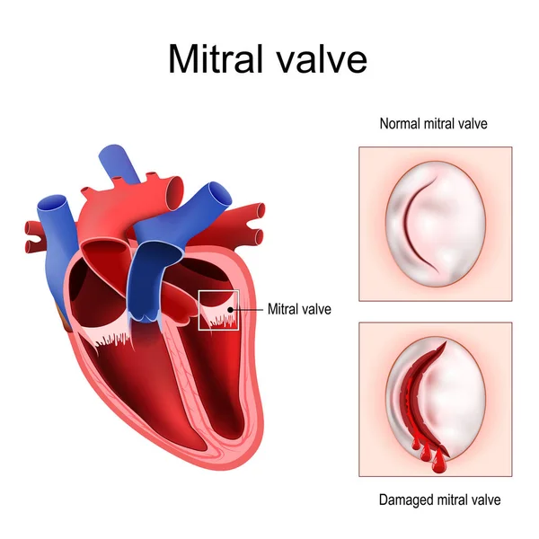
Heart Anatomy. Close-up Of Normal Mitral Valve And Damaged Mitral Valve. Cross Section Of Human Heart. Detailed Diagram. Vector Poster
Vector, 4.25MB, 4444 × 4445 eps

Structure Of The Heart Valves Anatomy. Mitral Valve, Pulmonary Valve, Aortic Valve And The Tricuspid Valve.
Vector, 10.89MB, 6000 × 4000 eps


Structure Of The Heart Valves. Mitral Valve, Pulmonary Valve, Aortic Valve And The Tricuspid Valve.
Vector, 9.69MB, 5000 × 3334 eps

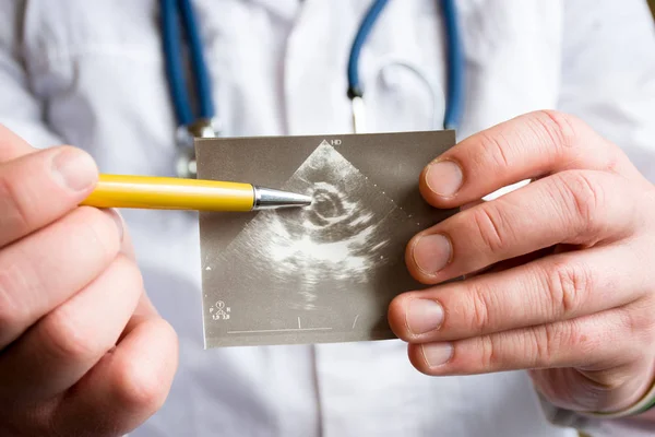
Doctor Holds Snapshot Of Ultrasound Of Heart And Indicates With Ballpoint Pen On Possible Pathology Of Heart Aortic Valve. Concept Photo Of Cardiac Ultrasonic Diagnostics Of Valve Apparatus In Adults
Image, 7.29MB, 6000 × 4000 jpg

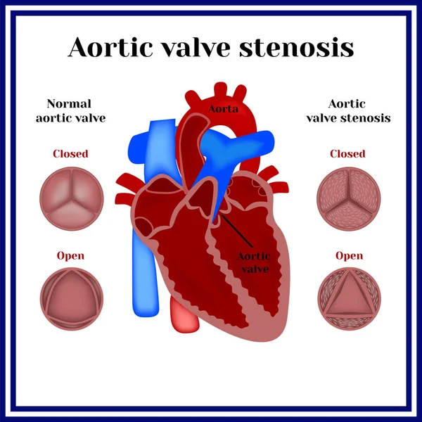

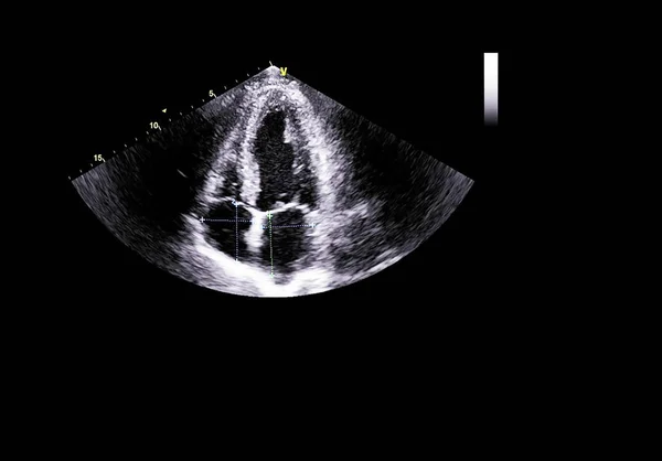
Image Of The Heart In Gray-scale Mode During Transesophageal Ultrasound.
Image, 1.07MB, 4000 × 2249 jpg


Lateral Ad Top View Of CTA Coronary Artery 3D Rendering Image Isolated O Black Backgroud For Finding Coronary Artery Disease.
Image, 10.15MB, 9290 × 6347 jpg
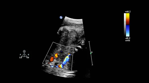


Image Of The Heart In Gray-scale Mode During Transesophageal Ultrasound.
Image, 1.13MB, 3500 × 1969 jpg


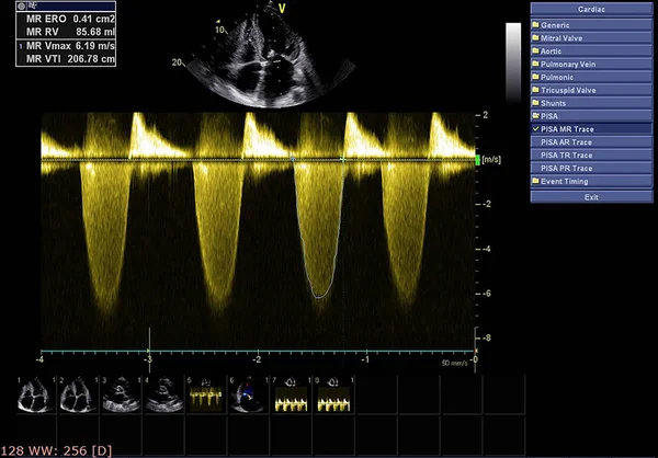
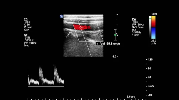
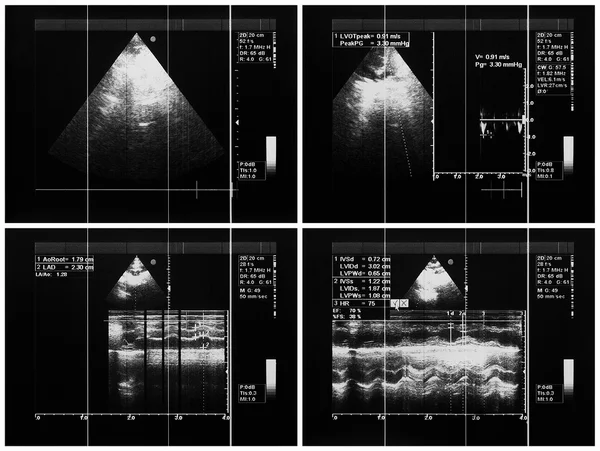
Image Of The Heart During Transesophageal Ultrasound With Doppler Mode.
Image, 0.61MB, 4000 × 2250 jpg
Angioplasty (or Balloon Angioplasty) Is An Endovascular Procedure To Widen Narrowed Or Obstructed Arteries Or Veins, Typically To Treat Arterial Atherosclerosis
Vector, 0MB, 4088 × 4264 zip

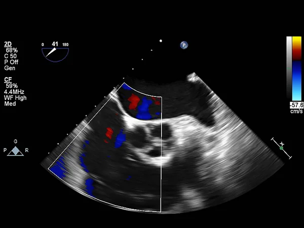


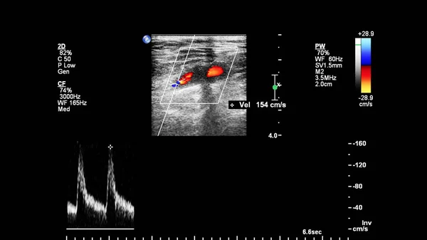
Image Of The Heart During Transesophageal Ultrasound With Doppler Mode.
Image, 1.66MB, 4000 × 2250 jpg
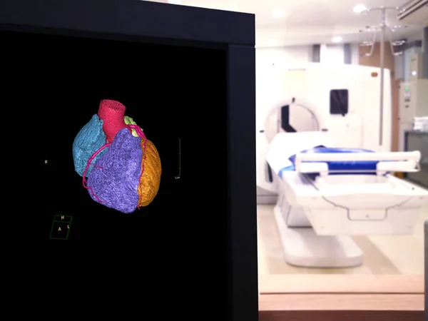
CTA Coronary Artery 3D Rendering Image On The Mornitor In CT Scanner Room At The Hospital.
Image, 8.55MB, 8192 × 6144 jpg

Image Of The Heart In Gray-scale Mode During Transesophageal Ultrasound.
Image, 0.86MB, 4000 × 2250 jpg


CTA Coronary Artery 3D Rendering Image For Finding Coronary Artery Disease.
Image, 1.54MB, 2430 × 2596 jpg
Page 1 >> Next